Are papillomas on the chest dangerous?
According to statistics, a large number of people suffer from HPV.
And you can live with this quite happily until it manifests itself. And he does this at the moment of weakening of the immune system, disrupting the measured order of our lives. Papilloma
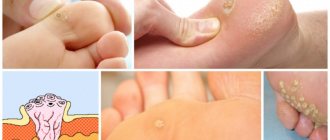
The presence of HPV in the body is indicated by the appearance of papillomas and warts. The development of viruses can cause the growth of many pathologies.
Papilloma on the chest
Papilloma on the chest is not always dangerous. In most cases, it manifests itself in the appearance of benign papillomas on the skin.
But there are also those that cause cancer. Breast papilloma can remain in a latent state for a long time (several years).
What to do if you notice changes and unpleasant symptoms? Growths can grow not only on the surface of the epidermis, but also inside the gland ducts. It is impossible to detect them in a timely manner through visual inspection. It is recommended to periodically conduct breast examinations to identify abnormalities and neoplasms.
The presence of a growth inside the mammary gland is indicated by a painful round nodule that is felt when pressed. Such neoplasms are found in large ducts near the nipple and in the area of the areola.
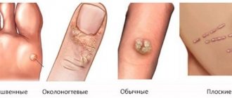
Papilloma on the chest can have different appearances and colors:
- dark brown to flesh-colored wart;
- thick base with soft round growth;
- a group of small “papillae” or single ones;
- growths of different diameters, attached to the skin with a thin stalk.
The location may also vary. Neoplasms grow on the side of the mammary gland, are observed under or above the breast, on the nipple and inside.
It is important to do this quickly if you notice dangerous changes:
- discharge from the nipple mixed with blood;
- changes in the density and structure of the growth;
- external changes in color, diameter;
- the appearance of pain at the site and throughout the mammary gland;
- increased nipple sensitivity.
Docteka
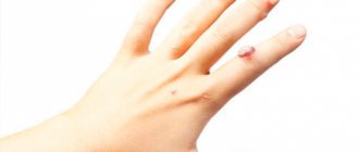
Wart on the chest: detect and treat!
The formation of true warts is promoted by the activation of the human papillomavirus (HPV). As a rule, the formation of skin formations occurs at the end of a long painful process (viral infections, postoperative period). At this time, the body experiences a “deficiency” state that requires immunomodulatory support.
The development of infectious activity of HPV does not have strictly defined boundaries. As a result, the appearance of a wart can be noted on any part of the body.
In most cases, a wart on the chest is a small, round lump that is gray, brown, or flesh-colored. It is noteworthy that the occurrence of a skin neoplasm has “gender” characteristics: in women, the area of the nipple areola is most often affected, while men can find a wart on any part of the chest.
This is interesting: How long does cryodestruction of a plantar wart take?
How does infection occur?
- Through sexual contact
- During pregnancy
- In public places (bathhouse, swimming pool, toilet)
- Household contact (the virus can remain on the surface of furniture or household products for a long time)
- Self-infection (shaving, depilation)
The cause of growths on the skin is the papillomavirus. Infection with this virus can occur during sexual contact with an infected person.
This often happens in public places, such as a bathhouse, swimming pool, or toilet. Or from simply touching an infected object.
The virus will remain dormant until a suitable environment for its progression arises.
It often happens that papillomas on the breast occur during pregnancy. Firstly, hormonal levels change. Secondly, during nine months of pregnancy, the body becomes very pregnant and the immune system weakens.
How are skin tumors treated?
Papillomas under the mammary glands occur after infection with HPV. Human Papillomavirus is widespread in the human population. According to statistics, about 60-70% of the world's population is infected with papillomavirus.
HPV is transmitted in three ways:
- Contact household (through towels, soap, toothbrushes, washcloths).
- From an infected mother to a newborn during the birth canal.
- Sexual transmission occurs after unprotected sexual contact.
The virus remains dormant for a long time. Under the influence of unfavorable factors, it is activated.
Papillomas appear on the neck, under the mammary glands, in the armpit; the following reasons contribute to HPV activation:
- Excessive sweating provokes a weakening of local immunity. Sweat irritates the sensitive skin under the mammary glands. A man scratches his chest area. Sores appear on it. The latter act as an entry gate for a dangerous virus.
- Wearing tight, uncomfortable underwear leads to chafing and damage to the skin. Through skin defects, the infection penetrates into the epidermal layer.
- Using someone else's bath accessories can result in a healthy person becoming infected with various diseases. Through washcloths and wet towels, the papillomavirus penetrates the skin and multiplies inside epithelial cells.
- Excessive passion for tanning and cosmetic procedures negatively affects the work of local and general defense mechanisms of the body.
- Bad habits (alcoholism, smoking, drug addiction), poor nutrition, hypovitaminosis affect the condition of the skin and suppress local immunity. Growths of various etiologies appear under the breasts.
- Problems with the endocrine and digestive systems are a favorable background for the activation of human papillomavirus infection.
- The appearance of papillomas can be triggered by hormonal changes in the body (during pregnancy, lactation, menopause, before each menstruation).
The appearance of external signs of papillomatosis under the breast requires consultation with a mammologist or oncologist. The specialist will examine the growth and prescribe appropriate laboratory tests and instrumental studies. A competent approach to treatment will allow you to find the cause of the disease and reduce the risk of developing complications of papillomatosis. The doctor will provide medical assistance in the early stages of the disease.
The list of methods for diagnosing papillomas under the mammary glands includes the following studies:
- OAC, OAM, biochemical blood test is carried out to identify inflammatory changes in internal organs.
- PCR is a modern research method that allows you to identify the type, type, and concentration of the causative agent of the disease. The material being tested is a scraping from the urethra or vagina. PCR is carried out in a special device - a thermal cycler. Works in two modes: heating and cooling. The material is placed in a thermal cycler and a certain temperature is set. Under the influence of specific substances, the hereditary information of the virus is copied. Experts compare the data obtained with the DNA in their database. The coincidence of the two materials indicates the relatedness of the viruses. In this way, doctors find out the etiology of the disease. PCR requires a highly qualified laboratory technician to reduce the likelihood of errors.
The main reason for the appearance of tumors on the skin is infection with papillomavirus. The infection enters the body at the moment of contact with the patient. This can be through sexual contact, through everyday contact, through touching a damaged area of the skin or the mucous membranes of infected body tissues. The papilloma virus does not manifest itself until a favorable environment for its reproduction appears.
The appearance of papillomas on the chest can be caused by various factors. Most often they begin to grow during pregnancy. The culprits are hormonal changes in the body. Over the 9 months of bearing a child, the immune system weakens and cannot resist infections. Papilloma occurs during pregnancy. During this period, you should observe her without resorting to radical treatment, which could affect the baby.
Growths begin to appear as a result of:
- neglect of hygiene standards;
- physical injuries to the chest;
- wearing uncomfortable, tight clothing for a long time;
- the appearance of microcracks in the nipples and between the mammary glands;
- polycystic ovarian dysfunction;
- increased work of the sebaceous glands;
- taking oral contraceptives;
- menopause;
- stress;
- period of puberty.
There are many reasons for the development of abnormal epithelial growth; to identify them, you must consult a doctor. For successful treatment of papilloma under the breast, it is important to use surgical techniques, take medications, and eliminate the provoking factor.
The growths appear due to a weakening of the immune system; patients need its strengthening and stimulation. After this, you can plan how to remove the growth.
It is worth noting that if there are neoplasms, you can breastfeed the baby if this does not cause discomfort to the woman.
All growths must be removed surgically. This measure is mandatory and helps eliminate the risk of cells degenerating into cancerous ones. It is prohibited to get rid of growths on your own. If the head becomes detached from the stem, serious complications may occur.
Manipulations to remove papillomas on the nipple, neck and under the breast should be carried out by a specialist using modern equipment.
- Laser technology is widely used for elimination. It is safe for health, does not cause pain and does not require a long recovery period. After treatment, there are no scars or marks left on the skin of the neck and other areas.
- To accurately eliminate the manifestations of papillomavirus, the electrocoagulation method is also used. It is based on the use of high-frequency electric current, which cuts out the growth and seals the treatment site, preventing bleeding.
- Flat growths are eliminated using liquid nitrogen, which burns the formation to the base without scars or scars.
- Radiosurgery is a popular modern method. To cut small papillomas and large neoplasms, a special scalpel is used that emits radio waves, which allows you to carefully, quickly and painlessly get rid of the papillomas that have appeared. At the same time, the incision is cauterized, which eliminates bleeding and further relapses of the disease in this area.
- The endoscopic method is used to remove skin lesions on the chest and intraductal nodules. The surgeon makes incisions along the areola and inserts an endoscope, which allows you to monitor every movement and remove the papilloma inside or under the breast in women without unnecessary trauma. The material removed during the operation is sent for histological examination.
Is it possible to remove papillomas on the chest using these methods only? These methods are not used independently, since it is important to undergo a course of drug treatment as a supplement. This will help put the virus into a calm state and prevent relapse.
At the first symptoms of HPV, contact a medical facility. Only a qualified doctor knows how to remove and treat papilloma on the delicate skin of the chest in each specific case. Remember that there are life-threatening strains that cannot be neutralized by traditional methods. It is possible to remove papillomas and eliminate the virus in a short time if you undergo the appropriate studies and adhere to the planned treatment plan.
Article verified
by the editors
Etiological and risk factors for the development and stimulation of papilloma in the mammary gland are divided into the following categories:
- hormonal imbalance in the body against the background of pathology of the endocrine glands or external influences: thyroid pathology, polycystic ovary syndrome, autoimmune aggression, diabetes mellitus, history of abortions, miscarriages, obesity;
- weakened immunity during pregnancy;
- prolonged depression, stress and emotional outburst contribute to the appearance of growths (juvenile);
- bad habits: alcohol abuse, smoking, drug use;
- long-term fibrocystic changes against the background of nodular or diffuse mastopathy;
- menopause;
- breast surgery;
- heredity.
Normally, there are no symptoms, but as the disease progresses and complications develop, clinical symptoms may occur:
- discharge from the nipple varies in color (depending on impurities, secondary flora), consistency (viscous, liquid, with flakes from desquamated epithelium of papillomas). They can be serous in nature, transparent in color or white - like milk, with admixtures of a pyogenic component, and have a yellow-green color. When the vessels of the ducts are injured, the cyst ruptures, or the formation itself is destroyed, blood is released. Trauma is possible when breastfeeding a child, which will complicate further lactation;
- when touched in the chest area, pain occurs that has a stabbing nature;
- feeling of fullness in the chest, swelling;
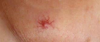
- local redness with signs of inflammation above the tumor;
- mastitis – when a secondary infection occurs, the pathological process provokes a temperature of up to 40 degrees, chills, severe shooting pain in the chest;
- general weakness, malaise.
What is the danger?
Human papillomavirus infection quickly spreads through the bloodstream throughout the body, which is fraught with multiple contamination - papillomatosis.
In addition, the pathological neoplasm causes discomfort in everyday life and spoils the aesthetic appearance. It is also easily injured by clothing or during hygiene procedures - bleeding occurs, and a secondary infection may occur with all the ensuing consequences (inflammation, suppuration, etc.).
The biggest danger that lies in common warts is the risk of degeneration of a benign growth into a malignant tumor.
Important!
Before removing a tumor, it is necessary to determine the type of pathogen, since some HPV strains initially have a high degree of oncogenicity. In addition, degeneration into cancer can occur as a result of ordinary trauma or inadequate treatment, remember this.
If characteristic growths appear in the breast area, immediately consult a doctor, mammologist or oncologist.
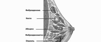
Papilloma on the chest and inside the chest
The formation of papillomas occurs with mastopathy and dilation of the ducts, with hormonal disorders. HPV has a tropism - it is targeted only at the outer layer of skin and mucous membranes that have contact with the external environment (air).
Mammary duct cystadenoma develops against the background of hormonal fluctuations. Localization can be unilateral, sectoral, or affect both breasts simultaneously.
Papilloma in the chest is represented by small round cavities inside the ducts. When several formations merge, volumetric papillomatosis occurs, which provokes multiple cystadenopapillomas.
Papillomatosis develops in the mammary gland mainly in adulthood, over 50 years. Growths more often occur in a group with blockage of the ducts (the proliferation of the epithelium closes the lumen of the duct); much less often, single types of small sizes are identified during diagnosis.
Solitary growths more often occur in the subareolar zone, and multiple ones - along the periphery of the mammary glands.
Groupings of lesions may contribute to the development of papillary cancer.
The appearance of gigantic, round-shaped growths due to papillomavirus is a reason for immediate contact with a breast oncologist. Such formations are dangerous with the risk of rapid malignant degeneration.
According to the ICD-10 classification, nomenclature code for the disease is D24.0 – benign breast formations.
Papilloma will not always be located on the surface of the skin. Sometimes it forms underneath, in the ducts of the mammary glands.
It is not always possible to visually check for the presence of malignant papilloma on the chest. Therefore, regular visits to the doctor are recommended to identify abnormalities.
Papillomas on the chest
The appearance of nodules in this area and an unpleasant sensation when pressing on the large ducts near the nipples also help to detect problems in the mammary gland.
The main symptom of a malignant tumor will be the release of fluid from the nipple. They can be abundant and small. Their color can be yellowish, milky, light green, brown.
Diagnosis of occurrence
The extent to which the treatment of papilloma that has arisen in the breast will be successful, and the patient will be able to completely get rid of all tumors, depends on the timeliness of diagnosis. Experts warn that the longer the growth remains in the chest or on its surface, the faster it will multiply and affect the healthy tissue around it.
The doctor will conduct a physical examination and order the necessary tests. If there is a suspicion that the tumor cells have degenerated into malignant ones, the patient will be advised to visit an oncologist.
Diagnosing papilloma inside the duct is not difficult; it is enough to use an ultrasound machine or perform a mammogram. X-ray examination is considered one of the effective methods for identifying nodules in the chest cavity. Thanks to it, you can confirm the presence of skin growths, accurately determine their diameter and location.
The woman must have a blood test taken. It will help identify the strain of papilloma on the chest. This step is important to rule out oncology and not waste time that could cost your life.
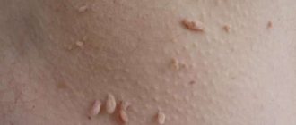
Locations
Neoplasms are localized on:
- nipples and halos;
- in the area under the breast;
- in the milk ducts.
Many papillomas on the nipples indicate a weakened body. They may cause discomfort when wearing underwear or not be felt at all. The appearance of such growths is also caused by disruption of internal organs or long-term use of antibiotics. Papillomas under the breasts in most cases cause pain.
Regular friction with clothing, underwear, sweat, and lack of air circulation leads to permanent injury. The situation is aggravated by poor hygiene and excessive sweating. Intraductal papillomas are difficult to identify by visual examination. They require the use of special equipment.
How to get rid of papillomas under the breasts using folk remedies?
When papillomas are detected on the chest, at the initial stage they try to cure them by using traditional medicine.
The main advantage of this treatment is that it does not harm the health of the body.
When treating at the initial stage of HPV, the following can help: garlic, onions, celandine.
They are crushed and compresses are made. Celandine can damage sensitive skin on the chest. In this case, it is recommended to mix its juice with baby cream or Vaseline.
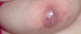
Complications
Possible complication
Sometimes papilloma on the chest can increase in size and become inflamed. The reason for this is stress, nervous shock, physical trauma to the chest, wearing uncomfortable underwear, and neglect of hygiene standards.
Very often this condition occurs after injury to this area. By touching or scratching, microcracks appear, which will be the place where microbes are attracted and the virus progresses in it.
After this, the papilloma may change shape and color, acquire an unpleasant odor and itching. Fluid may also begin to leak.
When to see a doctor:
- Possible complication
If you notice that fluid mixed with blood is released from the growth - Nipples become more sensitive
- Changed diameter and color
- There was pain when touched
- The structure of the growth has changed
With rapid progression and lack of adequate therapy, complications of breast disease are possible: degeneration into papillary cancer with metastases to internal organs (liver, ovaries, brain), blockage of the ducts with purulent melting of tissue against the background of a secondary infection.
The prognosis for treatment of breast papilloma depends on compliance with the following preventive measures:
- maintaining a healthy lifestyle without bad habits;
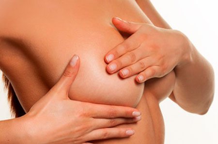
- regular annual examination by a gynecologist, the check can be carried out by a mammologist, breast self-examination is possible for preventive purposes;
- compliance with the treatment regimen for mastopathy;
- timely treatment of infectious diseases, gynecological disorders and hormonal imbalances.
If these rules are followed, the prognosis is favorable for life and recovery from pathology.
The article has been reviewed
by the site editors
The danger of human papillomavirus infection is the development of serious complications of the disease. These include the following conditions:
- Bleeding from papilloma under the breast occurs after damage to the formation.
- Tight underwear and mechanical combing provoke a violation of the integrity of the growth.
- Injured formations are an easy target for bacterial and viral flora. Suppuration of the growth is accompanied by unpleasant symptoms: pain, itching, local increase in body temperature.
Preventive measures help prevent infection with papillomatosis:
- Daily body hygiene prevents the colonization of the skin by viruses.
- Timely treatment of wound defects with antiseptic agents prevents HPV from entering the body.
- Treatment of chronic and acute diseases in the early stages prevents weakening of the immune system.
- Taking vitamin supplements as recommended by a doctor increases the body's resistance to infectious agents.
Reasons for appearance
The reasons for the appearance of growths under the breasts in women are varied. The main factor that provokes the appearance of tumors is a viral infection, which is transmitted sexually, through the use of personal belongings of an infected person, or through damage to the skin.
The appearance of rashes is also provoked by:
- lack of personal hygiene;
- weakened immune system;
- intraductal papilloma of the mammary gland;
- promiscuous sexual intercourse;
- unprotected sex;
- papillomas under the arms;
- Women wearing an inappropriately sized bra causes multiple papillomas under the breasts;
- frequent use of antibacterial drugs;
- regular stress;
- hormonal imbalance;
- Papilloma on the nipples in women appears during breastfeeding.
Most often, HPV is transmitted through contact with an infected person.
Removal methods
- Radiosurgery is a widely used method. The operation is performed using a special scalpel. The peculiarity of this device is that it easily helps to detect the growth. At the same time, he immediately cauterizes the incision. This reduces the risk of blood poisoning, as well as further growth of unwanted cells.
- Using a laser is a popular modern method. Does not require long recovery after surgery. Pain with this method is minimized. Does not leave scars on the skin.
- Endoscopy
Electrocoagulation. A high-frequency electric current is applied to the affected area. It cuts out the tumor and “seals” this area, preventing blood poisoning. - Endoscopic. This method is used to treat intraductal papillomas. The specialist makes incisions along the halo of the affected area. Then, using an endoscope, it penetrates into the tissues. This method allows you to accurately track every movement, as well as quickly and painlessly remove material. After it is removed, it is sent for histological analysis.
- Flat-type growths are cut out using liquid nitrogen. This method eliminates the appearance of scars.
Need advice from an experienced doctor?
Get a doctor's consultation online. Ask your question right now.
Ask a free question
Treatment of intraductal papilloma of the mammary gland includes several removal methods. Ductal formations are precancerous conditions. Depending on the size of the formations, the intervention can be partial (excision of gland lobes) or total (complete removal of the breast). Against the background of surgical intervention, conservative drug therapy is carried out in order to normalize hormonal levels and prevent the development of a secondary infection. Immunomodulatory drugs are prescribed to prevent relapse of papillomatosis.
The conservative method as the only form of treatment is permissible only in the presence of a single small formation without signs of growth.
The operation to remove papillomas in the area of the delicate tissue of the glands is carried out only under general anesthesia. For small growths, gentle surgical methods are used - resection along the periphery in sectors (incomplete excision with the application of a cosmetic suture). It is possible to preserve your own gland without losing the shape and size of the breast, without deforming the areola of the nipple, and an incision is made in the area around the nipple.
In the presence of voluminous growths with atypical growth of cancer cells, removal of the entire gland is used - mastectomy with excision of nearby tissue.
Among the methods of minimally invasive intervention for small growths of papillomatosis, laser destruction is used (rapid coagulation with a recovery period of up to 14 days). Hardware methods and surgery are aimed at preserving the functionality of the pectoral muscles responsible for the movement of the upper limbs.
The effect of various drugs, homeopathic medicines, including those for papillomas, is aimed at restoring the immune system in the body. Traditional methods of treatment are not used due to the high risk of malignancy and the lack of data on the clinical effects of home therapy. They use immunomodulators, antiviral drugs in the form of tablets, drops, ointments (Viferon, Ganaferon, Proteflazid, Immunoflazid).
Homeopathy and enzyme therapy in combination with traditional remedies for papillomas have a good effect. The effect of using a natural homeopathic enzyme is short-lived and the accumulation dose is not constant.
They take groups of vitamin-mineral complexes, antioxidants to strengthen the immune system and normalize metabolic processes.
Removal of papillomas under the breast is carried out in various ways: pharmaceutical preparations, folk remedies, hardware techniques. Any method should be discussed with your doctor. The specialist will prescribe the most appropriate therapy in each specific case.
Pharmacy drugs
- Supercelandine is a pharmaceutical preparation with necrotic properties. Cauterizes pathological growths. Papillomas under the breasts disappear a few days after using the drug.
- Papillectum consists of plant and synthetic components. The drug softens the top layer of skin and promotes the destruction of pathological structures. The medicine is available in the form of an emulsion. Apply to the formation using an applicator. After treating the growth, you need to wait until the emulsion is completely absorbed into the skin. The treated area should not be wet for 10 hours.
- Hydrogen peroxide helps to get rid of papillomas; it is necessary to treat the growth under the breast with a cotton swab dipped in the medicine.
- A drug called Duofilm is produced based on salicylic and lactic acids. Has a keratolytic effect. The solution is applied 1 time per day.
- Viferon ointment has two positive effects at once: it blocks the reproduction of papillomavirus and stimulates the body's defenses. You can lubricate the formation under the breast 4-5 times a day. Panavir, oxolinic ointment, has a similar effect.
- The drug Malavit acts primarily on flat papillomas. Reduces the symptoms of human papillomavirus infection.
- The medicine Stefalin eliminates birthmarks and warts. The product has a necrotizing effect. After a single application of the drug, a burn crust forms on the skin, which disappears over time, leaving a small wound.
- Surgical removal of papillomas in the chest area is performed with a scalpel under local anesthesia. Surgical intervention does not require special preparation. The surgeon cuts out the formation and sends it for histological examination. After removal, you should not sunbathe for 1-2 months. Otherwise, you can get an infection.
- Cryodestruction is a method of getting rid of papillomas under the breasts using liquid nitrogen. Cooling and thawing of the growth is accompanied by disruption of metabolic intracellular processes. After several procedures, the formations disappear.
- Laser removal of growths is carried out with a laser, which burns out the formations. The method does not cause pain, does not leave scars or other skin deformations.
- Radiosurgery is a universal method of removing formations with a radioknife. The device cuts off the growths. There is no bleeding or pain.
- Diathermoelectrocoagulation is a cauterization procedure using a needle electrode. Small crusts remain on the skin, which fall off within 1 week.
Folk remedies
- To treat papillomas under the breasts at home, a mixture of a solution of iodine, boric and acetylsalicylic acids is used. The listed ingredients are taken in a ratio of 1:1:1. Pour in 100 ml of medical alcohol. The resulting mixture is lubricated with the formations several times a day.
- An inexpensive lapis pencil is one of the proven remedies for warts and calluses. It contains silver nitrate, which cauterizes the formation. The product is applied 1-2 times a day.
- Celandine juice effectively rids the skin of papillomas. The growths disappear within a week. The pharmacy option is the drug “Superclean”.
- Benign growths can be lubricated with egg white. When it is completely dry, you need to apply another layer. In 5-7 days, the papillomas dry out.
- Grated garlic is mixed with rich cream. The resulting pulp is applied to the formation under the breast.
- The juice of wormwood and rowan has a cauterizing effect. The affected areas need to be treated 5-10 times a day.
- Some patients report a positive effect from treatment with banana peel. Removal occurs within 7-14 days.
- Dandelion cologne removes growths under the breasts due to its cauterizing and anti-inflammatory effects. The tincture is used to treat pathological areas every 4 hours.
- Tea tree oil promotes rapid drying of papillomas.
- Daily baths with chestnut nuts help get rid of formations in 2-3 weeks.
- You can apply aloe or Kalanchoe leaves to the papilloma. They are attached with a plaster for 4-5 hours.