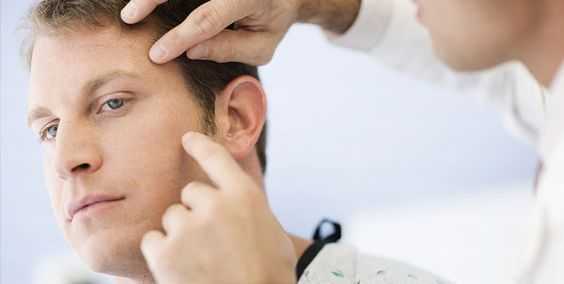
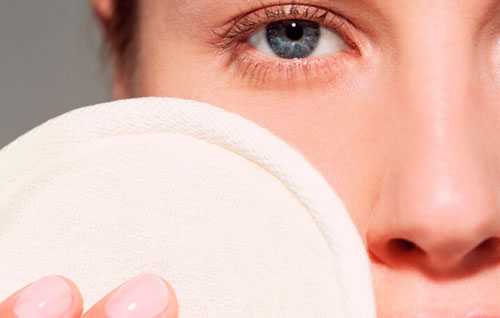
Red spots on the eyelids: allergic dermatitis and eczema
Allergic dermatitis (diffuse neurodermatitis) is a skin inflammation that occurs as a result of exposure to various allergens on the skin. Signs of allergic dermatitis are:
- redness of certain areas of the skin, in most cases it occurs on the eyelids;
- itching in the place where the spot appeared;
- bubbles with liquid appear;
- small ulcers may form;
- eyelids become swollen;
- It hurts to blink your eyes.
The disease can occur due to irradiation, exposure to temperature, exposure to chemicals (strong alkalis, acids), bacteria, and infections on the skin. That is, the allergen gets on the skin, which provokes an inflammatory allergic reaction. Substances that contribute to an allergic reaction can be found in cosmetics, hair dyes, washing powders, and detergents. Red spots on the eyelids, which indicate allergic dermatitis, can appear in people of all ages, including infants. There is a type of allergic dermatitis that occurs due to uncontrolled use of medications, and it is called drug dermatitis.
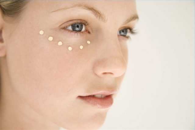
To treat red spots on the eyelids that are caused by this disease, third-generation antihistamines, a hypoallergenic diet, and desensitizing therapy are used. For local therapy and elimination of red spots above the eyes, corticosteroid ointments can be used.
Eczema very often occurs on the eyelids and is a skin disease. One of the signs of eyelid eczema is the appearance of red spots that itch. Both adults and children can get eczema. Over time, after the appearance of a red spot, peeling of the skin, itching, and inflammation appear. Eczema almost always occurs in an acute form, because people think that a red spot on the eyelid is a manifestation of an allergic reaction, so they do not seek help from a specialist. Eczema is not transmitted by touch. The causes of eyelid eczema can be the body's tendency to allergies, exposure to chemical elements on the skin, for example, nickel, chromium, dyes, fungal and bacterial infections, the fungus Malassezia furfur that has gotten on the facial skin, as well as impaired blood circulation in the eyelids.
Treatment of eczema is aimed at eliminating the cause of the disease and the red spots. For this purpose, hyposensitizing therapy, antihistamines, corticosteroids, and immunocorrectors are used.
If the red spots on the eyelids are very flaky and itchy, contact with water should be limited.
Treatment of red spots in the periorbital area
After identifying the source of the pathology, therapy is prescribed aimed at combating the “pest”:
- It is impossible to eliminate inflammation without surgery. It will help clean and rinse the abscess;
- If an allergic reaction or seborrheic dermatitis is detected, the doctor prescribes antihistamines (Gistan, Fenistil, Traumeel, etc.);
- Urolithiasis is treated by crushing stones or prescribing means to remove salts from the body;
- Increased pigmentation can be congenital or acquired. To combat it, cosmetic procedures are used.
| You will also need to take a vitamin course containing zinc, vitamins A and E. |
Treatment with medications
If you notice redness under your eyelids, immediately contact your doctor for a full examination. If it is not possible to visit the clinic, use several techniques to relieve unpleasant symptoms. First aid includes:
- Wash the damaged epidermis with chamomile decoction;
- Apply tea lotions to your eyes;
- Prohibit your baby from touching his face with his hands.
As soon as possible, go to the doctor so that he can make an accurate diagnosis and prescribe the correct therapy.
If you have been diagnosed with an allergy, take the following medications:
- "Elidel". Minimizes the manifestation of unpleasant symptoms, eliminates itching, relieves irritation;
- "Gistan". The product not only disinfects the skin, but also has anti-inflammatory properties;
- "Fenistil". Released in the form of a gel. Blocks histamine receptors, so allergies “die in the bud”, this will help avoid the appearance of red spots.
To combat dermatological pathologies, use the following medications:
- "Lokoid". Eliminates itching, swelling, inflammation;
- "Momat." The active substance mometasone has anti-inflammatory and antipruritic properties;
- "Advantan". Effective within a few minutes after application. Prevents the growth of pathology and thickening of the outer layer of the skin.
The following therapeutic drugs are also suitable for treating red spots:
- Antihistamines (Clariton, Telfast);
- Sedatives, they soothe irritated skin and block the spread of redness caused by stress;
- Enterosorbents. They have the unique property of “binding hands and feet” with allergens and toxins that can cause the appearance of red spots.
Traditional methods for treating red spots around the eyes
If the cause of the pathology lies solely in skin problems, you can try several recipes from your “grandmother’s” first aid kit:
- Potato mask. Wipe the tuber, place the resulting pulp on the damaged area, and leave for three minutes. The effect of the procedure is visible after the first session;
- Take dill seeds and place them in a cloth bag. Place in boiling water for five minutes, cool and apply to the reddened area;
- Combine ten grams of honey and wheat sprouts, put the resulting mass on the stains. After ten minutes, remove the composition and apply moisturizer;
- Pour twenty grams of chopped oatmeal with water and stir. Add some salt and apply to the damaged areas. Leave for ten minutes. If there are no serious abnormalities in the body, after three procedures the spots will disappear;
- Grate a raw cucumber, place the mixture on your eyes, and hold for fifteen minutes.
| You can use traditional recipes only after prior consultation with your doctor. |
Possible reasons
The main reason for the appearance of black dots on the eye is the destruction of the vitreous body. It occurs due to problems with the lens, insufficient blood circulation, which contributes to the destruction of collagen fibers with age and in serious diseases affecting the eyeballs. Fiber fragments become concentrated at one or more points in the lens, obstructing the passage of light, creating spots. Bad habits stimulate the manifestation of this pathology. Reasons why black spots appear in the eyes:
- crystalline formations;
- the appearance of malignant tumors in the eye;
- diabetic retinopathy;
- exposure to aggressive substances (acids, alkalis, etc.);
- entry of foreign substances and dirt particles into the eye;
- occurrence of injuries;
- migraine;
- long exposure to bright light;
- depletion of visual organs;
- other damage to the eye shell.
If black lines fly before your eyes, this may be a consequence of more serious pathologies. Such problems are not always immediately noticeable, but due to the fragility of the blood vessels they progress quickly. Peripheral vision deteriorates, areas of clouding in the visual organs grow, sparks and flashes may occur. It is important to see a doctor immediately and surgery may be necessary. Such symptoms may be the result of diseases:
- fungal or viral infections;
- hemophthalmos (bleeding into the vitreous body);
- vascular pathologies;
- tumors;
- retinal detachment or other damage;
- vitreous detachment.
To independently identify vision problems, look at a white or light-colored surface so that it occupies 100% of the visual area. If, in addition to the white color, there are defects, spots, or the appearance of some particles flying before your eyes, consult a doctor. You may not have a pathology, but it’s worth being safe. The doctor will make a diagnosis and prescribe treatment.
This symptom most often occurs as a result of aging of the body and worries people who have crossed the 50-year mark. The cause may be atrophy of the eyeball. If black spots in the eyes cause significant discomfort, you should visit an ophthalmologist: they can become a sign of retinal detachment.
Recently, ophthalmologists have noticed that the symptom has become “younger.” Young people are increasingly complaining about spots flashing before their eyes. This is due, first of all, to long-term exposure to electronic devices and gadgets on the visual system.
Other causes of blackheads include:
- Impaired blood circulation in the vessels. Intense physical activity, drinking alcoholic beverages, smoking, and frequent surges in blood pressure can lead to poor blood flow and damage to blood vessels. Bursted blood vessels often cause blood to thicken, which leads to dots flashing before the eyes.
- Lack of nutrients (especially vitamin A), metabolic disorders. Vitamin deficiency negatively affects all internal organs and is often the cause of black flies flying in the field of vision.
- Sometimes spots before the eyes can become a manifestation of cervical osteochondrosis and arise due to insufficient blood supply to the brain.
- Burns and traumatic damage to the organs of vision can lead not only to the death of vitreous cells, but also to retinal detachment, as well as loss of vision. Visual interference is also often observed when foreign objects enter the eyes.
- Black flies can be a result of exposure to pathogens that cause eye pathologies. This explains the presence of cloudiness before the eyes in some ophthalmological diseases.
- The appearance of black spots may be associated with overstrain of the visual system, while dots, circles and lines are seen by those people who constantly spend a lot of time at the computer or work with small details.
- So-called white spots before the eyes, as well as flashes of light, can be a sign of dangerous diseases of the visual system. These manifestations most often indicate pathologies of the lens, cornea or retina.
- Another reason is intoxication. When toxic substances enter the body, the patient has dots, spots or curved lines floating before his vision.
We invite you to familiarize yourself with the results of the analysis for some strains of HPV: what does it mean?
Also, provocateurs of an optical defect can be:
- migraine;
- diabetic retinopathy;
- tumors of the eye tissue;
- stressful situations;
- retinal disinsertion;
- vitreous hemorrhages;
- penetration of viruses and fungi into the retina;
- vitreous detachment;
- anemia;
- hemophthalmos;
- helminthiasis;
- diabetes;
- uveitis
Viral conjunctivitis
- Severe inflammatory reaction of the conjunctiva (swelling, redness due to vasodilation).
- Inflammation of the conjunctiva occurs almost simultaneously in both eyes
- Despite the pronounced inflammatory reaction, there is no abundant purulent discharge.
- As a rule, eye inflammation is accompanied by fever and inflammation of nearby lymph nodes.
There is currently no clear answer on how to treat viral conjunctivitis in adults. It should be remembered that treatment should be aimed at destroying the causative agents of the disease, which can be varied.
The basis of treatment is antiviral drugs intended for general and local use. Local medications include drops and ointments containing tebrofen or oxolin. And also an interferon solution.

Preventing the recurrence of milia
After treatment, it is very important to adhere to a number of rules. They minimize the impact of factors that may cause whiteheads to reappear. These requirements include the following:
Select and use hygiene products correctly. Cosmetics should be selected only in accordance with your skin type. Adjust your daily diet. In terms of nutrition, it is recommended to reduce the amount of foods with chemical dyes, sweeteners, fatty foods, smoked foods and foods with a lot of spices. Monitor the condition of the gastrointestinal tract. In most cases, white dots under the eyes occur precisely for this reason.

Take time to protect your skin from ultraviolet radiation during periods of intense sun. To do this, it is recommended to use creams with a filter of at least 30. Eliminate bad habits as much as possible. In some cases, whiteheads may appear due to excessive drinking or smoking.
Following these rules will not only help prevent the recurrence of milia, but will also improve your overall health. These measures will also help keep your skin in optimal condition.
Milia or white dot is a skin rash of a complex nature. It is important to remember that treatment of neoplasms should only take place under the strict supervision of a dermatologist and cosmetologist. It is the specialists in this field who will help you correctly determine why millet grass has arisen and how to get rid of the spots and prevent their reoccurrence.
Self-medication in this case can only aggravate the clinical picture and provoke the development of dangerous pathogenic processes at all levels of the dermis. Take care of your skin properly and be healthy!
How to warn
To prevent redness in the eyelid area, it is recommended to sleep at least 8 hours every day, and it is equally important to eat properly and nutritiously. In the summer, wear sunglasses to protect your eyes from ultraviolet exposure. It is worth controlling the time spent in front of the TV or at the computer, avoiding overstrain of the visual organs
You can avoid redness of the eyelids if you follow the rules of hygiene and do not touch your eyes with dirty hands. At the first unpleasant symptoms, contact an ophthalmologist
It is worth controlling the time spent in front of the TV or at the computer, avoiding overstrain of the visual organs. You can avoid redness of the eyelids if you follow the rules of hygiene and do not touch your eyes with dirty hands. At the first unpleasant symptoms, contact an ophthalmologist.
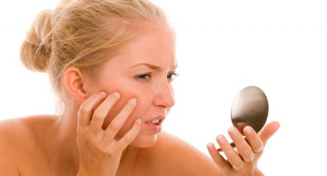
What to do if red spots appear under the eyes
Therapeutic tactics depend on the factors that cause red spots to appear under the eyes. If this is a stressful condition or lack of sleep, the person is recommended to normalize the daily routine, work and rest. Sedatives are prescribed, walks in the fresh air and sports are useful.
If the red spots do not go away, more serious treatment is required. It consists in the use of medicines and folk recipes. When red spots around the eyes in adults appear due to pathologies of internal organs, treatment should be carried out by therapists and specialized specialists.
Medication
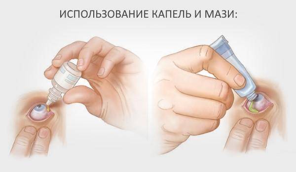
Requires the use of drops, ointments, and external agents.
- In case of an allergic reaction, oral antihistamines (Claritin, Zodak) must be prescribed. To eliminate external manifestations, you can use Gistan cream and Fenistil gel.
- Elimination of red spots near the eyes of a child can be done using Elidel cream. This is a non-hormonal drug that is effective for various types of dermatitis, including atopic.
- Intestinal sorbents are required - “Smecta”, “Enterosgel”. They help eliminate intoxication and improve skin condition.
- If helminthiasis is established, children are prescribed appropriate medications - “Nemozol”, “Pyrantel”.
Only a doctor can prescribe any medications.
Folk remedies
Effective in cases where changes in skin color are associated specifically with its pathology. Various wipes, lotions, masks are used:
- rubbing the skin of the eyelids with frozen parsley juice;
- eyelid mask made from grated raw potatoes;
- lotions from a decoction of dill seeds;
- applying slices of fresh cucumber to the eyelids.
Types of spots on the eyelid
Before prescribing therapy, each doctor will begin to differentiate the neoplasm. In this area, the skin is very delicate and incorrect diagnosis can provoke poor-quality treatment, which will lead to decreased vision and serious cosmetic defects. Spots on the eyelids are divided into several types, each of which has its own characteristics and requires an individual approach to treatment:
- freckles are small light spots that appear mainly in blue-eyed, fair-haired and red-haired people;
- chloasma – dark-colored spots that differ from freckles in their larger size;
- vitiligo – white spots caused by a lack of melanin;
- lentigo - convex dark spots that have clear boundaries;
- nevus - neoplasms of a round shape, can rise above the skin, and can differ only in color from the general skin;
- age spots – appear due to skin aging and prolonged exposure to ultraviolet radiation.
All of the above forms are classified as pigmented formations, but there are spots that can be caused by diseases and viruses; in medicine they are called acquired. This type of spots in the eye area can be characterized by swelling and severe redness of the skin.
Preventing the appearance of white pimples on the eyelid
To avoid the appearance of pimples on the face, as well as their spread, the following rules must be followed:
Cleanse the skin several times a day. Choose the right cosmetics. If necessary, it is better to consult with a specialist. It is advisable not to use foundation or greasy cream if you have acne. Consult a dermatologist if pimples appear and take the necessary tests.
Be healthy!
Probably everyone has encountered rashes on the face, and everyone knows several ways to solve this problem. In some cases, patients experience the appearance of an atypical white rash localized on the eyelids - milia.
The fight against these skin defects is somewhat more difficult than with typical acne, but it is still possible to solve the problem presented. In order for the treatment of white spots to be as effective as possible, it is necessary to understand what milia are and understand the specifics of therapeutic methods.
Treatment for itchy eyelids
Before starting therapy, it is necessary to eliminate all external factors that provoke peeling, itching and burning. Stop using cosmetics. If the doctor discovers that medications are the cause of the unpleasant symptoms, you will have to select analogues of the medicine or adjust the course of treatment.
Further therapy requires additional examination by several specialized doctors. Among the medications that are prescribed for the development of anomalies, medications based on panthenol have performed well.
| If it is determined that red, scaly spots on the eyelids are due to an allergic reaction, then the doctor will additionally prescribe antihistamines. In some situations, only hormonal medications can help get rid of unpleasant symptoms. |
Ointments and gels
If red spots on the eyelids itch and cause discomfort, then antiseptics and antibiotics are used to combat the anomaly. Most often, doctors recommend the following remedies:
- "Albucid". A solution intended for injection into the conjunctival sac of the organ of vision. Has antibacterial properties. The drug is universal; it is prescribed for the treatment of diseases of infectious and bacteriological origin, which are accompanied by itching and peeling of the eyelids.
- "Okomistin". Antiseptic drops. They effectively fight the majority of bacteria belonging to the gram-negative or positive class. They also work well against some viruses that cause itching and flaking.
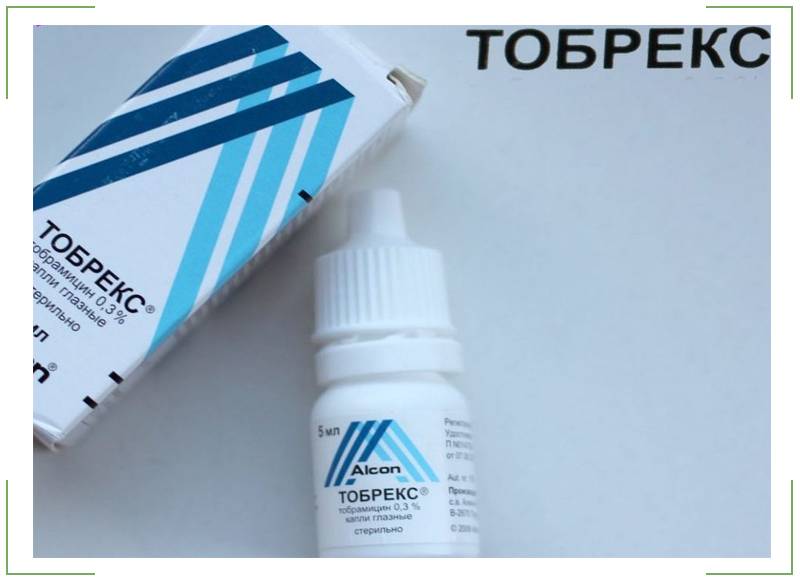
- "Tobrex". Broad-spectrum antibacterial drops. They belong to aminoglycosides. Effective in the fight against eye pathologies of an infectious nature. When taking the drug for a long time, its effectiveness decreases. This is explained by the development of resistance to the components of the product in pathogenic microorganisms.
- "Medeter." A combination drug that is both an antimicrobial drug and a corticosteroid. The drops are based on the active substance dexamethasone, which blocks inflammatory processes.
The main antimicrobial drug is Tobramycin. It is active against almost all bacteria that cause ophthalmic diseases.
Folk remedies
If a red spot appears on the lower eyelid, which is very itchy and flaky, then you can use “grandmother’s recipes” to relieve unpleasant symptoms. However, do not ignore drug therapy under any circumstances. Only with the help of medicines can you get rid of the pathology once and for all.
The following recipes will help eliminate peeling and itching of the eyelids:
Prepare a mask of honey, butter and bananas. The fruit should be overripe, with dark spots on the peel. In this state it has the maximum concentration of useful elements. Combine all ingredients in equal proportions and mix thoroughly. There should be no lumps in the finished mixture, so you should use a mixer to achieve a uniform consistency. Apply the composition to the eyelids and area around the eyes. Leave on for fifteen minutes, rinse off. Then apply moisturizer.

- Combine hot milk and oatmeal in equal proportions. Mix the ingredients well and cool to room temperature. Before using the composition, add butter to the mixture. Apply the mixture for fifteen minutes.
- Chop the parsley until the juice comes out. Then grind the grass additionally to obtain a mass of thick consistency. Apply the composition to closed eyelids and leave for fifteen minutes.
Treatment
Treatment of the disease consists of following a diet, namely limiting the consumption of foods containing animal fats. The main goal of therapy is aimed at eliminating the pathology that provoked metabolic disorders. The diet helps to correct the internal metabolism in the patient’s body. If the plaques do not go away with diet, your doctor may prescribe their removal.
For information! The formation of recurrence of white spots on the eyelids after removal is completely excluded.
Plaque removal methods include:
- Electrocoagulation - used together with the surgical method; after removing the white spot, its edges are connected to each other and cauterized with an electrode.
- Surgical excision - performed under local anesthesia, plaques are separated using tweezers and scissors. The edges of the spots are connected and lubricated with a solution of iron sesquichloride. The wounds heal within a week after the procedure.
For information! If the edges of the wound are peeled off, they are cauterized with electric current.
- The radio wave method is an absolutely safe procedure, carried out using high-frequency waves, where the instrument used is heated and tumor cells are evaporated with its help. This procedure is non-contact and painless.
- Cryodestruction - the method involves applying liquid nitrogen to the affected area. The temperature of the substance is -195C degrees, the time of application of nitrogen is determined by the attending physician. The main task of nitrogen is to destroy neoplasm cells.
- Laser therapy - the procedure is a gentle removal of formations without gross traces of intervention and injury to nearby tissues. Laser therapy eliminates infection of wounds, the formation of scars or marks after removal, and also eliminates the postoperative period.
READ MORE: Leg goes numb from hip to knee on the outside: causes, treatment
There is no specific treatment for xanthelasma. First of all, it is necessary to treat the underlying disease that caused the disease; special attention should be paid to disorders of fat metabolism, liver disease, and diabetes mellitus.
In medicine, there have not yet been any cases of yellow growths appearing on the eyelids disappearing on their own without any outside measures.
The key to successful treatment is to determine the cause of the pathology and eliminate it. This is the only way to guarantee that after removal the tumors will not appear again.
All drug therapy is selected according to the patient’s individual parameters and, as a rule, its main task is to normalize lipid metabolism.
Removing the yellow spots themselves can be done in the following ways:
- electrocoagulation;
- a liquid nitrogen;
- laser removal;
- surgical excision.
The treatment method is selected according to the characteristics of the patient’s clinical picture.
Regardless of the chosen treatment method, the recovery period, if everything was done correctly and the patient adheres to the doctor’s recommendations, passes quickly, since a minimally invasive therapy technique is used.
Among modern methods of removal, laser coagulation is considered the best. During its implementation, it is not necessary to use long-acting anesthetics, which significantly lightens the load on the body. The recovery process takes approximately 2 weeks.
The asymptomatic course of millet leads to the loss of valuable time when the process is still in the initial stage. A late visit to a specialist requires longer treatment and the appointment of other, complex treatment procedures. Therefore, we recommend that you pay close attention to the first white spots that appear under the eyes and study the following information in detail.
There are two directions. The first direction is traditional drug treatment, prescribed and carried out under the supervision of specialists, cosmetologists. The second direction is folk remedies.
Let's start with traditional drug treatment. Although the reasons that cause millet grass may be different, specialists have long had standards for its treatment in their arsenal. Usually the appointment is carried out in a beauty salon by a cosmetologist. Precisely a doctor. Under sterile conditions. Sterile instruments.
There are only three methods: invasive, non-invasive and hardware. The method, as well as the number and duration of procedures, is recommended and carried out by a cosmetologist.
The non-invasive method is usually used in the initial stages. This is an effect on the skin of the face in order to cleanse it and normalize the activity of the sebaceous glands. Cleaning the skin on the eyelids is done with weak solutions of fruit acids.
On other areas of the skin, more saturated formulations can be used. At the discretion of the cosmetologist, if the acne is located shallowly, a flash peeling is used. Single acne is removed mechanically, and for the treatment of multiple acne, a series of peels will be performed.
Paraffin masks have become widespread, helping to eliminate excess fat from the ducts of the sebaceous glands.
The invasive method acts directly on white lesions, that is, acne, with the aim of mechanically eliminating it. It is used at later stages. Mechanical removal of rashes is performed with a sterile medical needle.
The technique is quite understandable. By piercing the balls, the contents of the milia are squeezed out. Undoubted and strict adherence to hygiene rules and ensuring sterility is possible only in a beauty salon.
The surface of the skin is pre-treated with a special disinfectant containing alcohol, after which the procedure itself begins. The skin over the pimple is punctured. By pressing, its contents are brought out. Then the surface is disinfected again.
Electrocoagulation. Laser coagulation.
Electrocoagulation allows you to produce a targeted effect of alternating current on the problematic part of the face. The therapeutic effect is caused by cauterizing pimples with electric current using a special needle.
The procedure can only be performed by a doctor who knows the methods of hardware cosmetology. A thin needle acts directly on the pimple itself. In the same way, each nodule is processed and cauterized separately. The procedure is painless, cauterization marks quickly disappear.
A white pimple on the lower eyelid is less common. Self-removal of rashes in this area is not recommended.
Especially if the pimple is not milia. If the rash is of infectious origin, bacteria can be introduced into the conjunctival cavity.
When a specialist has confirmed the diagnosis of “millet”, the pimple can be carefully removed. To do this, the skin is treated with an alcohol solution and an antiseptic.
After this, it should be steamed in a warm room (sauna) and very carefully pierced the middle of the pimple with a thin needle. Then squeeze out the contents with two fingers.
This method is used if there are 1-2 rashes on the eyelids. If there are multiple milia, you should not remove them yourself.
Treatment for white acne should be prescribed by a dermatologist. Depending on the cause of the disease and the patient’s skin type, special cosmetics and antibacterial drugs are selected.
If milia quickly spread and increase in size, various ointments are prescribed (Tetracycline, Ichthyol, Zinerit). A cosmetologist can remove pimples.
For this, traditional methods (“cleansing” the facial skin) and laser procedures are used.
Usually, in order to get rid of white spots, it is enough to simply adjust the patient’s diet or replace hygiene and cosmetic products with more suitable ones. It is important to take vitamins throughout the treatment. They will help neutralize the impact of such causes as vitamin deficiency and maintain the general condition of the patient at an optimal level.
Electrocoagulation
Within the framework of this method, tumors on the eyelid and under it are excised by exposing them to high-frequency current. As a rule, the procedure is quick and painless under local anesthesia. After the procedure, a crust begins to form on the treated area, which disappears after 1-2 days.
After this intervention, small scars may remain in particularly sensitive areas. However, over time they will whiten and become less noticeable.
Laser excision
Removing milia using this method is similar to electrocoagulation. However, laser skin cleansing is considered less traumatic and painful. As a rule, after the intervention the skin remains completely clean and no scarring is observed.
Curettage
One of the most radical methods for removing white spots. As part of this intervention, the walls of the milium are carefully opened, and its contents (ball) are removed with a special spoon - a curette.
This method is one of the most traumatic and after it is carried out, obvious scars can form on the skin. This method is used quite rarely for the treatment of milia on the eyelids and facial skin.
READ MORE: Treatment of pinched sciatic nerve with drugs
Dysbacteriosis and demodicosis, hemangioma
If a person has an imbalance of beneficial and pathogenic flora of the intestinal tract, then red spots may appear on the eyelids near the eye. Dysbacteriosis occurs, which is characterized by stool upset, pain in the abdomen, and weight loss.
Typically, this disease develops during treatment of patients with antibiotics. But peeling before the eyes, along with redness and symptoms of intestinal damage, is characteristic of pathologies such as AIDS and blood cancer. Similar symptoms can also occur if there is a lack of beneficial bacteria in the gastrointestinal tract.
If you have this disease, you should immediately consult a specialist to make a diagnosis. Signs of eye damage will disappear after eliminating the causes that led to the development of dysbacteriosis. In this case, there is no need to treat the organs of vision separately.
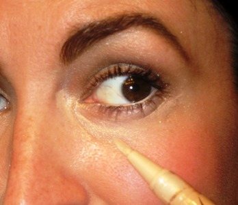
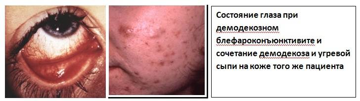
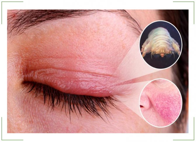
Demodicosis develops due to the strong proliferation of the skin demodex mite. The parasite is so small that it is almost impossible to see with the naked eye.
A tick can be identified because under its influence the eyelids become red and small dots appear on them. Along with peeling of the skin, small blood vessels dilate and bubbles develop in the affected area.
The patient complains of heaviness of the eyelids, and there is a disruption in the functioning of the sebaceous glands. The person feels burning and itching.
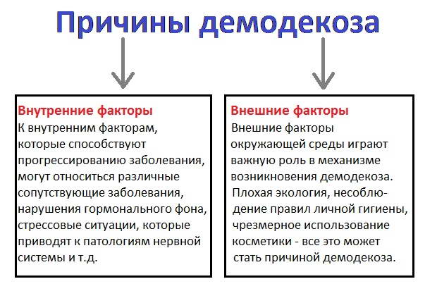
Typically, mites live peacefully in the sebaceous glands of 55% of people and do not cause any diseases. But under the influence of ultraviolet radiation, poor living conditions of the patient, a sharp increase in environmental temperature, and a decrease in human immunity, ticks can sharply increase their numbers.
If their number in one eyelid is no more than 1 copy, then the disease does not occur. When the number of parasites in the affected area reaches 4 units, then all the signs of the disease appear. The incubation period of the disease lasts from 2 to 12 months.
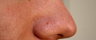
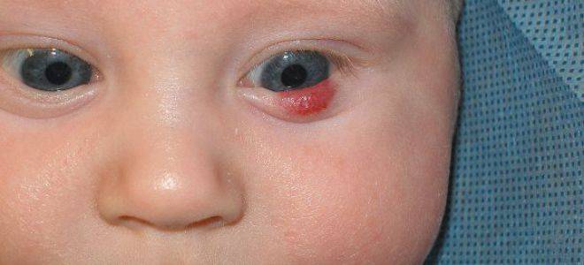
Hemangioma is another possible cause of the disease. This is a common disease that appears as red spots that look like moles. These spots are usually smooth, but sometimes have an uneven surface. These are benign vascular neoplasms that, under suitable conditions, can cause malignancy.
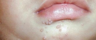
Definition of the concept of “milia” and the causes of their occurrence
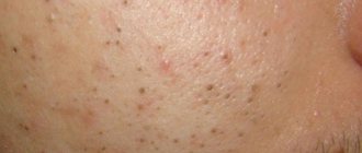
Before starting treatment for milia, it is necessary to find out the nature of this skin formation and the main reasons for its occurrence. Miliums (white dots, or popularly called “millet”) are a small neoplasm that is white in color and has a compacted structure resembling a ball. Visually, these pimples may resemble millet grains. Typically, milia are located in areas where the skin is most sensitive.
Basically, white spots under the eyes are not dangerous to the life and health of the patient. If the spot maintains its size for a long time, its location, color and structure do not change, it is not necessary to resort to an intensive therapeutic course. However, if any dynamics are noticed, you should contact a dermatologist immediately.
The mechanism of formation of milia is due to the fact that there is a disruption in the removal of sebum from the sebaceous glands due to clogging of the pores with keratinized particles of the dermis. Normally, excess fat should freely exit to the surface of the skin, transporting the secretion to the base of the vellus hair on the face. If this process is disrupted, the follicles become enveloped. As a result, white dots are formed.
Basically, the process of formation of white dots on the eyelids occurs for many reasons. They are individual in nature and directly depend on the general health of the patient and the quality of his skin. As a rule, if milia occur on the face, the reasons may be the following:
hormonal imbalance disturbance of the reactions of the sebaceous glands to hormonal surges autonomic disorders of the nervous system diseases of the gastrointestinal tract incorrect nutrition plan vitamin deficiency hereditary factor inappropriate hygiene and cosmetic products
In order for the treatment of white dots on the eyelid to be as effective as possible, it is necessary to correctly determine the cause of their formation. Unfortunately, it is impossible to do this correctly on your own.
To correctly determine the prerequisites for the formation of white spots under the eyes, it is recommended to consult a dermatologist or cosmetologist for advice. Doctors of this profile in an outpatient setting will be able to provide a comprehensive diagnosis of the dermis, identify milia on the face and the reasons for their formation, and prescribe the correct therapeutic course.
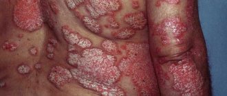
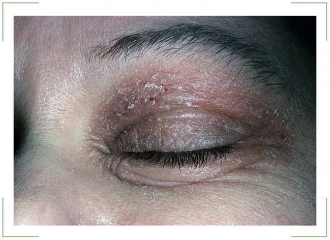
Psoriasis
Elements of this disease can be located on any part of the human skin, including the eyelids. A characteristic sign of the pathology is the formation of pink papules, the surface of which has a silvery-white surface and peels off. Psoriasis is accompanied by itching, but only the presence of characteristic signs can differentiate the disease from other dermatological problems. Such symptoms are the presence of a terminal shiny film, stearin stain, and pinpoint bleeding. The rash is accompanied by the release of a large amount of exudate, the surface of the wounds becomes weeping, which leads to the formation of crusts. Quite often, psoriasis first affects the scalp. The disease occurs over a long period of time.
If a red spot appears on the eyelid, treatment should be immediate.
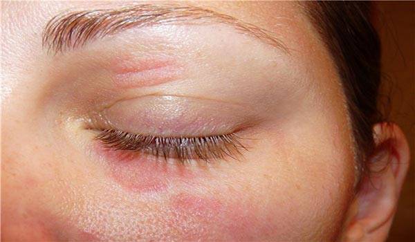
Demodicosis
This dermatological disease is also accompanied by the appearance of red spots in the eyelid area. Demodectic mange can appear when the skin is infected with a cutaneous glandular mite. The pathology is characterized by symptoms such as redness and swelling of the skin of the eyelids along the edges, the formation of scales at the base of the eyelashes, eye fatigue and itching. Examination reveals the presence of plaque covering the eyelid, as well as sticking of eyelashes, the appearance of crusts and slight hair loss. The disease is often accompanied by blepharitis, which affects the conjunctiva. And if a red spot on the eyelid peels off, what does it mean?
Traditional medicine to eliminate red eyes
In addition to treatment with traditional methods, you can consolidate the result with safe folk remedies.
At home use:
- Traditional medicine suggests getting rid of red spots that form on the eyes from fatigue with the help of any type of tea. Used bags are cooled beforehand. They are then applied to the eyes. This therapy lasts no more than half an hour. For the procedure, you can use loose leaf tea. It is brewed in a container to make it strong. Dip cotton pads into it and apply to the eyelids;
- To remove red spots, it is recommended to apply ice to the organ of vision. A cold object should be wrapped in a towel. This will not cause additional harm or hypothermia of the eye tissue. Afterwards it is necessary to evaluate the condition of the eyes. The red spot does not increase or small inclusions do not appear, then there is no cause for concern. Cold will stop bleeding from capillaries and blood vessels. Ice has no other medicinal properties. If other symptoms begin to appear and bleeding continues, you should consult an ophthalmologist;
- If red spots appear along with pain or pain, you can use potatoes. It is washed and cut into two equal parts. The peel is not peeled. The halves are applied to the eyelids. Stay in a lying position and carry out the procedure for 30 minutes. Before carrying out, you should prepare a towel. It is moistened in hot water and applied to the base of the neck;
- Any discomfort that appears can be eliminated with the help of pumpkin. It is not cleaned, but completely rubbed. The resulting pulp is wrapped in gauze. Then it is applied to the eye where the red spot is found. Therapy is carried out for 20 minutes. Then you can take a break and apply again;
- Kalanchoe leaves will help eliminate redness. Choose a young plant. The leaves are washed and ground. The resulting paste is applied to the eyelid area. The procedure is carried out within 20 minutes;
- To remove red spots from the whites of the eyes, use aloe juice. It helps to get rid of discomfort in a short time. The plant must be at least 2 years old. The juice is extracted from the leaves and 3 drops are placed in the eye with redness. If you don’t have such a plant at home, you can buy aloe juice at the pharmacy;
- At home, dill seeds are used. Use 1 teaspoon of raw material and brew with a glass of boiling water. Then leave for 40 minutes for the product to infuse. After time, filter through cheesecloth. This infusion is used as an eye lotion.
How to get rid of blackheads in eyes
To get rid of unpleasant rashes, several methods are used, but their essence is the same: opening the milia and removing its contents. The choice depends on the number and location of whiteheads, as well as their size and skin type. The effectiveness of all methods is the same, the difference lies in the cost and duration of the procedures.
- Mechanical removal. A dermatologist or cosmetologist, using a thin long needle or curette (a surgical spoon for scraping out soft tissue, removing suppuration, etc.), opens the milia and removes its contents. Before the procedure and after it, antiseptic skin treatment is carried out. At the site of each removed formation, small wounds remain, which soon heal. This method has its pros and cons. Of course, the advantage is the low cost of the procedure, but at the same time, mechanical removal is not suitable for people with thin or overly sensitive skin. Another disadvantage of this method is that in one procedure it is possible to remove only 10-15 milia, after which a break of several days is taken. Treatment can take quite a long time. Also, the mechanical method is not suitable if the white dots are located on the eyelids very close to the roots of the eyelashes. The skin in this area is extremely delicate, and exposure to it with a needle is undesirable, and noticeable redness will remain. Currently, this method is used only for medical reasons when other types of procedures are not suitable.
- Electrical coagulation is the destruction of milia by cauterizing them with high-frequency electric current. This procedure has much greater advantages compared to the first method. Using electrocoagulation, you can get rid of fairly deep formations, which is difficult to do with a needle or curette. But this method has its own characteristics: after cauterization, a dense crust forms at the site of the milium, which must be treated for 10 days until it falls off. Also, shallow scars may remain at the spots - small, but still noticeable.
- Laser coagulation is the most modern and safe method of getting rid of milia. It is suitable for large clusters of points, as well as when they are located too close to the organs of vision. To carry out the procedure, a CO-2 (carbon dioxide) laser is used, which has been successfully used in aesthetic cosmetology for almost three decades. It eliminates pathological formations layer by layer through thermal action. After its use, there is a lack of redness and suppuration, which can be observed in the case of a mechanical procedure, since the laser beam also has a bactericidal effect. In one or two procedures you can completely get rid of milia. Laser coagulation has the highest cost among the described types of treatment.
We invite you to familiarize yourself with Pityriasis versicolor in humans: photos, treatment, causes and symptoms

Vision is a key resource of the body, and you should not treat it with disdain. Treatment of black spots and dots with folk remedies is impossible; the problem itself also does not go away. You are allowed to drink herbs approved by your doctor; they will help you recover faster and prevent the recurrence of vision problems. Do not self-medicate - it is dangerous.
If you suspect the presence of black spots and dots, contact an ophthalmologist who will conduct an examination and confirm or deny the symptom. Contact a public clinic or a trusted private clinic. It happens that black spots arise due to another disease (tumor, infection, etc.), and in a private clinic you can be treated only for symptoms, without paying attention to the pathology itself that caused this condition.
During treatment in the hospital, existing defects are eliminated and the source of the pathology is sought. In most cases, it is possible to get rid of the disease that led to the appearance of black spots and dots, but it is not possible to clear the eye of defects. Dead cells are not removed from the vitreous body on their own.
In rare cases of floaters appearing before the eyes, for minor problems, vitamin drops are prescribed: Taufon, Quinax, ethylmorphine hydrochloride solution. Effective removal of dark spots is possible with the help of potassium iodide drops. If it is necessary to accelerate the regenerative component of the vitreous body, Wobenzym and Emoxipin are used.
If the disease is detected in the initial stages, it is recommended to take multivitamin complexes. The doctor prescribes examinations and procedures that eliminate the causes of vision problems. After getting rid of them, the black spots on the eyes completely or partially disappear on their own. If the disease is advanced, serious measures may be needed.
- Vitrectomy is a surgical procedure in which the vitreous humor is partially or completely removed. It is being replaced by an artificial environment. This extremely dangerous operation can lead to cataracts, retinal detachment, and hypotension. It is used only in cases where other methods have not produced any results.
- Vitreolysis - using a laser, the threads are broken, that is, clusters of dots are destroyed. The eye is completely reanimated. The operation is complex and is performed only by experienced doctors.
Possible causes of stains
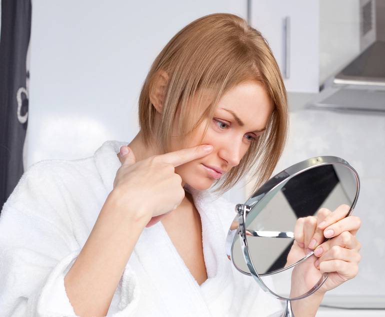
The reasons for the formation of spots on the eyelids can be very different. Among the common provocateurs for the development of atypical formations on the skin in the eye area are:
- avitaminosis;
- hereditary disposition;
- hormonal imbalance in the body;
- reaction to medication use;
- insect bite;
- reaction to cosmetics and hygiene products.
Very often, neoplasms itch, itch and are characterized by severe swelling. This significantly disrupts the patient’s usual life, and he strives to get rid of the tumor as quickly as possible, but this should not be done at home. Only if you are sure that you have been bitten by an insect, you can take antihistamines and delay visiting the doctor.
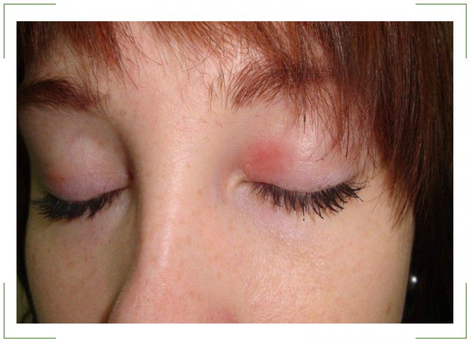
Red spots on the eyes
Red spots may indicate seborrheic dermatitis, allergies, demodicosis and hemangioma. Each of these diseases has accompanying symptoms by which you can recognize it. An allergic reaction, or, as it is commonly called, eczema, can occur due to irradiation, exposure to low temperatures and exposure to chemicals on the skin of the eyelid. The inflammatory process can also occur as a result of contact with aggressive substances, which are found in cosmetics, washing powders and detergents. Red spots can appear in both children and adults; eczema has no age preferences and manifests itself equally in everyone. It is painful for a person to blink, the eyelid swells greatly, waters, and the white gas turns red, vision may decrease, but the pathology has a short-term effect.
Violation of the intestinal microflora can manifest itself in the formation of red spots. It would seem very strange, but everything in the body is interconnected and pathogens that enter the intestines can manifest themselves in places with increased susceptibility. The skin on the eyelids is very delicate and thin, it is characterized by low local immunity, and accordingly turns red in the presence of a pathogen.
Seborrheic dermatitis can develop due to excessive activity of the sebaceous glands and lack of hygiene. The disease is most often observed in infants and men. You can recognize this dermatitis by the spots that are very flaky and peel off in whole layers. The skin around the tumor is very itchy, but does not swell.
Hemangioma is a benign neoplasm. Externally, it resembles a pigment spot or a mole of an unnatural shape. You can recognize a hemangioma by applying pressure. It turns white, then gradually returns to its original color.
If this factor is present, it is very important to visit an ophthalmologist, because hemangioma can degenerate into a malignant tumor, which requires intensive therapy
Yellow spots
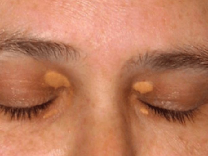
White and yellow spots are medically called xanthelasma. They are formed mainly in older people. Xanthelasmas are formed against the background of dysfunction of the stomach, liver and intestines. They can be recognized externally by their raised tubercles, soft and smooth to the touch. The formations have different sizes and shapes, it all depends on the intensity of the disturbance of fat metabolism in the body. The larger the tumor, the more complicated the situation. People with diabetes, obesity and food lovers with high amounts of cholesterol are at risk for the formation of xanthelasma.
Brown spots
Brown-colored formations are called chloasma; they can be transmitted genetically or develop against the background of diseases of the endocrine system, weak immunity, hormonal imbalance and chronic inflammatory processes in the body. This type of tumor is characterized by severe itching and irritation.
If a spot appears on your eyelids, regardless of its color, you need to go to the doctor, because this is not a healthy signal from the body about a malfunction. Treating such a delicate area at home will not lead to anything good. It’s stupid to lose your sight and become disabled because of your passion for traditional therapy.
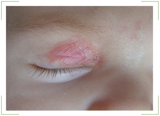
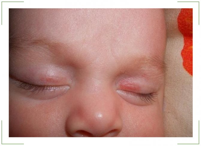
Any deviations from the norm in the condition of the newborn make parents wary. Their visual organs – fragile and sensitive structures – are especially worried. It happens that babies have red spots on or under their eyes.
Sometimes this is a normal physiological feature, sometimes it is a symptom of disease. In any case, this phenomenon cannot be ignored.
Causes of the disease
In most cases, the disease is diagnosed when the following pathologies develop in the human body:
- Conjunctivitis. In addition to redness of the eyelid, erythema of the mucous membrane of the visual apparatus is observed.
- Allergic dermatitis and eczema lead to damage to the skin of the eyelids.
- Kidney failure.
- Dysbacteriosis.
- Urolithiasis. In this case, not only redness is observed, but also dots on the surface of the eyes.
- Hemangioma.
- Lack of vitamin D in the body.
- Blepharitis. Staphylococcus aureus is considered the “initiator” of the pathology. Less commonly, the anomaly develops when the visual apparatus is damaged by other infections.
- Demodecosis.
| An allergic reaction to medications can also trigger the appearance of unpleasant symptoms. In this case, peeling and redness of the eyelids is observed in both eyes. All other ailments affect only one eye. |
Conjunctivitis
The most common cause of redness and peeling on the epidermis of the eyelids. The disease is accompanied by inflammation of the mucous membrane of the visual apparatus. The disease can be triggered by an allergy, infection by a virus or bacteria.
Characteristic symptoms of conjunctivitis:
- Swelling and redness of the mucous membrane of the eye.
- Itching and burning.
- Sensation of the presence of a foreign object in the visual apparatus.
- Intolerance to bright light.
- Difficulty opening the eyelids after sleep, as they are stuck together with purulent discharge.
- Decrease in visual acuity.
Additionally, patients complain of increased lacrimation and the appearance of exudate of purulent origin.
Allergic dermatitis
It develops when an infection enters the body or as a result of exposure to chemicals. When the irritant gets on the skin, it causes an allergic reaction. Girls often suffer from pathology due to the use of low-quality cosmetics. However, men and children are also not protected from the disease.
If red dots appear above and below the eyes, we can talk about the development of drug-induced dermatitis. Its appearance is due to the regular use of a certain group of medications. In addition to redness of the eyelids, the disease is accompanied by:
- itching and burning,
- the appearance of small ulcers,
- swelling of the lower eyelid,
- difficulty blinking,
- the appearance on the epidermis of vesicles filled with clear liquid.
Eczema and red spots
If a dark spot appears on the skin of the eyelid, there is a high risk of developing eczema. The cause of the disease can be dust, medication, or hormonal imbalance.
Clinical picture of the pathology:
- unbearable itching of the epidermis,
- swelling,
- pain when touching the eyelids,
- the formation of small red spots that are very itchy and flaky.
Return to contents
Dysbacteriosis
The occurrence of an imbalance of beneficial and pathogenic microflora of the gastrointestinal tract is often accompanied by the formation of a convex spot inside or outside the visual apparatus. The anomaly can be provoked by oncology, AIDS, or long-term use of antibacterial drugs. In addition to the appearance of redness, dysbacteriosis is characterized by:
- diarrhea,
- severe weight loss,
- the occurrence of discomfort in the stomach area.
Demodectic mange and red spots
Damage to the epidermis by subcutaneous mites is called demodicosis. According to scientific research, such parasites are present on the skin of almost every person. However, pathology develops only as a result of hormonal imbalance and a malfunction of the immune and endocrine systems.
Also, the cause of the disease is hidden in regular stress, the presence of chronic pathologies, and metabolic problems. Constant washing in public baths and saunas can also “awaken” the tick.
Demodicosis is accompanied by the following symptoms:
- swelling of the upper and lower eyelids,
- the appearance of red spots,
- itching and burning,
- eyelash loss,
- formation of pustules,
- peeling of the epidermis of the eyelids,
- inflammation of the conjunctiva,
- the eyes get tired quickly even with minor loads.
Hemangioma
A benign neoplasm, which in appearance is a red dot. The disease is dangerous because it is prone to rapid growth and causes functional discomfort.
At the initial stage of development of the disease, a slight swelling forms in the eye area and a change in the shade of the skin is noted. Touching the affected area causes pain. As the tumor progresses, it grows, which leads to a malfunction of the eyelids. The anomaly also causes cosmetic discomfort, since it is visible to the naked eye.
Red spots on the eyelids as a sign of hemangioma
Hemangioma is a benign neoplasm of vascular origin, which is localized anywhere, and can be located on the eyelid. If a hemangioma is present, it is imperative to consult an ophthalmologist, since it can grow through the eyelid. In some cases, some types of hemangiomas require mandatory surgical intervention in a specialized clinic. In other cases, vascular neoplasms require only observation and disappear on their own over time.
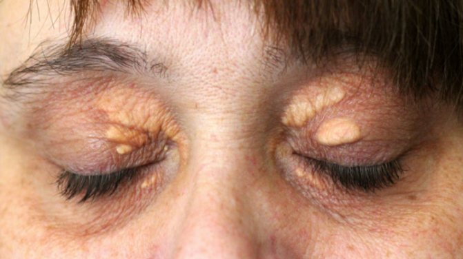
For quite a long time, medical specialists considered spots on the eyelids (xanthelasmas) as a cosmetic defect of the skin, and only recently their connection with heart health has been confirmed.
Yellow spots on the eyelids appear in middle-aged and older people.
Most often, this cosmetic defect can be found in women. The yellow color is due to the extensive accumulation of subcutaneous lipids.
Removing such stains is not difficult for cosmetologists.
For this purpose the following is used:
- electrocoagulation (electrical influence);
- laser coagulation;
- surgical removal.
However, even after surgery, the spots may appear again. Therefore, patients after cosmetic removal of xanthelasma are recommended to eat a diet of low-calorie foods with a low content of harmful substances and cholesterol. For such patients, sudden weight loss is recommended.
Xanthelasmas often signal diseases:
- intestines;
- stomach;
- liver;
- about the onset of atherosclerosis.
There is a direct connection between the level of cholesterol in the circulatory system and the presence of xanthelasma.
Such cosmetic disorders can be a symptom of various malfunctions in the body, especially malfunctions in fat metabolism.
A red spot on the eyelid may appear as a consequence of allergic dermatitis.
In such cases, patients are concerned about:
- weeping skin;
- cracks in the skin;
- severe itching;
- peeling;
- swelling;
- redness.
You can relieve inflammation for a short time with hydrocortisone. True, then everything starts all over again.
- do not eat chocolate;
- coffee;
- canned food;
- smoked meats;
- spicy dishes.
You can take the course:
- lactofiltrum;
- calcium pantothenate;
- digestive enzymes.
Externally, you can use tea lotions from repeated brewing. Dexamethasone eye drops can be applied to inflamed eyelids.
Xanthoma can sometimes have an orange tint or resemble straw in color.
Usually two or three small spots appear on the upper eyelid, near the inner edge of the eye. In rare cases, they can creep onto the bridge of the nose. And sometimes a yellow stripe with irregular boundaries may run across the entire surface of the eyelid.
Xanthomas appear quite often on the lower eyelid, but this inflammation rarely occurs in an isolated form. Quite often, the spot on the lower eyelid is located at the inner edge of the eye or is a continuous ribbon of spots. The surface of the skin on the spot is normal and smooth.
Xanthoma develops very slowly but steadily.
Xanthoma and xanthelasma are essentially the same. Their appearance is associated with disruptions in fat metabolism.
Xanthelasma of the eyelids is a flat xanthoma of the eyelids - a benign pale yellow skin plaque that appears on the upper eyelid from the inner corner of the eye. The size of the plaques can reach the size of a pea, the color varies from light yellow to orange, the plaques are soft to the touch, often merge to form a continuous spot, and sometimes extend to the bridge of the nose. Xanthelasma of the eyelids is often confused with wen or blisters, but xanthelasma is completely smooth, does not cause pain and practically does not protrude above the skin.
The word “xanthelasma” itself has Greek roots: “xanthos” - golden yellow and “elasma” - plaque, plate. Xanthelasma is equally common throughout the planet, but women suffer from it more often than men. In children, this disease is extremely rare; most often, xanthelasma of the eyelids occurs in middle-aged and elderly people and is considered a sign of atherosclerosis or a pre-infarction condition.
Symptom Definition
Flat single and multiple plaques of yellow color, located on the eyelids, the size of a pea to a bean, soft consistency; tend to merge and form lumpy elements. They occur in middle-aged and elderly people, more often in women. Appearing suddenly, they remain unchanged for a long time.
Xanthomas are straw-like in color, sometimes have an orange tint and protrude slightly above the surface of the skin. They are soft to the touch. Sometimes there are two or three small spots on the upper eyelid near the inner corner of the eye; in other cases they also extend onto the bridge of the nose; finally, in some cases, the entire eyelid is crossed by a yellow stripe of irregular shape.
- Yellow spots on the upper or lower eyelid, usually on the inside of the eye.
- The size of the spots ranges from a small pea to multiple continuous plaques throughout the entire eyelid with transition to the bridge of the nose. Typically the size of the plaque is 0.5-1.5 cm.
- The spots do not hurt, do not itch, or cause discomfort.
Flat multiple or single yellow spots, localized on the eyelids, have different sizes (from a pea to a bean) and a soft consistency. Often they merge, forming lumpy elements. As a rule, spots appear for no reason in women (less often in men) of middle or old age and remain for a long time without obvious changes.
The color of the spots resembles straw, sometimes with an orange tint, and protrude slightly above the skin level. Usually, 2-3 small bumps are found on the upper eyelid in the inner corner of the eye. In other cases, the spots may extend to the bridge of the nose or even cross the entire eyelid in the form of a yellow stripe of irregular shape.
Yellow spots may also appear on the lower eyelid, although in this case the lesion is not isolated. On the lower eyelid, yellow spots also appear at the inner corner of the eye, in some cases forming a continuous stripe. Their surface is completely smooth, with a normal epidermis and stands out only for its yellowish tint. The development of xanthomas is extremely slow and imperceptible, does not cause any subjective sensations, but continues steadily.
The formation of such spots is associated with lipid metabolism disorders. At the same time, in the case of their localized form, it is usually not possible to detect a violation of fat metabolism, although patients, as a rule, suffer from metabolic syndrome, diabetes or hypertension.
Xanthelasma plaques are soft and have a yellow or orange tint. Their surface is usually smooth, but can also be uneven. Several plaques may coalesce into a band that runs along the upper eyelid or lower eyelid. There is no pain or other unpleasant sensations from the appearance of plaques.
xanthelasma is a benign formation. It does not degenerate into a malignant tumor.
The appearance of xanthelasma may be associated with disorders in the body, especially in lipid metabolism. Plaques form slowly, without any accompanying symptoms. From a small pea they can sometimes grow to the size of a large bean.
If xanthelasmas are a manifestation of xanthomatosis, then it happens that they also affect the lower eyelid, on which xanthomas form. In this case, xanthomas are localized on the face, neck, knees and elbows, buttocks, and so on.
It should be noted that plaques persist throughout life, increasing in size and quantity.
Yellow spots can also form on the conjunctiva of the eye, and are called pinguecula and pterygium, depending on the location and growth of the tumor.
If the sclera of the eye does not have the usual white or pink-white surface, but a shade of yellow or even pronounced yellow spots, you should immediately be examined to identify internal diseases of the body.
In addition to changes in shade, often appearing spots are accompanied by pain in the eyeball area, decreased visual acuity, discharge, itching and photophobia. In addition, if the disease is general in nature, then the symptoms include lack of appetite, nausea, very rapid fatigue, body heat and chills.
As you can see, there are plenty of reasons for concern. In order for the cause of yellow eyes to be detected in time and eliminated, it is necessary to find time to contact a specialist at the early manifestation of such symptoms, since the causes of the disease can be hidden in any part of the body. To identify them, it will take quite a long period of time, tests, a full examination and determination of the treatment process.
Why do red spots appear under the eyes?
Common reasons why red spots appear under the eyes are as follows:
- Diseases of the kidneys and genitourinary area. Kidney failure and the presence of stones are often accompanied by swelling around the eyes; a person has swollen eyelids, which are most noticeable in the morning. The skin in this area is darkish with redness.
- Insufficient activity. Sitting for a long time at the computer, poor diet, lack of routine, bad habits (smoking, alcohol) - all these actions are reflected on the face in the form of redness and sagging skin around the eyes.
- Nervous tension, stress and chronic fatigue often lead to dark red spots under the eyes and can be complicated by conjunctivitis.
- Allergy. The appearance of red spots under the eyes, as well as on the cheeks, may be the result of an allergic reaction to exotic foods (citrus), pollen, medications, etc.
- Avitaminosis. Often, insufficient intake of vitamins leads to the formation of red spots in the eye area, which are itchy and flaky.
- Skin diseases. With some skin diseases, for example, eczema, dermatitis, etc., peeling, dryness, redness and cracking of the facial skin appears.
- Metabolic disease. Often appears in a child during adolescence, in women during pregnancy, menopause; accompanied by slight redness under the eyes.
- Inflammatory processes in the orbital area, such as abscess and cellulitis, can manifest themselves in the form of redness and increased body temperature.
Redness can also be a symptom of seborrheic dermatitis, the symptoms of which can be observed in the form of redness and peeling of the skin on the lower eyelid.
Red spots in children
If red spots appear on your child, you should immediately consult a pediatrician. Indeed, in this case, the appearance of spots or dots cannot be attributed to an incorrect lifestyle or overwork. In children, the appearance of spots under the eyes most often indicates that an infectious process or inflammation of the eyes has begun.
If, in addition to the appearance of red spots, the face swells, snoring appears, breathing through the nose becomes difficult, then this may indicate the growth of lymphatic tissue (adenoids) in the nasopharynx area. In this case, surgery can help. This cannot be left untreated, since lack of nasal breathing can lead to brain hypoxia.
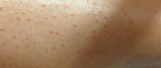
In young children, red dots similar to freckles may appear on the face. Such dots may appear in a child after a strong tantrum or after vomiting. They usually go away on their own and do not require any special treatment.
Sometimes redness under the eyes can be caused by an individual characteristic of the subcutaneous tissue. The child can eat well and sleep peacefully. In this case, there is no need to worry.
The reason for the appearance of spots may indicate a serious malfunction in the body or be a signal that the person is simply overtired.
Most often, this problem appears in children and adolescents, as well as in young women under 30 years of age.
We invite you to familiarize yourself with Drops for stye on the eye - 10 effective remedies
If red spots appear on your child, you should immediately consult a pediatrician. Indeed, in this case, the appearance of spots or dots cannot be attributed to an incorrect lifestyle or overwork. In children, the appearance of spots under the eyes most often indicates that an infectious process or inflammation of the eyes has begun.
Hemangioma and dysbacteriosis
The cause of spots on the eyelid is hemangioma, which is a benign formation on the skin of a vascular nature. It should be borne in mind that untimely treatment or its complete absence can lead to the hemangioma turning into a malignant formation. The presence of the disease is indicated by bright red spots with jagged edges, which appear most often on the skin of women. The disease develops due to the presence of various types of infections in the human body or unfavorable environmental conditions in the region in which he lives. During the treatment process, hormonal drugs are used, radiotherapy is used, and in exceptional cases, surgical intervention is possible. In this case, it is mandatory to undergo an examination procedure in an ophthalmology office.
The area around the eyes is quite sensitive to various influences, so red spots on the eyelids can form as a result of an imbalance in the ratio of beneficial and pathogenic microorganisms present in the intestines. In addition to abdominal pain and problems with stool, age spots appear that begin to peel off. The disease can be caused by:
- taking antibiotics for a long time;
- insufficient amount of beneficial bacteria in the intestines.
The reasons for the appearance of flaky spots in the eyelid area can only be determined by a specialist based on studying the results of relevant tests. The peculiarity of the treatment is to restore the balance between intestinal microorganisms. In order to avoid the appearance of dysbacteriosis, along with bactericidal drugs, it is necessary to take drugs whose action is aimed at restoring normal intestinal function.
Treatment of demodicosis
This disease develops as a result of damage to human skin by Demodex mites. The parasite is so small that it is almost impossible to see it without the use of optical instruments. The reproduction of ticks is facilitated by weakened protective functions of the human body, high air temperature, and the presence of bright ultraviolet radiation. At the same time, the eye vessels dilate, bubbles appear in the eyelid area, and the skin begins to itch. It takes an average of 3 months to fight the disease. During the treatment process, disinfectants are used, the action of which is aimed at relieving inflammation, and means to enhance immunity.
It should be borne in mind that treatment with medications must be supported by compliance with certain rules. The patient needs to make certain adjustments to his diet and stop drinking alcoholic beverages. Stressful situations that could negatively affect the treatment process should be avoided whenever possible. In addition, you need to fight inflammation by periodically moisturizing the skin in the eye area.