Cause for concern
In most cases, nevi are not dangerous if they are not subject to mechanical damage and do not change in size. Reasons for concern and contacting a doctor are:
- change in the size, color or structure of the nevus;
- bleeding from a nevus;
- pain, itching and discomfort of the skin around the spot;
- formation of erosion at the site of the nevus;
- hair loss;
- dryness and skin changes.
Stains located in the folds of the skin and areas of friction with clothing are often damaged. Such exposure can lead to discomfort and bleeding from the nevus. In this case, it is important to treat the wound in a timely manner, as well as make an appointment at a clinic where moles are diagnosed.
The damaged nevus must be removed for further examination of the cells for oncology.
Features of large moles
Large birthmarks in themselves are not dangerous to humans, but they are much more easily injured. This can happen due to negligence, and systematic damage to skin growths increases the risk of their malignant degeneration. It is the owners of large birthmarks, the diameter of which exceeds 6 mm, who complain that over time they begin to grow and itch.
Therefore, people with a similar problem need to contact the appropriate doctor, constantly monitor the condition of the formation and take care of it. In some cases, it is advisable to remove the birthmark, especially if it has increased in size or has become itchy.
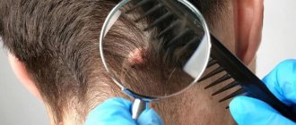
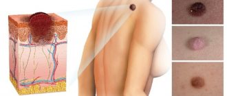
How is the verification carried out?
Damage to a nevus or spot does not always mean a risk of developing cancer, but it is impossible to predict what changes will follow in the cells. To protect yourself from possible risks of the growth of malignant cells, you need to consult with two doctors - a dermatologist and an oncologist.
The dermatologist will conduct an examination and, using special medical equipment, carefully examine the characteristics of each spot on the patient’s body. If necessary, the doctor will refer the patient to an oncologist who will perform epiluminescence dermatoscopy. This method allows you to visualize all the features of the birthmark cells.
Which moles cannot be dangerous
Removal of not all moles is necessary, because as such their potential danger may well be absent. This does not depend on the type and location of the nevus.
Pigment spots that are safe for humans can be identified by the following characteristics:
- the mole is completely flat;
- the diameter of the formation is small – up to 5 mm;
- color – dark brown or black with uniform shade;
- hair grows;
- the relief is identical to the skin.
Please note that even if one of the listed criteria does not fit, this does not mean that all types of moles should be removed immediately, because they will definitely develop into melanoma.
Please note that when neoplasms do not acquire changes for a long time, there is no cause for alarm.
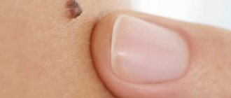
A normal mole is flat and small
Self-diagnosis
At home, it is recommended to carefully monitor the dynamics of changes in the structure of the birthmark. You can independently either check a mole for cancer or draw conclusions about the need to see an oncologist.
Thus, there are five signs on the basis of which a conclusion can be drawn about the degeneration of cells:
- asymmetry of the body of the spot;
- contour violation;
- color change;
- increase in size;
- dynamics of change.
A healthy, harmless nevus is always symmetrical. When cells degenerate, this symmetry is broken. For self-diagnosis, it is recommended to take a photograph and from time to time compare the mole on the body with the photograph taken.
A healthy birthmark or nevus always has a clearly defined, slightly rounded contour. A sudden change in the severity of the contour and its color is a reason to contact the clinic.
Signs of a benign course
It can be difficult to notice that moles have begun to grow on the body. This is possible when skin lesions have changed their size or shape over a short period of time. But usually nevi are capable of adding 1-2 mm in diameter per year, which is very difficult to notice.
The following signs indicate the benign nature of a birthmark:
- The skin formation is uniformly colored over the entire area. Its color can be anything from light flesh to black or red.
- The shape of the neoplasm is flat or slightly convex.
- Hair may grow on the surface of the mole, which is a reliable sign that it is benign.
- The surface of the nevus may be warty.
- Absence of any pain, discomfort or itching in the area of the skin near the birthmark.
Factors that provoke the degeneration of moles
Melanoma, or skin cancer, most often develops due to genetic predisposition. However, when moles are exposed to negative factors, there is a possibility of degeneration of skin cells. These factors include:
- mechanical damage to the nevus;
- exposure to ultraviolet radiation;
- exposure to aggressive chemicals.
By avoiding such negative effects, you can minimize the risk of developing cancer.
In cases where nevi are often damaged, for example, during shaving, it is necessary to consult a doctor about the possibility and methods of removing the birthmark. If for some reason removal is impossible or undesirable, it is recommended to cover the body of the mole with a bandage during depilation or showering.
You can protect yourself from the negative effects of sunlight by using special protective equipment with UV filters.
Moles located on the hands are often exposed to household chemicals, which can negatively affect the condition of the birthmark. Wearing protective gloves will help avoid this.
What types of moles are there?
Nevi are classified into different types according to different criteria. The following types of moles can be distinguished:
- borderline - spots of a uniform dark shade, do not react to exposure to direct sunlight;
- epidermo-dermal - common birthmarks;
- complex moles – especially dark and raised ones;
- dysplastic - refers to formations that are transmitted hereditarily, have
- asymmetrical appearance, large diameter up to 12 mm;
- blue - dense formations with a smooth surface, without hair: in appearance
- nevi look quite unusual.
Formations that appear on the face are called intradermal nevus. Typically, this is a pigment spot with a diameter of 2 mm to 2 cm. It can be a flesh-colored mole or with a characteristic whitish tint. If such pigmentations do not bring obvious discomfort to the owner, then they do not need to be removed.
Complex moles are dark in color and have a noticeable bulge
Security measures
Any damage to a mole should not be ignored. If you notice blood, itching, or any other discomfort, the patient should consult a doctor to check the nevus for the risk of developing cancer.
Under no circumstances should you remove moles yourself at home. If the spot causes discomfort or changes, you should consult a dermatologist about its removal. The doctor will offer the patient several options for removing the nevus:
- surgical excision;
- laser excision;
- cryodestruction.
How moles will be removed depends on the size and structure of the nevus. As a rule, after removal of a mole, the tissue is submitted for histological analysis to eliminate the risk of cell degeneration.
If a patient has a large number of moles, it is recommended that he undergo an oncology examination and check-up with a dermatologist every year. The doctor will also help you choose sunscreen products for the skin to protect moles from degeneration under the aggressive influence of ultraviolet radiation.
Knowing how and where to check moles for oncology, it is important to carefully monitor the dynamics of changes in the structure of nevi and spots, and promptly consult a doctor for advice.
It is important to remember that any changes in nevi require urgent oncological analysis and examination of moles for the likelihood of cell degeneration.
Innovations in medicine: remote diagnostics
Some modern clinics, meeting people halfway who do not like to visit hospitals again, have developed a new method for diagnosing nevus using photography .
Thus, by sending a photograph of a mole that causes concern, you can receive an initial consultation about possible next steps for the person contacting the clinic.
But everyone who uses such a service needs to understand that a personal appointment with a doctor will give the doctor much more information in order to make a correct diagnosis. And diagnosis by photograph may become just an advertising ploy, so that you still visit a doctor and become a patient of this clinic, which has found such a non-standard marketing approach.
Having learned the entire list of specialists that may be needed for problems with moles, you should not try to treat them yourself. Timely assistance from specialists will help avoid sad consequences that no one needs. Monitor your health and at the first dangerous symptoms, visit a specialist in your field.
Subspecialty specialists
What to do if there are malignant moles on the body? Which doctor should I contact in this case? Most often, they are referred for consultation to an oncodermatologist. This doctor specializes in skin diseases caused by pathology. The most dangerous and fairly common disease is melanoma. After a thorough examination, the specialist decides whether to remove or leave the mole.
If the nevus is located in the breast area of women, then they should be examined by a mammologist. He also conducts all necessary studies, including histological ones. A consultation with a dermatologist and mammologist allows you to determine whether a mole is dangerous. However, it should be remembered that not every nevus can degenerate into malignant. However, a woman should be examined at least once every 6 months.
Self-examination of moles
You can check a nevus for a malignant nature yourself. Doctors advise conducting an independent examination once a month to record even minor changes. Inspection measures should be taken within the walls of a room with good lighting, where there is a large mirror. You need to arm yourself with a ruler, a small powder mirror, a notepad or tablet to write down the results of the check.
First you need to take off your clothes and examine your entire body for the presence of age spots and moles.
The following deserve special attention:
- feet,
- groin area,
- breast,
- armpits,
- hairy area.
If neoplasms are detected, data about their location, size, and external data are recorded in a special notebook. Every month a similar event is held to check for cancer.
40% of the fairer sex suffers from cancer in the lower extremities, and 50% of men suffer from cancer in the torso.
Causes of moles
Modern representatives of the medical field identify several causative factors for the formation of birthmarks:
- predisposition at the genetic level (representatives of several generations are likely to form these spots in the same place),
- hormonal changes (most often these are puberty, pregnancy, lactation, abortion, menopausal syndrome, each of the conditions requires careful and detailed monitoring),
- interaction with the rays of the sun (the effect on the skin of our luminary contributes to the variable stimulation of the formation of birthmarks, that is, in some situations they may not appear, and sometimes they may form in large quantities),
- undergoing diagnostic procedures (especially X-rays and fluorography),
- injuries in the skin (they themselves do not act as provocateurs of birthmarks, however, the solar radiation to which they are exposed entails the activation of processes that give rise to the appearance of moles on the surface of the skin),
- light skin along with an impressive number of freckles also increases the tendency to form brown, pinkish, red spots on the skin,
- The age factor also plays a role, because growths form in youth, but the most active phase is observed in old age, the skin during this period is most susceptible to various factors.
The treatment process begins strictly after determining the causative factor of the phenomenon.
Which doctor should I contact?
If you find a suspicious tumor on your body, you must seek medical help. In recent years, cancer has become widespread. Melanoma is an insidious skin cancer. The tumor develops from small pigmented growths, within which cells have degenerated into cancerous ones. To prevent the oncological process, professional diagnosis is required.
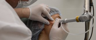
To begin with, the patient goes to a dermatologist. The specialist will conduct a physical examination without surgical or invasive intervention. The key procedure is dermatoscopy. Allows you to study the structure of the formation, determine its nature - papilloma or birthmark. The examination allows you to decide: remove or treat.
Thanks to dermatoscopy, it is possible to identify skin diseases and prevent dangerous processes on the skin. The microscope makes it possible to study the deep layers of the epidermis.
Doctors recommend examining suspicious growths on the body once every six months.
If negative dynamics are detected, the dermatologist refers the patient to remove the nevus.
Mole removal specialists
A mole is a harmless formation, but if there is a high risk of injury and if signs of malignancy are detected, removal is recommended.
Do not go to cosmetology clinics for this purpose! A cosmetologist is a specialist who has acquired knowledge about the types of skin and their health, and he did not always graduate from medical school!
A dermatologist surgeon or oncologist surgeon can provide qualified assistance. These specialists are qualified to destroy skin growths and have the equipment necessary to diagnose tissue obtained during surgery. To remove moles and papillomas, a strictly surgical procedure is required.
Depending on the indications and location (back, neck, groin, head, intimate genital papillomavirus), one of the following methods is selected:
- Laser. It is used in cases where the question of aesthetics arises. Allows you to keep your face and other open areas of the body clean, without scars. The beam evaporates the mole in 2-3 minutes, while simultaneously sealing the vessels. As a result, there is no long-term rehabilitation period. Painless, without blood and consequences.
- Diathermoelectrocoagulation. A special device emitting high-frequency electric current cuts and cauterizes the tumor at the base. The excised tissue element is sent for biopsy. The suspicious element is thoroughly investigated.
- Cryodisruption. The growth is exposed to liquid nitrogen, which causes cell death. Low temperatures cause the formation to darken and dry out. The problem may arise with large papillomas and moles. Repeated procedures are required.
- Surgical excision. It is prescribed in cases where the birthmark, papilloma, nevus is large or has begun to become malignant. The surgeon excises the tumor and applies cosmetic stitches. After healing, a trace remains.
Removing moles and other growths on your own is dangerous! Entrust the work to the doctor!
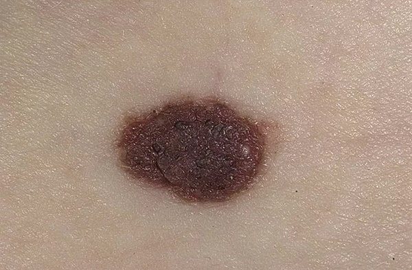
How can you recognize a dangerous mole?
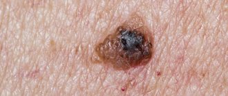
Bad moles can signal a possible risk of degeneration into melanoma. Melanoma is a malignant tumor characterized by intensive development at the site of nevus formation.
In order to determine whether a nevus should be removed or not, you should know which moles are dangerous to human health. So, moles with the following signs can be dangerous:
- with a diameter of more than 1 cm;
- skin lesions appeared over the age of 25;
- Over time, the shade, size and shape change.
A special technique has been developed with the easy name ABCDE to make it easier to determine how dangerous moles look and manifest themselves. A certain letter from the abbreviation corresponds to the symptom on which you need to focus your attention.
- A (assemmetry) – asymmetry of the mole. The nevus grows unevenly in different directions.
- B (border irregularity) – uneven edge. A nevus may have unclear, as if jagged, boundaries. Moles with smooth outlines are safe.
- C (color) – color. Normal pigment formations have a uniform shade. If color disturbances appear (splashes of red, blue, or a colorless nevus), then this is a signal of transformation into melanoma.
- D (diameter) – diameter. Pigmented areas with a diameter of 7 mm or more require examination by a doctor.
- E (evolving) – variability. You should pay attention to changes in all external features: color, shape, size. An atypical nevus is a clear symptom for further medical examination.
If you find these signs of dangerous moles in yourself, then do not delay visiting a specialist. It happens that the nevus begins to lighten or become soft, which indicates the start of the process of degeneration, then in this case it is better to talk with a professional in this matter.
Nevi that a person has had since early childhood are considered absolutely harmless. Other neoplasms must be approached with trepidation in order to notice the stage of degeneration of a mole into melanoma.
In addition to the characteristic features, you should pay attention to the parts of the body where the mole appeared. Nevi that arose in an “unfortunate” place are also a risk factor. This place could be the groin area, lower back or neck. It is here that a birthmark can cause bleeding, deformation, and subsequently the development of melanoma.
Changes in a mole should make a person immediately wary
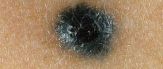
How dangerous moles appear
There are symptoms that indicate that nevi are turning into problematic ones:
- there is a shiny highlight on the surface of the mole;
- the process of deformation, which leads to asymmetry;
- hair loss;
- the beginning of the inflammatory process;
- the formation of a dry crust or obvious peeling;
- with minimal touching, blood begins to flow;
- unpleasant sensations: burning, itching;
- moisture appears at the site of formation.
In addition to melanoma, a mole can also signal that a nevus is developing. This is a small pigmented area, 3–10 mm in diameter, uniformly dark or black in color with uniform edges.
Why are large nevi on the skin dangerous?
Quite often, people develop moles of significant diameter, mostly red in color. In medicine, there is still no clear opinion about the causes of large-area nevi. Such age spots can serve as a symptom of a malfunction:
- large intestine;
- pancreatic secretion;
- lipid metabolism.
Similar formations appear on various parts of the body. The formation of a mole of significant diameter on the back can lead to significant discomfort. And pigmentation of areas on the head is often accompanied by unpleasant itching. In such cases, consulting a doctor is simply necessary. In addition to removal, the course of treatment also involves a number of preventive procedures that will consolidate the result.
The pancreas can signal the presence of a disease with moles
Diagnostic complex
The examination is carried out by one of the presented doctors. This could be a dermatologist, oncologist, surgeon. Regardless of which doctor is chosen, he must conduct a detailed survey specifying complaints, medical history, and also prescribe a set of auxiliary procedures. Through this tactic, it becomes possible to make a correct diagnosis and determine the correct treatment tactics.
- Dermatoscopy. This is a non-invasive research method characterized by increased safety. It involves the use of a specialized device - a dermatoscope. The doctor applies oil to the surface of the growth, and then carefully examines the nevus through the device.
- Computer examination. The technique involves taking an image of the tumor using a digital camera, as well as subsequent detailed processing of the data using a computer program. The system provides automatic verification and subsequent comparison of data with the existing database and displays the result.
- Histology. This study is mandatory and is aimed at studying cellular structures and tissues taken from the surface of the spot. Through analysis, it is possible to determine the nature of the formation and the degree of malignancy.
- Biopsy. Research involves taking material and further studying it. The doctor can carry out this tactic using an electric knife or taking a puncture using a thick needle. The material itself is examined under a microscope.
You should go to a doctor for diagnosis immediately, since only a competent physician is able to study the degree of danger, prescribe further therapy and provide assistance in getting rid of the nevus.