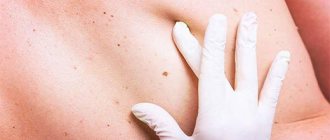
Consultation with a doctor
It is imperative to go to see a doctor if the birthmarks have increased in diameter so much that they only cause pain and inconvenience to the person when they come into contact with clothing.
Sometimes the pain and redness is only temporary, but don't let your body confuse you. After some time, all the unpleasant symptoms will return again, but at this time they will become much stronger and more unpleasant. Light skin, which has a large number of birthmarks, requires a more attentive and serious attitude. Deaths often occur when melanoma is detected late. Its main danger lies in the fact that metastases occur almost immediately, affecting the internal organs of a person.
Currently, modern diagnostics includes methods for identifying dangerous moles that are completely safe and painless for humans (good procedures are: x-rays, computed tomography, laboratory tests, biopsy). Large moles must be removed without fail to prevent any oncology from occurring. At the moment, their elimination occurs surgically with the use of radiation therapy.
Many of us think that melanoma is something terrible and scary, it is necessarily noticeable. However, in fact, even a small speck can develop into a dangerous tumor. However, it does not have to be huge. Very often its size does not exceed 5 mm and resembles a dark spot on the surface of the skin. In this case, there may not be any pain in this area.
Of course, most often melanoma begins to bleed and hurt. But usually this happens in the later stages, when treatment is not particularly effective, and the likelihood of a complete cure is minimized. Thus, only regular examinations, as well as constant monitoring of one’s own health and timely consultation with a doctor, contribute to recovery.
Melanoma
Thus, a specialist will be able to almost immediately identify a malignant neoplasm or a common nevus. The fact is that under a microscope, as well as a magnifying glass, all the bends and fractures, as well as the relief of the mole, are much better visible. Thus, melanoma can be detected quite quickly and easily at an early stage, which guarantees recovery.
Diagnostic options
Why do new moles appear?
Most of these formations appear due to stress, excess hormones or even unfavorable ecology in the city
The formation of neoplasms depends on many factors. Therefore, it is necessary to ensure that the causes that cause such a problem are eliminated. If you notice that there are a lot of moles on your body, you need to wear closed clothes, hats and, in general, hide in the shade on sunny days, especially in summer. Why do moles appear? Most of these formations appear due to stress, excess hormones, or even unfavorable ecology in the city. But a person begins to notice such spots only when it causes some discomfort: it hurts, itches, there is redness, or there is a sharp increase in growth. Consult a doctor who will conduct a full examination of such a formation and tell you how dangerous it is.
How to determine the oncogenicity of a tumor
Let's look at the main signs of malignant moles. First of all, various changes on the surface of the growth indicate the degeneration of birthmarks. Peeling, increase in diameter and change in structure are the main symptoms of nevus transformation. Symptoms such as itching, burning and tingling sensation in the area of moles are also important. Less commonly, various allergic reactions occur in those parts of the body that are adjacent to the growths.
Hair loss, color changes and the formation of papillomas are one of the clear signs of metamorphosis. Subsequently, new small-sized moles are added to the above-described symptoms, located close to the main growth. The appearance of small wounds and hemorrhages is also a negative symptom.
Skin cancer is a deadly disease!
Above are photos of moles that need to be removed in order to prevent the development of cancer. Each of the symptoms indicated in the list may be the beginning of pathological processes that have a destructive effect on the body
That is why it is very important to seek qualified help in a timely manner.
Potential danger
The reasons for the appearance of new moles on the body are related to hormonal levels, the amount of solar radiation and even stress.
In most cases, nevi do not pose a health risk. On the other hand, it is necessary to protect yourself from situations that provoke damage to birthmarks. The same goes for excessive sun exposure.
Blue moles are considered the most dangerous. They are the ones that most often degenerate into tumors. Doctors also say that many cases of degeneration occur with brown nevi.
You should make an appointment with a specialist if the mole:
- changed its appearance, shape;
- acquired unclear boundaries;
- found itself surrounded by a ring-shaped formation, which represents inflammation;
- changed in color;
- became more dense and increased in size.
Only a detailed examination will make it clear why moles constantly appear on the body. An unfavorable sign is the appearance of cracks, itching, pain and burning. Sometimes you can notice pinpoint hemorrhages on the surface. Atypical tissues develop quite quickly, so a person needs to remain vigilant.
What is a mole?
A mole is a skin growth filled with cells capable of forming the pigment melanin. These elementary units that produce melanin, located in the lower layer of the epidermis, are called melanocytes. The creation of melanin by melanocytes occurs under the influence of natural or artificial ultraviolet rays.
People have approximately the same number of melanocyte cells, but each person produces melanin individually. Based on the amount of suspended polymer compounds produced that color the fabric, humanity is conventionally divided into four classes.
Representatives of the first class have snow-white skin, light-colored eyes (blue or green) and red hair. Often their face is covered with freckles. Little melanin is produced, so the skin almost never tans.
The third class is represented by dark-skinned people with gray or almond eyes, coffee-colored or dark-brown hair. Due to the high amount of melanin produced, their skin quickly darkens in the sun, and damage to the skin during prolonged exposure to sunlight is extremely rare.
And the participants in the fourth grade are not sensitive to ultraviolet radiation, they are dark-skinned, brown-eyed, with dark hair.
Methods for removing flat moles
Removal is carried out by a dermatologist or oncologist surgeon. The course of the operation is determined strictly according to the indications and examination results. Pigment formations should not be injured to avoid dangerous consequences.
Serious reasons for surgical intervention are:
- degeneration into a malignant tumor;
- begins to enlarge, become convex, causes discomfort, increases the risk of injury;
- The mole is located on an open area of the body and does not look aesthetically pleasing.
Removal cannot be carried out during viral diseases, acute viral infections, during inflammatory processes, or in people with mental disorders.
Modern techniques:
| Name | Advantages and disadvantages | Contraindications |
| Cryodestruction. The birthmark is treated with liquid nitrogen. The result is gradual destruction. |
|
|
| Laser. The high temperature beam excises the tissue layer by layer. |
|
|
| Radio wave therapy. The doctor uses a special instrument - a radio knife. The tissue is cut by thermal energy without contact, which preserves the health of the surrounding skin. |
|
|
| Diathermo-electrocoagulation. The fabric is burned away by electric current. |
|
|
| Surgical excision. A flat nevus and a healthy area around it are cut out with a scalpel to avoid dangerous processes. |
| There are no contraindications; it is used when identifying prohibitions on other techniques and in complex cases of precancerous and cancerous conditions. |
The choice of method is made by a dermatologist, oncologist or surgeon, based on the location (head, lower back, face, other areas), shape, size, type of formation
It is important to choose an effective and safe option for the patient.
Type of malignant neoplasms
At the moment the baby is born, there are no pigment spots on his body. They actively appear on the skin of children by the age of four.
The types of moles in children and adults differ.
Intradermal, Various in size and color. Some species resemble blackberries, others grow on a stalk.
Congenital. The most common pigment formations among babies. They develop as a result of a failure in the process of converting the skin of the embryo into melanin.
To prevent an ordinary mole from causing trouble, you must follow some simple rules:
- sunbathe at the right time - before 10 am and after 6 pm
- use high-quality creams and lotions that protect from bright sun rays
- do not visit the solarium
- do not injure the mole with belts, straps, chains and bracelets
- visit a skin doctor
- if necessary, visit a medical institution specializing in the removal of skin tumors and perform surgery
Carcinoma has two distinct forms, called basal cell and squamous cell. The reason for the appearance of this pathology is prolonged exposure to sunlight on the skin. According to experts, this disease is most often observed in patients whose age exceeds forty years. Carcinoma is considered to be a curable disease, provided that the pathology is detected in a timely manner.
Basal cell carcinoma is a disease better known by its abbreviation basal cell carcinoma. This type of cancer is considered one of the most common. Tumor formation occurs under the influence of atypical dermal cells. Basalioma has the appearance of nodules with a dense structure.
Over time, benign neoplasms can change, grow, change color, thereby becoming substandard
Most often, neoplasms appear at the site of accumulation of individual branches of the vascular system. These areas of the body include the upper torso, neck and face. Basal cell carcinoma does not metastasize to healthy tissue. The development of the disease is accompanied by inflammatory processes and the formation of ulcers on the affected tissues.
Squamous cell carcinoma spreads deep under the skin, attacking healthy cells. This type of disease is formed against the background of the activity of certain cells that are located deep under the epidermis.
Melanoma is the most aggressive type of cancer. Let's find out what a malignant mole of this type looks like. This type of neoplasm looks like a small nodule that rises above the surface of the skin. Less commonly, melanoma is flat in appearance and literally pressed into the skin.
The degeneration of education is accompanied by certain modifications, which are easy to determine visually
The development of a tumor leads to the fact that cancer affects the lymph nodes and individual elements of the vascular system. The disease is formed under the influence of pathological processes occurring in the cells responsible for the pigment. If pathology is detected early, melanoma can be cured. However, this type of cancer is considered to be the most dangerous, since cancer metastases spread at high speed. In order to make an accurate diagnosis, a specialist will need data obtained through laboratory testing.
Wine stains
A child's mole is a plaque-like, spherical formation, delimited from adjacent tissues, diameter: 0.5-0.7 cm. A characteristic sign is angiomatosis (severity of capillaries). The skin on the surface of the formation is thinned
Requires extremely careful handling, because trauma can activate the mechanisms of malignant degeneration
Prevention of occurrence
A person who has many nevi on the surface of the body thinks about preventing the appearance of new ones. Experts abroad claim that DNA contains information about the number of such neoplasms and their other features. It turns out that it is simply impossible to prevent their formation on the body.
There is another opinion, according to which the action of ultraviolet rays from the sun can not only provoke the degeneration of mole tissue into atypical ones, but also adversely affect the skin as a whole. That is why there is no point in sunbathing with a taped nevus, as some people do.
If any changes are detected and the number of nevi increases, you should not delay the examination. There are various ways to remove them. The most popular today is the use of laser, cryodestruction, and electric current. The radiosurgical method is also in demand. The most suitable option is determined by an experienced doctor. To do this, he first studies the mole and draws appropriate conclusions.
Even when we are talking about a mole that is not dangerous, it can undergo unfavorable changes if it is located on the hands, hips and lower back, as well as the chest. Nevi located here are most often damaged. Before removing them, you should get tested and consult a doctor.
A nevus can also appear after the body has experienced a hormonal shock. Small spots usually do not pose a danger unless they have an atypical color. But sometimes a whole scattering of barely noticeable pale moles may appear.
In such a situation, you can undergo an examination, but it is not a fact that this will indicate melanoma. The onset of this disease is often spontaneous, but only on a separately located nevus.
Is it possible to remove moles?
Often the removal of flat spots is associated with a cosmetic defect. For example, a rash of moles on the face will not look piquant, unlike one small spot near the mouth a la Marilyn Monroe. In this case, it is recommended to carry out cosmetic procedures for lightening. Moles need to be removed only when they have reached the stage of malignancy.
How to get rid of it at home?
It is difficult to independently distinguish a “good” mole from a “bad” one. A specialist will help with this problem, because doctors know the criteria by which they recognize danger and give recommendations for removal. Removal at home is dangerous: if an injury occurs, irreversible cellular changes may occur, not for the better. After treating the wound, we urgently make an appointment with an oncodermatologist.
Removing flat nevi at home should not involve mechanical methods.
True, you can “disguise” small scattering with folk recipes. Some natural ingredients have a “lightening” effect. Among the safe ones are:
- celandine juice - lubricate the tumor 3 times a day;
- apple cider vinegar - apply one drop for 5 days;
- vinegar essence - a drop per day is enough, use for a month;
- garlic and lemon juice - use in turn, half a minute every day (period - 2 weeks);
- honey and flaxseed oil - compresses are made from this mixture for 3 minutes;
- castor oil - has a whitening effect, 2 times a day is enough.
Removal by a specialist
The doctor will prescribe correct removal based on the reason:
- surgery is the only way to remove malignant melanoma, which leaves scars and scars on the body;
- removal with liquid nitrogen (cryodestruction) is used to remove nevi located on the skin;
- exposure to radio waves (electrocoagulation) provides a good result with minimally noticeable scars, but can cause burns;
- Laser removal of flat moles is considered a popular method because it is safe and leaves almost no marks.
Vascular
A red mole on the skin can appear at absolutely any age. The color of the neoplasm is due to the accumulation of a large number of blood vessels. The medical name for such moles is angiomas. These are vascular tumors of benign origin. These types of moles do not require special attention; they do not affect the functionality of the body.
The appearance of a red mole is a consequence of impaired vascular formation
Vascular moles can be located at any level of the epidermis. The most common are capillary neoplasms, but venous and arterial ones can also occur. Sometimes you can find small convex red spots from which red rays extend. Because of their peculiar shape, such moles are called spider-shaped angiomas. They also do not pose a health risk.
Large, flat, vascular growths (red spots) often appear in people suffering from pancreatic diseases. Therefore, if such a skin defect appears, it would not be superfluous to undergo a full examination of the body. Why are moles needed? In fact, nevi have no function.
Medical classification
Nevi are diverse and changeable - the variety of their shapes, structures and degree of danger requires different approaches to classification. Let us describe the types of moles that are identified by practicing dermatologists when making a diagnosis.
Lentigo
Somewhat reminiscent of freckles. The main difference is a more saturated color. In addition, under the influence of ultraviolet radiation, the number of such spots and the intensity of their color do not change. Border formations located in the basal layer between the epidermis and dermis.
Epidermo-dermal
They are non-convex nevi of small size up to 1 cm. Like lentigo, they are based in the border area. Color from flesh to black. Localized on the skin of the feet, palms and genital area.
Intradermal nevi
The melanocytes that form this type of nevus are located in the thickness of the dermis. The deeper the melanocytes lie, the more convex the nevus. In this case, the plaques necessarily protrude above the skin. Color - from beige to dark brown.
Complex nevi
Melanocytes of such a nevus are located in both the dermis and epidermis. These moles always protrude above the skin and are very dark in color.
Sutton's nevi
A characteristic feature of this type is the ability to spontaneously disappear and appear. They are easily distinguished from others by the presence of a ring of unpigmented skin around the nevus.
Dysplastic nevi
They have a number of characteristic features:
- They first appear in people over 35 years of age.
- Diameter - up to 12 mm.
- They are located in areas hidden from sunlight (buttocks, chest, scalp).
- Often these are numerous clusters.
- Passed on by inheritance.
- Paradoxically, these irregularly shaped plaques with blurred edges and uneven coloring, although they contain signs of cancer, extremely rarely turn into malignant forms.
Blue nevi
The color range of such nevi is varied - from gray-blue to blue and dark blue. A distinctive feature is color variations within the blue palette. These are towering formations with a smooth surface and a clear boundary. The size does not exceed 2 cm, hair does not grow in their area. Located on the face, limbs and buttocks.
Cellular blue nevus
Visually, this type of nevus is indistinguishable from a simple blue nevus. Histological examination shows that this type of mole is distinguished by its ability to rapidly divide melanocytes. This is an unfavorable sign indicating the possibility of developing melanoma.
Giant pigmented nevus
It is a flat spot, the color of which can be either brown or dark gray. It can reach impressive sizes, since it is a congenital formation that tends to grow with the child.
Nevi of early childhood
In addition to the giant pigmented nevus, there are other congenital pigmentation disorders. As a rule, these are vascular types of moles, namely hemangiomas, port-wine stains and pigmentation at the base of the skull. The latter goes away on its own within 1 - 1.5 years.
Hemangioma
Benign formations that developed as a result of abnormal formation of a blood vessel. They appear during the first week of life and look like a pink-red spot the size of a needle point. For some time they may increase slightly in size, but in most cases they turn pale and disappear by 3 years.
Wine stains
Another name for them is flaming nevi. They occur due to dilation of blood vessels on the face and scalp. With age, they do not disappear or fade, but grow with the child. If such a spot appears, it is better to immediately contact a dermatologist and begin treatment as early as possible in order to prevent it from growing and turning into a serious cosmetic defect.
Be attentive to yourself and your loved ones! Visit a dermatologist, study your moles - this will give you peace of mind and confidence in your health.
A mole (nevus) is a benign neoplasm on the skin, which is caused by an accumulation of pigment cells - melanocytes. When there is an excess of melanin, the substance that gives color to the skin, dark growths form. When the production of this substance decreases, a person may develop a white birthmark.
There are several classifications of birthmarks on the body:
- Depending on the depth at which they formed, they are divided into epidermal, intradermal and borderline;
- Based on their appearance, they are divided into melanocytic (flat), noncellular (convex) and organoid (warty);
- Also, moles vary in size and are conventionally divided into small, medium, large and giant;
- By color they can be divided into red (vascular), dark (pigmented, non-vascular) and white.
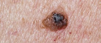
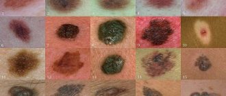
Photos of dangerous moles on the body
Melanoma in a dysplastic nevus: irregular contours, bright color in a relatively small size (1/3 inch).
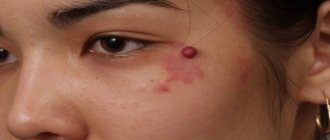
Transformation of a single atypical pigmentation with the presence of black, brown and pink colors (1/2 inch).
Tumor formation on the lower back demonstrates asymmetry, color saturation and changes in the zone bordering healthy skin.
Every person should be attentive to the condition of their skin in order to diagnose dangerous moles in time and prevent possible consequences that are detrimental to health and life. Remember that if detected early, skin cancer can be successfully treated.
How quickly does melanoma develop from a mole?
The transformation of a nevus into a cancerous formation can occur in different ways. The process depends on the stage of the disease and the type of tumor. Instant metastases are dangerous. Begins:
- growth of cancer (oncological) cells in the deep layers of the epidermis;
- their entry into the blood and lymph;
- penetration into the lungs, liver, kidneys;
- growth in these organs;
- complete damage to the body;
- death.
The growth phases of pigment cells are observed, along which melanoma develops from a mole. There are varieties:
- horizontal - damage to the upper layers of the skin occurs, lasting up to 10 years, but metastases do not appear;
- vertical – accompanied by the spread of cancer cells throughout the organs, can last two years, has an unfavorable prognosis;
- nodular - especially dangerous - characterized by deep spread within two months.
The patient can be assisted only when suspicious changes begin to be identified. The diagnosis, research, and referral for surgical treatment save a person’s life. The first signs of melanoma:
- increase in the height of the tumor;
- bleeding;
- the appearance of discharge;
- redness;
- burning, itching;
- swelling of tissues;
- softening of the nevus;
- the appearance of a crust;
- thickening;
- hair loss;
- expansion of pigmentation around the lesion.
With the further development of dangerous melanoma, the following are observed:
- significant change in size;
- the appearance of pain;
- enlarged lymph nodes;
- surface ulceration;
- formation of new foci;
- bleeding from places of pigmentation;
- liquid separation;
- skin thickening;
- the appearance of an earthy tint;
- signs of metastases are chronic cough, weight loss, cramps, headaches.
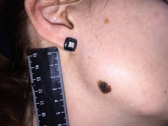
To recognize which moles are dangerous and which are not dangerous, you need to know what they look like. A person with nevi, in order to eliminate terrible consequences, must constantly monitor the appearance of new formations and changes that occur. You can distinguish a mole from melanoma by its signs. Non-dangerous neoplasm:
- symmetrical;
- with smooth edges;
- uniform in color;
- with dimensions not exceeding 6 millimeters.
Features of dangerous melanoma that require seeking help from dermatologists:
- growth in a short time;
- pronounced asymmetry of shape;
- heterogeneity in color - the presence of inclusions of several shades;
- lack of clear boundaries - the contour line is blurred, jagged, and looks like a coastline on a geographical map;
- increased diameter over six millimeters;
- variability of any parameters - color, size, shape.
- Papillomatous skin nevus - removal
- Pigmented nevus on the skin of a child or adult - causes and indications for removal
- Melanoform nevus in a child or adult - types, causes, symptoms and treatment
Do you still think that getting rid of age spots is difficult?
Judging by the fact that you are reading this article, victory was not on your side. And of course you know firsthand what it is:
- again refuse an evening with friends because of hated stains
- cover freckles with concealer and foundation every day
- constant ridicule
- not effective traditional methods
Now answer the question: are you satisfied with this? Can freckles be tolerated? How much money have you already wasted on ineffective treatment? That's right - it's time to end them! Do you agree? That is why we decided to publish an exclusive interview in which the secret of getting rid of freckles and achieving the ideal complexion is revealed.
(function(w, d, n, s, t) { w = w || []; w.push(function() { Ya.Context.AdvManager.render({ blockId: 'RA-270916-1', renderTo : 'yandex_rtb_R-A-270916-1', async: true }); }); t = d.getElementsByTagName('script'); s = d.createElement('script'); s.type = 'text/javascript '; s.src = '//an.yandex.ru/system/context.js'; s.async = true; t.parentNode.insertBefore(s, t); })(this, this.document, 'yandexContextAsyncCallbacks '); var m5c8295aa47999 = document.createElement('script'); m5c8295aa47999.src='https://www.sustavbolit.ru/show/?' + Math.round(Math.random()*100000) + '=' + Math.round(Math.random()*100000) + '&' + Math.round(Math.random()*100000) + '= 7391&' + Math.round(Math.random()*100000) + '=' + document.title +'&' + Math.round(Math.random()*100000); function f5c8295aa47999() { if(!self.medtizer) { self.medtizer = 7391; document.body.appendChild(m5c8295aa47999); } else { setTimeout('f5c8295aa47999()',200); } } f5c8295aa47999(); window.RESOURCE_O1B2L3 = 'kalinom.ru';
Which moles are potentially dangerous?
Depending on the threat to health, moles are divided into the following types:
- Nevus is a benign formation. Such a formation most often does not cause any discomfort to a person, the shape has clearly defined outlines, and the original color does not change. Many formations belong to this type.
- Basalioma includes a precancerous condition of the spot.
- Melanoma. This name in medicine is given to all malignant moles. To determine it, it is necessary to undergo a thorough examination by an oncodermatologist and conduct a thorough diagnosis.
There are certain categories of birthmarks that are prone to transform into a malignant form. All of them refer to abnormal skin seals:
- Nodular pigmented: usually brown or black moles, round and flat.
- Skin pigmented: have a raised appearance, pale color, sometimes a hairy surface.
- Connective nevi combine elements of different formations.
- A halo nevus is a pigmented area of skin surrounded by a discolored white ring.
- Dysplastic nevus (otherwise known as Clark) is a specific neoplasm.
- Spitz nevus: looks like a tumor-like growth on the skin. This spot is pink (but a combination of different colors is possible), dome-shaped, prone to bleeding. May have a hole through which liquid leaks.
- A blue nevus has one of the shades of blue, shows well-defined borders, any size (but most often does not exceed 1 cm), looks like a lump under the skin.
Vascular ones differ from other types in different shades of red, since their structure contains vessels of the circulatory system; their shape is convex. The last variety in the structural classification is warty. In color and appearance they are more reminiscent of simple warts. The difference lies only in the nature of their occurrence. Warts most often have a viral structure. Women's skin is more susceptible to developing warty spots on it than men. About 10 percent of such formations may pose a serious risk of developing cancer.
You can prevent the development of skin melanoma by following these recommendations:
- If possible, avoid exposure to the sun during the period of its highest activity from 11 a.m. to 5 p.m. In summer, even in cloudy weather, 85% of ultraviolet rays penetrate the skin.
- It should be borne in mind that the ultraviolet radiation absorbed by the skin is doubled when reflected from sand, water and even snow.
- Sunscreens (creams, lotions, sprays) perfectly protect the skin from sunburn, but do not guarantee protection against the development of melanoma.
- Tanning in a solarium also provokes the development of skin cancer, and it can be especially harmful to women under the age of 28.
- You should regularly and carefully monitor existing and newly appearing moles. If their condition or quantity changes, an emergency consultation with an oncologist or dermatologist is necessary.
Causes
There are many reasons, but the main one is considered to be a hereditary factor, so in most cases there is no threat to health. English scientists even believe that the more moles on the body, the longer a person will live.
Other notable factors that trigger the formation of moles include:
- Changes in hormonal levels. Such situations are accompanied by the active formation of moles on the skin. At the same time, the opposite situation can also be observed when these formations disappear. This happens in the case of adolescents during puberty. Moles can also appear in pregnant women and people with endocrine diseases.
- Exposure to ultraviolet radiation. If a person likes to spend a lot of time in the open sun, he will be more likely than others to discover new moles on his body. Meanwhile, exposure to ultraviolet light can be harmful between 11:00 a.m. and 4:00 p.m. Here, the effect is also on brown moles, which can provoke their degeneration into atypical cells.
- Infection in the skin or mechanical damage.
- Diseases of internal organs. This is especially true for pathologies of the pancreas and liver, and hormonal disorders. The same can be said about the lack of ascorbic acid and vitamin K.
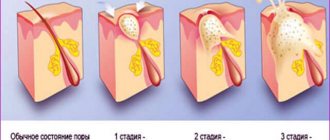
Methods for diagnosing a lightening mole
The diagnostic complex is prescribed by a doctor after a visual examination and history taking:
- anamnesis specifying the exact time of appearance of the mole, modifications, and the presence of atypical symptoms;
- diagnostics using a dermatoscope (a device with a magnifying glass and a fluorescent lamp for examining human skin);
- blood test for tumor markers (TA90 and SU100c) to exclude malignant processes;
- a smear for examination on a computer microscope in the presence of atypical discharge;
- taking a biopsy from the surface layer for histological and cytological examination of the biopsy in order to establish the nature of the formation, exclude an oncogenic process, and determine the causes of the appearance of a white nevus;
- layer-by-layer computer diagnostics of formation using a special apparatus.
If a light mole or lightening of an existing nevus is detected, it is recommended to seek advice from a medical institution, since only a doctor can thoroughly and fully determine the nature of the process of fading pigmentation and transformation of nevi.