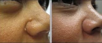
Brown dot - what is it?
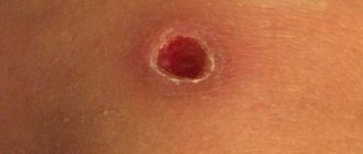
Sometimes, some time after the mole has been successfully removed and the skin has healed, a dot appears in the same place.
Most likely, such a spot is a symptom of a relapse - the reappearance of a nevus. It may take 1-2 months from the moment of surgical removal to the visual appearance of a new mole, sometimes more. When you carefully examine the brown spot that appears at the site of the removed mole, under a microscope you can see cells that are typical for all moles. They are capable of producing a brown pigment known as melanin. It is thanks to him that the nevus turns brown.
Why did it appear again?
The main reason for the reappearance of a mole after removal lies in the insufficient quality of surgical intervention. If a previously existing nevus was not completely removed, part of its cells (a microscopic fraction is enough) will remain in the skin, and after some time will begin to produce brown pigment.
However, some doctors claim that even when a mole is removed, including a significant amount of healthy surrounding tissue, there is a risk of relapse. In this case, it is not yet possible to determine its causes.
What to do if it is not completely removed?
If some time after the mole was removed, it suddenly grows back, do not panic. The first thing you need to remember is how the nevus was eliminated. If it was removed according to all the rules with a histological examination of the tissues, and the tests did not find any pathological (cancerous cells), you can calm down and not worry. Of course, it would be a good idea to show a new (recurrent) mole to a doctor and make sure it is safe.
When is pigmentation dangerous?
A benign nevus, which for some reason appears again, is considered a benign phenomenon. However, there are typical symptoms that require special attention. They are, in principle, similar to similar signs of malignancy of any mole and include:
- Increase in size. If a new nevus grows before your eyes, you should immediately show it to a dermatologist-oncologist.
- Ulceration (appearance of cracks, erosions on the surface of the nevus).
- Bleeding.
- Soreness.
- Permanent injury.
It is worth noting that the risk of developing melanoma after incomplete removal of a benign mole is quite small. Nevertheless, this possibility is worth considering.
Recurrence of dysplastic nevus
Dysplastic nevus has an increased tendency to malignancy. Such a mole can have different sizes, bizarre borders and heterogeneous coloring. Sometimes it rises above the skin level. Doctors strongly recommend removing such single moles with mandatory histological examination. But even after surgical resection, they can appear again, which is considered a rather alarming symptom.
If a dysplastic nevus recurs, it is better not to hesitate and go to the office of a dermatologist-oncologist. Most likely, such a mole will need to be excised again, followed by a comprehensive examination.
What if there was no histology?
If the nevus was removed in a regular beauty salon or evaporated by a laser (electric current, etc.) without a histological analysis, there is already something to worry about. After all, there is a high risk that malignant processes of degeneration have already begun in the removed mole, and accordingly, the tumor may continue to develop. Moreover, the time for timely diagnosis and early treatment may already be lost. Tumors that develop in the skin after a mole is removed can grow especially quickly and metastasize.
If the nevus recurs, it is best to remove it again and conduct a histological examination. Only in this case will you be able to remain calm about your health.
In place of the removed mole there is a new one, what to do?
If a removed mole has grown, there is no need to panic; first of all, you need to adhere to some rules:
- Be sure to ask your doctor to make sure that the treatment chosen is correct. Only in this way can you completely get rid of the nevus along with its root.
- Learn how to treat a wound after treatment. It is especially important to monitor her for the first two weeks - this will help prevent infection from occurring. Otherwise, not only will a new mole begin to grow, but inflammation will also begin to develop.
- A crust always forms on the wound after treatment; under no circumstances should you remove it yourself. Wait until the dead cells fall off, a pink spot will remain under them - this is healthy skin.
- In the summer, you need to protect the wound from exposure to the sun; sunbathing in a solarium is also prohibited.
These are the main activities that must be carried out without fail. This way you can protect yourself and your health from complications.
Repeated excision
Usually, to remove a recurrent mole, doctors resort to excision of such a formation with a scalpel. It is believed that this method allows one to reliably eliminate all nevus cells, capturing the surrounding healthy skin, and also obtain suitable material for histological examination. Of course, using a scalpel is considered a rather traumatic method of removal, and it leaves scars on the skin.
In some cases, it is possible to remove a mole using a laser, electric current or radio wave method. But at the same time, the doctor should not evaporate the nevus, but cut it off in order to again obtain material for histological analysis.
The red spot does not go away
Sometimes, even after successful removal of a mole by laser or other methods, a red or pinkish spot remains on the skin, which is not a recurrence of the nevus. This mark may appear:
- If the skin healing process is still ongoing. It may take about six months or even more until the color of the epidermis is completely evened out.
- Soon after the crust falls off. In this case, a pinkish spot is observed on the skin - an area of young, delicate epidermis.
- If you tear off the crust ahead of time (accidentally or intentionally). Such a dark spot at the site of a removed mole can remain for life; to get rid of it, you must definitely use means to activate regenerative processes (silicone ointments and patches, Contractubex, Solcoseryl, etc.).
- With improper skin care. In particular, reddish spots may appear when the young epidermis is exposed to sunlight. In this case, they indicate a skin burn and can subsequently lead to pigmentation of such an area.
- Due to the penetration of infectious agents. In this case, the problem area may swell, itch, and even be noticeably painful. The formation of an abscess is also possible.
If strange spots appear on the skin after removing a mole, it is better to play it safe and consult a specialist. This will help reduce the risk of developing melanoma, a skin cancer.
There is nothing wrong with the appearance of moles on the human body. The process of mole formation is a mechanism that depends on various factors - internal and external. A qualified doctor will be able to determine the clear reasons for the formation of pathology. The specialist will identify nevi that need to be removed. In the future, a person must adhere to a number of rules and lead a healthy lifestyle.
The process of removing a mole is a complex procedure that requires high-quality execution from a physician. The structure should be eliminated through surgery if it has multiplied in size, hurts or changed color. After surgery, the mole may grow back. Relapse is formed under the influence of a large number of reasons. The development of education may be due to the fact that it was not completely removed. Some of the cellular structures of the nevus remain in the skin, which affects the production of brown pigment. Melanin is restored some time after the operation.
Histological examination allows us to assess the quality of the operation and possible risks of complications. When benign formations are removed, the re-development of the pathology will not be dangerous. Intervention without histology often leads to melanoma.
A relapse after a mole removal procedure can form from melanocyte cells contained in the nevus. Pathology should be treated in specialized clinics with specialized equipment. The operation is preceded by a systemic diagnosis of the body. With the help of an examination, you can determine the depth of the mole and the prospects for enlargement. Removal of inflammation of the formation should be gradual. Sometimes complications develop.
How to prevent the appearance of a new mole
First of all, you need to carefully select a method for eliminating pigment spots together with your doctor. Proceed with the procedure only after making sure that all cells included in the nevus can be reliably removed. After surgery, avoid infection of the wound, follow all doctor's instructions for treating its surface. Under no circumstances should you remove or scratch the crust covering the healing tissue.
Until the wound has completely healed, you should not be in the bright sun or visit a solarium.
The delicate area of skin that opens after the crust falls off must be protected from exposure to ultraviolet rays. To do this, you can cover it with clothes when you are outside or lubricate it with a special cream with an SPF factor of 50 or more.
Mole grows after removal
5 (100%) 1 vote
There is nothing wrong with the appearance of moles on the body, but sometimes the reason for their occurrence is fraught with many mysteries. The first nevi appear in infancy, and the rest can be acquired throughout later life. Sometimes it happens that you have a mole removed, and it appears again, but why does this happen?
Causes of relapse after primary removal
The occurrence of relapse is due to a number of reasons and complications. The pathological process is based on the human factor. Incorrect determination of the depth of pigment spots affects the outcome of the operation. It's easy to get an infection into an open wound. The use of antiseptics and medications is a prerequisite for rapid healing of the area.
Some people, after removing a mole, remove the resulting crust. You should wait for the keratinized cells that cover the pink skin. After surgery, select effective wound treatment options. The main task is to prevent inflammation and tumor growth.
Incorrect determination of the depth of pigment tissues
Epiluminescence microscopy magnifies images of pigments 40 times. The procedure allows you to visually study the depth and structure of the formation. The computer records information on the camera, comparing it with the data in the database. In practice, dermatologists do not often resort to cryodestruction for help. Controlling the depth and level of tissue freezing is problematic. If the operation is performed poorly, part of the nevus in the epithelium remains viable. As a result, a relapse will occur and the previously removed mole will grow back. The danger is that damaged structures can transform into atypical cells. It will be difficult to eliminate the pathological process!
Incomplete removal of a mole is fraught with complications and additional disorders. To prevent degeneration, systematically monitor the pigment spot on the skin at home every day.
Incorrect removal method selected
Determining the causes of melanoma development underlies the choice of therapeutic regimen. Options for nevus removal are surgical. Modern technology and equipment allows operations to be carried out through fine tissue dissection. Effective methods and procedures for removing formations:
- surgical intervention;
- use of laser (including during transformations);
- carrying out electrocoagulation;
- radiosurgical approach.
The choice of the appropriate option is carried out personally for each person. Brown structures and cancerous growths are removed with a scalpel. Using this manipulation, nevus tissue is cut out from the deep layers of the dermis. Other moles are removed with laser and liquid nitrogen. The manipulation is carried out carefully, without large losses of blood.
An incorrectly selected treatment regimen is a source of additional disorders. Treating an area of skin with liquid nitrogen can cause severe burns. As a result, wound healing will be prolonged and painful. A person has an increased risk of infection.
Degeneration of nevus
The mechanism of degeneration of a mole is the body’s response to external stimuli. The main activators for a person in this case are prolonged exposure to the sun and daily visits to the solarium. If a person touches the structure with a fingernail, melanoma may develop.
The pathology is dangerous for pregnant women. Hormonal changes affect changes in the structure and cells of the epidermis.
Poor quality surgery is also a source of nevus degeneration. An inaccurate incision or infection entering the wound can cause complications. At the first sign of additional complications, seek help from your doctor.
Self-removal of a mole
At home, it is almost impossible to maintain complete sterility. This factor increases the risk of infectious agents entering a weakened human body. In practice, relapse of the nevus occurs if the growth is not completely removed. Melanoma-prone and malignant structures should not undergo cosmetic procedures.
The formations are eliminated by surgical excision, which is performed in the area of healthy tissue. The disadvantages of scratching or damaging a nevus are:
- a multiple increase in inflammation in size (will grow gradually);
- severe infection;
- repetition and dangerous consequences may occur;
- development of pathologies and complications.
What may cause the appearance of a tubercle?
Sometimes it seems that a new mole has appeared due to tissue swelling as a result of damage to the lymphatic vessel during excision of the nevus. Usually their condition recovers within a few days. If an infection gets into the wound, an inflammatory process may begin, accompanied by an increase in temperature to 39 °C or more. To eliminate it, antibiotics are prescribed.
A dense tubercle may also grow if the removed nevus has degenerated into a malignant tumor. In this case, examination and treatment by an oncologist is necessary. If necessary, a new operation is performed. The nature of the neoplasm can be confirmed by histological examination of its tissues.
It also happens that the mole has grown due to the fact that during the operation the deeply located melanocyte cells that make up the nevus were not removed.
As they grew, they caused pigmentation to appear in the same place. The reason for incomplete removal of mole tissue is sometimes the choice of an insufficiently effective procedure method - the use of laser radiation, cryodestruction. To correct the error, the spot is re-excised using a scalpel. Not only the tissue of the mole is cut out, but also the adjacent layers of the epidermis are captured at a short distance (3–4 mm). After healing, a scar may remain at the surgical site.
What to do if a mole reappears after removal
If a removed mole re-grows on the body, you should carefully monitor the color and size of the mole. Inspect the operated area daily. If a large scar or scar forms, you should seek help from a doctor. After analyzing the patient, the specialist will select the optimal medications. Medicines will remove the crust on the wounds.
If the recurrence of the nevus is malignant, the following signs are observed:
- pain in the area of the new mole;
- the formation of severe itching of the skin;
- the surface of the body peels off and becomes covered with small cracks;
- blood oozes from the growth;
- education takes on a different color and shape.
If you experience the above symptoms, seek help from your doctor. A medical professional will determine the risks of cancer development and select effective therapy. The main task is to neutralize the effect of metastases, which dynamically spread throughout the human body. Otherwise, complete recovery will become unrealistic.
Possible complications
After surgery, you need to carefully monitor all changes occurring in the wound. As a rule, the postoperative period proceeds well, but sometimes complications may occur. If unusual symptoms appear or you suspect that the healing process is not going well, you should consult a doctor. An examination by a specialist will help determine whether there really are grounds for concern or whether the concern is caused by imaginary reasons.
It is necessary to visit a doctor if:
- severe pain is felt at the site of the postoperative scar;
- the wound bleeds or discharges liquid with an unpleasant odor;
- body temperature increased significantly;
- a lump appeared at the site of the removed mole.
Consultation with a specialist is required in cases where even the use of painkillers does not help eliminate the pain.
It must be remembered that immediately after surgery you should not take Aspirin and Ibuprofen, as these drugs thin the blood, increasing the risk of bleeding.
High temperature is evidence of an inflammatory process
Is it necessary to remove it again?
Diagnostic methods allow us to objectively study the condition of a person’s skin. Effective options are:
- examination in a specialized clinic;
- performing dermatoscopy;
- digital analysis of tumor growth (in dynamics);
- formation of a “map” of structures at a specific location;
- performing a skin biopsy.
Dermatoscopy is the process of examining the skin using a specialized apparatus. A physician can examine the internal structure of the growth and classify it. Afterwards, an effective treatment plan is selected. An equally informative diagnostic method is a puncture biopsy. The research mechanism involves manipulation of a special needle. The material is collected under sterile conditions using painkillers. Diagnostics allows you to study the state of neoplasms. If melanoma or tumor is detected, you should immediately consult a doctor.
Signs of a mole healing after removal are:
- a dry dark crust remains on the operated area;
- the size of the keratinized structure is 1 mm;
- after a couple of weeks the growth will peel off from the skin;
- the formation of a small red pit, the depression does not exceed 1-2 mm;
- after 28 days, the bottom of the fossa gradually rises to the epithelial level;
- 90 days after the intervention, the inflammation disappears, and an invisible unevenness or scar remains from the mole.
If the rules of the operation are not followed, the incision becomes suppurated. As a result, the inflammatory process drags on for months. The wound swells and accumulates a lot of pus. The patient experiences pain and body temperature rises.
If a light-colored liquid flows out from under the crust, this is a good sign, which means the treatment is going according to plan. To keep complications to a minimum, follow all doctor's instructions. The incisions are treated with high-quality antiseptics.
Oncologists and dermatologists agree that recurrence of nevus is only in rare cases dangerous to human health. The risk of melanoma formation increases markedly if the operation was performed without dermatoscopy. Taking a scraping or puncture will allow you to clearly determine the causes of the development of the pathology. Qualitative histological examination in this case is the main indicator of a person’s health status.
It is not recommended to remove tumors using cosmetic surgery. Unskilled impact on the structure leads to complications. It will be a shame when a person faces cancer due to negligence or inattention. It is better to perform procedures in specialized institutions. Before the mole removal process, a high-quality skin diagnosis should be performed. Otherwise, you increase the likelihood of additional complications.
This group includes pigmented nevi
, which have reappeared at the site of previous removal by laser, electrocoagulator or other method. The risk of relapse is due to the lack of clear criteria for assessing the adequacy of the impact during these procedures. Most often, relapses occur within the next few weeks after removal of the primary formation and look like irregularly shaped foci of pigmentation located within the scar.
Clinical manifestations
Such nevi are very diverse, but the unifying point can be considered the presence of cicatricial changes around the nevoid tissue. Accumulations of melanocytes are immured in the scar, and they are located at the level of the dermis and the dermo-epidermal junction. The pathohistological picture of recurrent nevi may resemble the structure of melanoma in situ, which was the reason for the appearance of the name “pseudomelanoma”.
During dermoscopic examination of recurrent pigmented tumors
there is also a great similarity between these formations and melanoma. They may contain an atypical pigment network, foci of uneven homogeneous pigmentation and radial radiance, irregular pigment dots and atypical globules. The formations are usually asymmetrical, polychromy is possible. Options for combining dermoscopic structures are very diverse. The difficulty of differential diagnosis with melanoma determines the recommendation to perform surgical removal of recurrent nevi with pathohistological examination.
In addition, there is an area that strongly resembles regression structures along the right edge, and a blue-white veil. Individual small foci of brown pigmentation are visible in the scar. In this case, most of the signs were in favor of melanoma. Pathohistological examination after excision - recurrent nevus.
On the left is an area of pale brown pigmentation with small dark brown and gray dots evenly distributed in it. The whitish spots are the mouths of the excretory ducts of the glands (pseudonetwork).
A fresh scar with an abundance of blood vessels has a pink-brown zone in the center, against which there are dark structures of irregular shape and size, with a gray tint and blurred boundaries, resembling globules.
Dermoscopically - uneven structureless pigmentation against the background of whitish scar areas, polychromy (pink, brown, gray). Upon careful examination, the pink areas contain an abundance of thin linear vessels.
Taking into account the patient’s reluctance to remove the recurrent nevus surgically, the tactics of active observation were chosen.
- Uneven contours
, asymmetry of formation, polychromy, focal radiance of the pattern and the presence of a gray hypopigmented zone interspersed with pigment grains in the center - the picture does not exclude melanoma. This recurrent nevus must be excised. - The black spot in the scar
appeared after hyperinsolation and gradually increased. Recurrent nevus looks like a cluster of large dark brown dots against a background of homogeneous gray-brown coloring. Pathohistological examination after excision of the relapse revealed no signs of malignancy. - Recurrent nevus
, similar in structure to a Spitz nevus, is a radiant brown structure with a small number of globules located in the very center of the scar left after laser removal of an intradermal pigmented nevus. - A classic example of a relapse
that occurred at the site of a non-radically removed intradermal pigmented nevus.
The formation formed within the first two months after laser exposure. An irregularly shaped black-brown zone of hyperpigmentation in the center and a white-blue veil, atypical globules and areas with radial radiance are all signs of melanoma
. Such nevi are subject to surgical removal with mandatory pathohistological examination. - In this case, we are dealing with a borderline nevus
, simulating a recurrent nevus. This situation became possible due to the formation of a scar at the site of a large boil that appeared in the skin adjacent to the nevus. - The area of black-brown pigmentation in
the center of the scar after laser removal of an intradermal nevus has radiant contours during dermatoscopy. Within this area there is a chain of round whitish inclusions (hair follicles?).
All people on Earth have moles. One person can have up to thirty moles on his body. Scientifically, medical experts call them nevi. In the old days, people were afraid of brown spots, they were afraid to touch them with their hands, for fear of harming themselves. They even came up with a proverb: “The more moles, the happier the person.” Modern people are not so superstitious, but there is some truth in the fact that the disappearance of such a formation may not be good for the body.
Relapse Prevention
After diagnosis and confirmation of the benign quality of the formation, you should consult a doctor with experience in this field. The country's leading dermatologist-oncologist Igor Evgenievich Sinelnikov faces similar problems every day. He offers various ways to remove the growth without complications. To avoid consequences and re-growth, you must follow these rules:
- In the first week, do not allow water to enter the area after removing the growth.
- Read the instructions for proper wound treatment. Wash your hands thoroughly before handling and monitor the area for two weeks to avoid infection. Will help avoid inflammatory processes and relapse.
- After the procedure, a crust will appear on the wound; it is not recommended to remove it yourself. Over time, it will disappear, leaving healthy pink skin.
- It is not recommended to sunbathe in the sun or in a solarium. Ultraviolet radiation negatively affects the skin, stimulates the growth of old and new formations, and the appearance of birthmarks. If you cannot hide from the sun, cover the area with an adhesive plaster or cover it with clothing.
Simply following the rules and carefully caring for the skin area is the key to a quick recovery. preventing relapse. The use of medicinal ointments and creams will hide cosmetic defects.
Classification
Many people suffer from their unaesthetic appearance, spoiled by a mole in a prominent place. Nevi come in different shapes, sizes and colors, which also does not speak in their favor. Moreover, they can be located not just in “prominent places”, but in inconvenient places: in the folds of the skin, for example. In short, they cause inconvenience for a long time.
By size they are distinguished:
- small (up to 15 mm);
- medium (1.5-10 cm);
- large (more than 10 cm);
- huge (neoplasms that occupy part of the body).
The form is:
- flat;
- convex;
- hanging;
- warty;
- smooth;
- hair
By color they are distinguished:
- black;
- brown;
- bodily;
- blue;
- red.
Today they are not afraid to touch them and there are different ways to remove them from the body and, it would seem, forever forget that they once existed. However, a common problem is that after the nevus is removed, the mole grows back.
Methods for removing tumors
In order to eliminate moles, modern medicine uses the following methods:
- Laser removal. It is considered the most effective method. When working with a laser, it is possible to regulate the depth of penetration, which allows you not to touch nearby tissues and, accordingly, not damage them. This method is used to remove nevi on the most delicate areas: face, neck, chest;
- Cryodestruction (removal with liquid nitrogen). Nitrogen is brought to a temperature of -180 degrees. Under the influence of this substance, tissues undergo necrosis, which leads to their rejection by the body. After the procedure, a new, healthy layer of epidermis begins to form. This method is not so popular, since there is a high probability of tissue injury, and there is also the possibility of relapse: after removal, a new melanoma may appear in the old place again;
- Electrocoagulation or radioknife. The method involves burning out the formation with an electric current. You can get excellent results in one procedure. A significant disadvantage of this method is the appearance of a scar at the site of the nevus;
- Surgical method. Most often used for cutting out large forms of tumors. Antibacterial drugs are administered to the patient, and the procedure is performed under local anesthesia. What’s good: the formation does not appear again after removal, but an unsightly scar may remain.
Removal of tumors should only be carried out by a qualified specialist. One person may have 3-4 types of nevi on his body, each of which may require a personal approach.
What can happen after a nevus is removed?
Often, after the removal of moles, complications arise: either it grows back, or the patient does not follow the rules of caring for the skin from which the tumor was removed. Therefore, before the operation, a complete examination of the person must be carried out, and after this, the nevus must certainly be sent for histological examination.
Consequences of laser removal:
- In the case of a flat and small tumor, with proper care and compliance with medical recommendations, no traces remain;
- If a raised, deep nevus is removed, it is likely that a small depression or small scar will remain on the skin;
- Sometimes after the procedure, rashes on the skin are observed.
Consequences of cryodestruction:
- A significant disadvantage of this method is that it is impossible to guess to what depth liquid nitrogen will penetrate, and whether it will thoroughly remove the tumor or only damage it. If the subcutaneous cells of the formation are intact, there is a high probability that the nevus will appear again;
- A skin burn is likely, which will damage healthy tissue next to the nevus;
- If the mole is not completely removed, a compaction forms in its place.
Consequences of electrocoagulation:
- No pain during the procedure;
- The skin is not damaged;
- Most likely there will be a scar. For this reason, removal of formations on the face and neck by this method is not practiced.
Consequences of the surgical method:
- The healing process takes longer compared to other methods;
- There is redness on the skin after surgery;
- Scars remain, which fade over time and become less noticeable. The size of the scar will depend on the size of the tumor and the skill of the surgeon;
- There is a possibility of infection getting into the open wound during the procedure;
- If the mole is not completely removed, it will appear again. Most likely, this will no longer be a benign formation. There is a high risk of its transformation into melanoma.
The most common consequence of mole removal is recurrence. A nevus appears in the same place, of the same or larger size. If the formation appears again and again, then an urgent need to do a biopsy to prevent the onset of a malignant condition.
If the elimination procedure went well and the mole was definitely removed, then there is nothing to worry about. There will be no complications.
It all depends on the characteristics of the person’s body who underwent the operation:
- Does he have a predisposition to relapse;
- Hereditary factors;
- Is the required balanced diet being maintained?
- Ecological situation.
Why you can’t eliminate education yourself
Eliminating nevi at home is a dangerous undertaking. The most optimistic prognosis after such a procedure will be the appearance of a new mole, the worst - the development of cancer. There are formations that cannot be removed under any circumstances.
Carrying out such an operation at home, you are at great risk. At home, it is impossible to ensure the necessary hygiene, which increases the likelihood of wound infection.
Despite the apparent simplicity of the procedure, you absolutely, without special equipment, will not be able to determine to what depth the nevus cells go. So after cutting off the upper part at the count of “one”, a new mole will soon form.
A common occurrence after cutting out a nevus at home is scars or growing scars.
What is surgical excision of a nevus? Is it worth removing this formation? It is best to discuss such questions with your doctor, since the answers to them depend on the characteristics of the formation itself, which only a specialist can study.
A nevus is a growth on human skin. Its other name is mole. Nevi can be either congenital or acquired.
The result of nevus removal is impressive!
- if excision of the mole is recommended by a medical report;
- if the nevus causes physical discomfort to a person and there is a threat of damage.
Today, the most effective method of removing a nevus is with a laser, but such an operation is performed only if there is indications from a dermatologist.
Getting rid of nevi on your own at home can result in cancer. If the formation brings discomfort, as, for example, if it is located in the lip area, then you need to find a specialist who will conduct an examination and prescribe removal of the nevus.
The mole is growing again
Whether to remove moles or not is a personal matter for everyone. This depends on many reasons: aesthetic and medical.
What to do if a mole grows after removal? Is this hazardous to health? People who have encountered similar troubles often ask such questions.
In any case, if the nevus recurs, you should consult a dermatologist who will help you deal with this problem.
The growth of a new mole at the site of a removed one is not a rare phenomenon that can be avoided with proper postoperative care.
Mole removal
There are moles on every person's body, some have more, some have less. They do not always decorate the body with their presence. Their ability to degenerate into malignant tumors leads to severe cancer. Therefore, when they discover new birthmarks or convex growths, people run to the doctor, demanding that the suspicious mark be removed.
Removing moles is a serious procedure that requires a high level of professionalism from the doctor.
A mole needs to be removed if it bothers a person, for example: it has increased in size, hurts when touched, changes color and structure.
If the tumor is located in a place that is often subject to friction or clinging, then it is better to remove it too, so as not to damage it or cause infection.
Reasons for relapse
If a new mole appears at the site of a removed mole, you need to look for the cause, since sometimes relapse becomes a real problem. Causes of secondary occurrence of moles on the human body:
- The surgeon incorrectly determined the depth of the pigment tissue, which is why the mole was not completely removed.
- The wrong method of mole removal was chosen. For example, the use of cryodestruction often leads to complications. Liquid nitrogen can cause a burn to the skin, which after surgery will prevent its healing, and an open wound is a direct access to infection.
- Infection. In most cases, the infection enters the wound after the mole has been removed. The reason is insufficient care of the affected area, as well as non-compliance with medical recommendations.
- Degeneration of nevus. Occurs as a consequence of improper removal of the tumor. In some cases, it leads to melanoma.
- Self-removal of a mole. At home, it is impossible to maintain complete sterility, which is why the affected area can become infected. Again, there is a risk of not completely removing the growth. As a result of all this, a relapse of the nevus occurs.
Is it dangerous?
When a mole appears at the site of a removed one, is it dangerous? The answer lies in studies conducted before the operation on the nature of the malignancy of the growth.
If histology, dermatoscopy or other diagnostic methods were carried out before surgery and confirmed the benign nature of the neoplasm, there is no need to worry. If there is no research before the removal procedure, there is a health risk.
In some cases, secondary foci of malignant tumor growth may appear.
What to do if a new spot appears at the site of a removed mole?
If a brown spot or bump appears again at the site where the mole was removed, nothing needs to be done. The main thing is to examine the problem area on the skin every day. If a scar or scar forms at the site where the nevus was removed, you can consult a doctor who will prescribe medications that promote rapid healing of the wound and resorption of the skin defect.
In the event that a recurrent formation causes concern and manifests itself with unpleasant signs, you should immediately consult a doctor. When a malignant nevus recurs, the following signs appear:
- if a mole appears and hurts when touched;
- severe itching of the skin appears;
- the surface peels off and becomes covered with small cracks;
- blood oozes from under the growth;
- the mole grows in size;
- the growth changes color and structure.
Recurrence of a nevus at the site of a previously removed one requires repeated removal.
If a person has all of the above signs, this can lead to unpleasant consequences. The doctor will conduct the necessary studies to determine the nature of the malignancy. When cancer is detected at an early stage, it is important to start therapy on time.
The operation carried out at the initial stage will be successful. The surgical treatment method involves excision of melanoma and nearby tissues.
In the case when the metastases have managed to spread throughout the human body and infected vital organs, alas, the treatment will be ineffective.
If the removal took place without histology
Before removing a nevus, the doctor is required to conduct research into the nature of the neoplasm. This is not necessarily an expensive histological examination. You need to remember what procedure the diagnostician performed - dermatoscopy or puncture.
Any of the methods evaluates the nature of the tumor with no less accurate results than histology. Therefore, if a mole appears again, there is no need to worry; it is important to monitor it daily. If desired, you can delete it again.
Preventing the appearance of a new mole
Before surgery to remove a mole, it is recommended to undergo a histological examination, which will determine the nature of the neoplasm. This diagnostic method is considered the most accurate.
After confirming the benignity of the nevus, you should choose the best clinic and doctor who has sufficient experience in this field. Next comes the selection of the removal method. There are many of them, so it is necessary to choose the safest method that guarantees complete removal of the tumor.
To avoid unpleasant consequences after removing a mole, you must follow the recommendations that the doctor must give:
- Do not wet the area where the growth is removed for the first 7 days.
- Properly treat the damaged area of skin. Before handling, wash your hands thoroughly to avoid infection.
- After 1 week, a dry crust will appear on the wound, which under no circumstances should be removed. The dead skin will fall off on its own over time.
- If the operation is performed in the summer, you should avoid prolonged exposure to the sun, do not sunbathe, and do not visit the solarium. If you cannot avoid direct sunlight, the wound should be covered with clothing, and a bactericidal patch can be glued on top. Exposure to ultraviolet radiation can cause the appearance of pigment spots in areas of skin damage.
Compliance with careful care of the affected area after removal of a mole is the key to successful recovery and prevention of recurrence of the tumor. If scars begin to appear on the skin, it is necessary to use ointments and creams, the therapeutic effect of which will help hide such cosmetic defects. Properly done independent and medical work will lead to a good result.
Source: https://StopRodinkam.ru/rodinki/udalenie/opyat-rastet-rodinka.html
Removal of nevus by surgical and radio wave methods
Surgical removal of nevus is the oldest method. This method helps with deep and large formations on the skin. However, this procedure has a drawback: it leaves scars and scars on the skin.
When resection takes place, the growth is cut out, including healthy skin. The manipulation itself takes place under local anesthesia and lasts 30 minutes. Once the formation is removed, the wound is sutured so that the scar is less noticeable.
After the operation, the person goes through a recovery period. The main thing is to care for the place where the manipulation was performed. It should be treated with anti-inflammatory drugs for a week after the stitches are removed.
Completing the process of surgical removal of a mole
Surgical removal of the growth is an outdated method; it has been replaced by more modern options. For example, removing a mole with Surgitron. Surgitron is a radio wave surgical device for getting rid of tumors. Its use is considered the safest to date. The procedure takes place without the electrode coming into contact with the skin. The main advantage is that there are practically no postoperative complications, and the rehabilitation period is easy.
This method is well suited for removing nevi, for example, in the lip area, since there are no scars after it. If you remove a mole near the lips surgically, you will be left with an unsightly scar that causes aesthetic discomfort.
After surgery, you should avoid bright sun for a couple of weeks. It is not recommended to visit the sauna and swimming pool for 3 days. Wound care is necessary. The area where the birthmark was removed should be treated with an anti-inflammatory agent for a week. This time will be enough for complete recovery after surgery.
Features of the wound healing process
The duration of tissue healing at the site of mole removal depends on the area of the damaged area of skin and the individual characteristics of the body. On average, the duration of the process is 20 days; discomfort may be felt at the removal site for another 2–3 weeks. It is recommended to continue special skin care in this area until it acquires the same shade as the surrounding skin.
The wound heals better not when open, but when a bandage is applied with pre-treatment with brilliant green or a solution of potassium permanganate. The attending physician may also prescribe special ointments to apply to the wound. When using medications, specialists take into account a number of factors, including the size of the mole and what removal method was used.
After surgery, nevus tissue is sent for histological analysis. At the next visit to the clinic, the patient is informed about the results of the study. If the mole was on the face, the sutures are removed 3–6 days after surgery. For other localizations of the nevus, the sutures are removed after 7–20 days. The doctor gives recommendations on how to care for the wound surface to prevent infection from entering it. It is necessary to protect it from contamination, damage, and squeezing.
Zelenka will help in wound healing
Nevus removal with laser
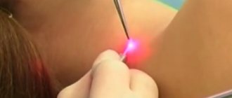
One of the most effective methods for eliminating moles is also laser removal. The laser removal surgery is performed under local anesthesia. This method is also one of the safest.
The procedure takes no more than 20 minutes, and healing occurs within 2 weeks. After this time, any traces that indicate the operation being performed disappear, there are no scars. This is very important if the operation is performed on the face, for example in the same lip area.
One of the features of the laser procedure is that the likelihood of the nevus reappearing is excluded.
It is also possible to remove a mole with a neodymium laser. This method is used only in aesthetic medicine and cosmetology. It is effective if a mole needs to be removed from the face, such as the eyelids, nose, lips or cheeks.
Despite the fact that neodymium laser is safe, it has a number of contraindications. It should not be used in certain cases:
- during pregnancy and breastfeeding;
- for purulent diseases;
- if the moles are malignant;
- if there are cracks, scratches or cuts at the impact site.
Consequences of nevus removal
Sometimes, after removal, a nevus recurs, that is, it reappears. This happens if the nevus cells were not completely removed during surgery. At the site of excision, when the crust falls off, a small dark spot may appear. It is worth knowing that this does not lead to cancer, but medical supervision is necessary. If the patient wishes, the nevus can be removed again. But in this case, mandatory histological examination will be required.
The person decides for himself whether to remove a mole or not. And if a decision is made about surgical intervention, it is necessary to take care of choosing a specialist who will carry out this manipulation.
The main thing is that the doctor has experience in carrying out such manipulations. You should always remember that removal that was carried out incorrectly can cause serious consequences, in particular, the risk of developing cancer increases. And if a mole does not bother a person and does not cause him aesthetic or physical discomfort, it should not be removed.
If a mole still bothers you, be sure to consult a doctor. Don't try to remove it yourself. The specialist, after examining the formation, will suggest surgical, laser or radio wave removal. Be sure to listen to the recommendations.
The course of the postoperative period
Careful selection of the medical institution where the procedure will be performed will help minimize the possibility of complications after mole removal. You should trust clinics that have been providing services in this area of cosmetology for a long time, equipped with modern equipment. Contacting an experienced specialist will significantly reduce the risk of nevus recurrence after removal.
After completing the procedure, you must follow all medical recommendations when caring for the skin in the surgical area. A brown crust will soon appear at the wound site. It must be protected from contact with soap solution, shampoo, cosmetics - cream, powder, etc. Do not allow the wound to come into contact with ordinary water - the microorganisms it contains can cause inflammation of tissues not covered with a protective layer of epithelium.
It is impossible to remove the crust that has healed over the wound: a new skin epidermis is formed under it. If it is torn off before it falls off on its own, a rough scar may appear in this place.
After tightening the area where the mole previously grew with new skin, the crust easily comes off on its own. Since at first the skin is very delicate and thin, it must be protected from damage and solar radiation.
Face powder should never be applied to the site of a removed mole.

