April 28, 2020 Warts
It is very rare to meet a person with perfectly clear and smooth skin.
Under the influence of certain factors, various formations may appear on it, which sometimes have a similar appearance, but often differ in the nature of their origin. Skin growths in the form of warts
can appear on the face, arms, legs, neck, etc. It is very important to correctly differentiate formations, doing this in a hospital setting, using a comprehensive examination. This will ultimately help you decide on the choice of treatment for growths, which are not always benign.
Types of seals on the back
- Atheroma, it is called a sebaceous gland cyst. It can occur under the shoulder blade or elsewhere on the back. Atheroma is often diagnosed. Occurs due to blockage of the sebaceous duct. If the sebaceous gland is not functioning properly, the ducts close. As a result, an accumulation of purulent fluid occurs. The ball is located under the skin. In advanced cases, the size exceeds a chicken egg; at the initial stage, the size of the atheroma does not exceed a pea. The sebaceous gland expands in size. This is a dense and mobile formation, its diagnosis does not cause difficulties. The danger is that with increased size, the atheroma can burst and infect surrounding tissues.
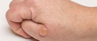
- A ball under the skin on the back means a lipoma or wen. This is a harmless growth of adipose tissue. Does not develop into a malignant formation. Causes cosmetic discomfort to patients. Wen appears anywhere on the skin where there is fatty tissue. The lump on the back under the skin does not hurt when pressed. On palpation, a soft and mobile tumor is felt.
- Fibroma is a benign tumor formed from connective tissue. It occurs on the back and any other part of the body. A compaction with clear boundaries that can be felt when pressed. Usually the color of fibroma does not differ from the skin, but sometimes it can be bluish-red. There are two types of fibroids: hard and soft. The first looks like a hard lump, and the soft fibroma hangs over the skin.
- A hemangioma is a red bump on the skin. Can be big or small. It is a collection of small blood vessels. Hemangioma injures surrounding tissues and in some cases can grow rapidly.
- A furuncle is a purulent and inflammatory process in the hair follicle. It develops as a result of bacteria entering the layers of the skin through microtraumas. The boil grows quickly. The lump on the back hurts when pressed and is bright red. Swelling of the surrounding tissues is pronounced. To the touch it is a hard, spherical and hot seal. It is dense and mobile. With severe inflammation, throbbing pain, fever, and deterioration in general health are possible.
- Myogelosis is a disease usually found in novice athletes. For beginners, the back muscles are poorly trained. During high physical activity they experience a lot of pressure. The result is myogelosis or growth on the spine.
- Malignant neoplasms have recently become more common. They can appear in people of any age. A tumor appears on the back due to osteosarcoma. This is a growth on the bone tissue that causes a lump to appear. The main cause of occurrence is chronic osteomyelitis.
The lump can grow alone or several compactions form at once. They can merge with each other. If the tumor is subcutaneous, then the doctor will not be able to determine with one palpation how many lumps are growing at the same time.
Treatment
Dangerous or not?
Should you worry if a wart grows on your back? They are not dangerous in themselves, and there is no need to treat them. But sometimes it is necessary. If the papilloma on your back itches, you will have to go to the doctor. These warts cannot be touched, since mechanical stress leads to their growth. Or the growth will degenerate into a malignant tumor. This probability is not great, but it exists. About 10% of keratomas contain cancer cells. Only a doctor can determine whether a tumor is malignant or not, after a histological diagnosis.
It is not always possible for a person without specialized education to independently determine that he has seborrheic warts. Therefore, a consultation with a dermatologist will not be superfluous.
Sometimes the growth becomes the cause of infection entering the body and the appearance of an inflammatory process. Particularly dangerous are warts that are voluminous, convex, and have a loose structure. If you injured it or scratched it, bleeding will appear and the formation will become infected, which will lead to serious consequences. Due to mechanical stress, it begins to grow.
How to treat
How to remove warts on the back? If it doesn't bother you, you don't have to touch it. It doesn't cause any harm. But patients sometimes ask to remove it, because it disfigures the appearance. On the back the growth is not so noticeable. But if you are worried about itching, it starts to itch, you should visit a dermatologist. Another reason for going to the doctor is the risk of the growth degenerating into an oncological tumor. Or the growth gets in the way, is constantly injured and there is a high risk of infection.
After examining the wart, the dermatologist will recommend a treatment method.
Treatment
How is the treatment carried out? If a person has one wart on his back, and it is small, the doctor will suggest using creams or ointments that contain sulfur, retinoids, and salicylic acid. They will either completely remove the growth or make it invisible and smooth it out. Other medications are not used. But there is evidence that large doses of ascorbic acid stop the disease. Long-term treatment with vitamin C is required, up to 2 months. Thanks to ascorbic acid, new spots and nodules will not appear, but old ones will not disappear.
You need to take vitamins under the supervision of a doctor, who will choose the dosage for you. Otherwise, you will develop additional health problems, such as kidney stones.
Main features and differences
Before you sound the alarm and run to the doctor, you can figure out the reasons for the appearance of unwanted lumps yourself. And if all the signs completely coincide with the descriptions, and the cause turns out to be insignificant, then you can get rid of the swelling yourself
But, pay attention! If the red bumps itch and enlarge, if their localization grows, and they appear for no apparent reason, it is better to consult a doctor immediately
Diagnosis by a dermatologist
We also recommend that you consult a medical professional if you notice similar problems in your child. He will not be able to accurately answer whether the tumor itches and when exactly it appeared.
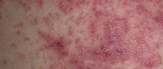
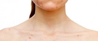
One of the most common causes is an allergic reaction of the body to some irritant. This is especially common in young children. When you notice small red spots on the body that cause itching (see photo), you should take an antihistamine in accordance with the age of the victim. Allergies can be caused by numerous external irritants, the most common being:
- household chemicals, detergents, washing and cleaning products;
- food products, most often sugar for a child;
- medicines;
- cosmetics and perfumes;
- pollen of flowering plants;
- dust and mold;
- animal fur.
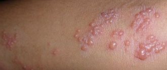
Allergic rash in a child
Note! Anything can be an allergen
It is important to identify him as quickly as possible and completely limit contact with him. A rash alone may not be enough
More severe forms may include swelling and difficulty breathing. In this case, you should immediately call an ambulance
The rash alone may not be enough. More severe forms may include swelling and difficulty breathing. In this case, you should immediately call an ambulance.
Many people have small lipomas on their bodies, but some don’t even know what they are. Benign tumors made of fat cells can be located where there is most fat tissue. They are dense and quite elastic; they can be quite small in size, or can reach 2-3 cm in diameter. As a rule, lipomas do not cause discomfort or itch, see photo. But if there is an inflammatory process in the body, the bumps on the body itch. There may be a lot of them on the arms and legs. It is recommended to periodically undergo examination by an oncologist.
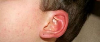
When an infection enters the human body, the lymph nodes are most often the first to react to its appearance. They become inflamed and red, and quickly increase in size. If the lump on the head itches and is located behind the ears, then most likely it is a lymph node. When pressing on it, pain occurs. These are quite dense formations that can change their location, causing discomfort. These red bumps are a filter for the blood; they trap the infectious agent. They should be treated strictly according to the doctor’s recommendation. Lymph nodes can also be located on the arms, legs, neck, and head.
Insect bites are the least dangerous, but they also require attention and additional treatment of damaged skin. If the bumps on your legs itch from bites, then you should take any antihistamine and treat the bite area with special means.
Important! Without knowing the exact cause of the appearance of tumors, you do not need to start treating them yourself!
How the virus spreads
Method of transmission of the virus: any bodily contact. People often do not even realize that they are infected. The incubation period of the disease does not show the reasons for its existence. Only after 3 weeks and up to 6 months, the first symptoms appear on a person’s skin - small neoplasms. They are called papillomas. These can appear on different areas of the skin. Papillomas often attack: the face, neck, arms, genitals, fingers and feet. The localization of neoplasms is related to the condition of the skin. If there are scratches, abrasions or wounds, the risk of neoplasms occurring in this area increases. And if the method of transmission of the virus occurs through sexual intercourse, then papillomas arise precisely in the intimate area. Each sexual contact is accompanied by several microtraumas, abrasions and stretches of the skin. When the virus is transmitted sexually, such places are the first to become infected.
If there are other causes of infection, then it is often a household factor. Using someone else's hygiene products: washcloth, soap, towel, etc., can play a cruel joke on a person. The broken rule “use only your own” often causes the virus to get on the skin.
Many cases have been recorded where the appearance of papillomas is associated with visiting public places: gyms, swimming pools, saunas, etc. Virus cells can be located on furniture surfaces and on the floor, as well as the walls of the room.
Such places include showers and locker rooms. Virus-carrying bacteria, once on the skin, quickly find a way to penetrate the blood.
Public swimming pool, habitat of the virus
About the subcutaneous boil
A boil is a purulent inflammation of the skin. There are many reasons for its appearance:
- obesity;
- diabetes;
- metabolic disorders;
- intestinal disorder;
- abrasions and cuts.
Boils are painful subcutaneous pimples on the face that are accompanied by pain at the slightest touch. At the base of this formation there is a purulent rod, and at its tip there is a head filled with purulent fluid. Often they try to remove it to remove the pus, but this only leads to worse consequences: the boil begins to increase in size, damaging more and more tissue. Many, not understanding the danger, try to squeeze out the unfortunate pimple, enduring extreme pain when pressed. But the inflammatory process can only deepen into the skin with such actions.
Manifestation of furunculosis
Under no circumstances should you apply compresses; they only create excess moisture, which contributes to the development and enlargement of acne. To reduce the size of the formation, it is necessary to dry the skin; even heating helps to reduce the inflammatory process and its faster maturation. An abscess will gradually form, which should be lubricated with a special ointment with the addition of an antibiotic; you must first consult a doctor. He will analyze the condition of the pimple and give recommendations for its removal.
Important! Subcutaneous boils on the face are treated comprehensively. It is mandatory to prescribe a course of medications for external and internal use, as well as a complex of vitamins to cleanse the blood
A preliminary examination is carried out, which is aimed at finding out the cause of the disease.
Reasons for appearance
Subcutaneous neoplasms appear for various reasons. They are classified according to the etiological factor.
The following types of formations may appear under the skin:
- Abscess. It looks like a hard lump filled with pus. Hyperemia (redness) appears around the perimeter of the neoplasm. Local temperatures are rising. In advanced cases, an abscess threatens to spread the infectious process throughout the body.
- Intradermal cyst. It is a compaction on the face in the form of a ball under the skin of varying sizes. It can also appear on the jaw.
- Hemangioma is a painless lump under the skin on the face in the form of a hard or soft red lump. As a rule, hemangiomas go away on their own without treatment.
- Lipoma is a painless and harmless growth under the skin. It looks like a ball and moves freely when pressed. This seal does not pose a danger to life; it is removed solely for aesthetic reasons.
- Cancerous bumps look like a slight compaction. In the early stages of development they do not manifest themselves at all.
- Hygromas are dense, inactive balls under the skin. Very rare on the face. Hygromas do not hurt, do not cause discomfort, and can very rarely itch. The main problem in their occurrence lies in the presence of psychological discomfort. Sometimes the hygroma disappears after an accidental blow or strong pressure.
- Atheroma can occur as a result of a hormonal imbalance or a deterioration in the outflow of sebaceous gland secretions. This cone is mobile and has a dense structure. The swelling can quickly increase in size. The purulent form of the pathology causes severe pain, deterioration of health, and increased body temperature.
- Fibroma is a benign growth inside the skin. It is a small compaction that does not contain pus. Most often, fibroma appears as a result of prolonged stressful situations or hormonal imbalance. It can only be removed as part of aesthetic treatment.
Fibroma on the face
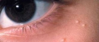
- Milia is a type of acne. These neoplasms look like small bodily pimples or bumps on the skin. They cause mainly cosmetic inconvenience. They do not cause pain or itching.
Reasons why subcutaneous lumps appear in the facial area:
- hormonal disorders;
- cold;
- poor diet with consumption of large amounts of harmful foods;
- consumption of alcoholic beverages;
- smoking;
- allergies;
- pathologies of the nervous and endocrine systems;
- injuries;
- unfavorable heredity;
- prolonged exposure to heat or cold;
- penetration of infection;
- improper use of cosmetics;
- failure to comply with the rules of hygienic skin care.
What are neoplasms and what are they like?
By their structure, all skin neoplasms (they are also called “tumors” or “neoplasia”) are the result of uncontrolled cell proliferation
In terms of their structure, all skin neoplasms (they are also called “tumors” or “neoplasia”) are the result of uncontrolled proliferation of cells that have not yet reached maturity, and therefore have lost the ability to fully perform their functions. Depending on the clinical picture, they are usually divided into 3 types:
- Benign (atheroma, hemangioma, lymphangioma, lipoma, papilloma, mole, nevus, fibroma, neurofibroma) Do not pose a threat to human life, but if poorly placed or large in size, they can cause disruption in the functioning of other systems and/or organs of our body. Under external influences they can sometimes transform into malignant neoplasms.
- Malignant (basal cell carcinoma, melanoma, sarcoma, liposarcoma) grow quickly and aggressively, penetrating into surrounding tissues and organs, often with the formation of metastases . The prognosis of such diseases is often unfavorable, given the difficulty of curing them and the tendency for frequent relapses, and in some cases, active metastasis leads to death if vital organs are irreversibly damaged.
- Borderline or precancerous skin conditions (senile keratoma, xeroderma pigmentosum, cutaneous horn, Bowen's dermatosis) Formations whose tissues, under the influence of hereditary or current causes, have changed, having the potential to degenerate into malignant tumors.
Benign neoplasms
The cells of these formations partially retain their original functions and have slow growth rates. Sometimes they press on nearby tissues, but never penetrate them. In their structure, such neoplasms are similar to the tissues from which they originated. As a rule, they respond well to surgical and other hardware treatment and rarely recur.
Atheroma
A tumor of the sebaceous gland formed after its blockage. Most often it occurs on the scalp, neck, back, and groin area, that is, in places with a high concentration of sebaceous glands. It looks like a dense formation with clear contours, elastic and mobile during palpation, and does not cause discomfort. When suppuration occurs, redness and swelling of the tissues, pain, and increased body temperature appear.
The inflamed atheroma can break out on its own, releasing purulent-sebaceous contents.
This epithelial cyst has a tendency to transform into a malignant form - liposarcoma. Atheroma can only be removed through surgical excision.
Hemangioma
Hemangioma is a benign vascular tumor formation
Benign vascular tumor formation. It can be simple capillary (on the surface of the skin), cavernous (in the deep layers of the skin), combined (combining the two previous forms) and mixed (affecting not only blood vessels, but also surrounding tissues, mainly connective).
Capillary hemangioma can reach large sizes, its color varies from red to bluish-black, and grows mainly to the sides. The cavernous variety is a limited subcutaneous nodular formation covered with bluish or normal-colored skin. Most often, these tumors appear in newborns, literally in the first days of life, and are located in the head and neck area.
If the geangioma is located in a complex area of the body (for example, on the face in the orbital area) or occupies a large area, it is removed by radiation. Other treatment methods are sclerotherapy, cryotherapy, hormonal drugs. When the tumor is deep and conservative treatment is ineffective, surgical excision is required, including the underlying layers of skin.
Lymphangioma
A benign formation from the walls of lymphatic vessels that occurs in children even at the stage of intrauterine development.
Most of these tumors are detected before 3 years of age.
It is a thin-walled cavity ranging in size from 1 mm to 5 cm or more (cystic lymphangioma, consisting of several isolated or communicating cysts). It grows very slowly, but in some cases there is sudden growth to a significant size - in this case, surgical removal is required. Also, lymphangiomas located in close proximity to the trachea, larynx or other vital organs are necessarily removed.
Lipoma
Lipoma structure
A tumor of the fatty layer (often called a “wen”), located in the subcutaneous layer of loose connective tissue. It can penetrate deep into the body to the periosteum, seeping between vascular bundles and muscles. Most often found in areas where the fat layer is thinnest - the outer surface of the hips and shoulders, shoulder girdle, upper back. It looks like a soft formation, mobile and painless on palpation.
Lipoma grows quite slowly and is generally safe for the body, although in rare cases it can degenerate into a malignant formation called liposarcoma.
At the same time, if the wen grows and begins to put pressure on surrounding tissues, surgical removal is indicated. It is better not to wait for this moment, since the larger the tumor, the more noticeable the postoperative scar will be.
But small-sized wen are easily removed using laser, radio wave or puncture-aspiration methods, after which there are practically no traces left on the skin.
Papillomas and warts
Papillomas and warts are formations in the form of a nodule or papilla that have a viral origin
Formations in the form of a nodule or papilla, having a viral nature. They are caused by various strains of human papillomavirus (HPV), usually due to decreased immunity, stress and vegetative disorders. Externally they are very diverse, most often they look like growths of various shapes and sizes, coloring from light to dark brown and gray.
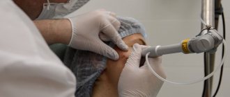
Some types of warts can develop into cancerous tumors, but for the most part they are relatively harmless to health. Complex treatment includes the prescription of antiviral and immunomodulatory drugs, as well as the removal of the growths themselves; almost any method is suitable for this: treatment with chemically active acids, interferon injections, cryodestruction with liquid nitrogen, electrocoagulation, radio or laser exposure, surgical excision.
Moles and nevi
Benign skin tumors, congenital or acquired. They are a cluster of cells overflowing with the pigment melanin. They can have different sizes, shapes, colors and surface textures (for more details, see the article “Types of moles”).
Some of them have a high potential for degeneration into a malignant form - melanoma. For example, a pigmented border nevus, a flat nodule of dark brown or gray color with a dry, uneven surface.
Such formations must be removed, and only surgically (for more details, see the article “Malignant moles”).
Melanomic moles and nevi do not require treatment, but experts recommend getting rid of those that are constantly injured or are located on open areas of the body and are often exposed to sunlight in order to avoid complications. The method here is not so critical: in addition to a scalpel, a mole can be removed with a laser, cryodestruction or radio waves.
Fibroma (dermatofibroma) is a formation in the connective tissue that is most often found in women at a young and mature age.
Fibroma (dermatofibroma)
Formations in connective tissue, which are most often found in women at a young and mature age. They are small in size (up to 3 cm), look like a deeply sealed nodule, spherically protruding above the surface of the skin, the color is gray to brown, sometimes blue-black, the surface is smooth, less often warty. It grows slowly, but there is a possibility of oncological complications: in rare cases, fibroma can degenerate into malignant fibrosarcoma.
Neurofibroma
A tumor that grows from nerve sheath cells. Most often located in the skin and subcutaneous tissue. It is a dense tubercle measuring from 0.1 to 2-3 cm, covered with depigmented or highly pigmented epidermis. Multiple neurofibromas are caused by hereditary or genetic causes and are considered a separate disease - neurofibromatosis.
This tumor rarely transforms into a malignant one, but in itself it is quite dangerous - it can cause constant pain and cause serious functional disorders in the body, and therefore requires treatment, at least pharmacological (retinoids). In difficult cases, surgical excision or radiation therapy is indicated.
Malignant neoplasms of the skin
Formations of this type quickly grow, penetrate into surrounding tissues, and often form metastases even in organs distant from the lesion due to the transfer of pathological cells through the circulatory and lymphatic systems. In these tumors, the body's control over cell division is completely lost, and the cells themselves lose the ability to perform their specific functions. Malignant neoplasms are quite difficult to treat; they are characterized by frequent relapses of the disease even after surgical removal.
The main signs of the degeneration of a benign tumor or a stable borderline skin condition into a malignant formation are:
- pigmentation changing in color or saturation;
- sharp and rapid increase in size;
- spread of the tumor to neighboring tissues;
- bleeding, ulceration, etc.
Metastases of malignant neoplasms can occur in any organs and tissues, but most often the lungs, liver, brain, and bones are affected. At the metastasis stage, the prognosis for treatment is often negative, including death.
Melanoma
One of the most common types of cancer. In most cases, it is the result of malignancy of moles and nevi after severe trauma or excessive ultraviolet irradiation. Gives metastases to almost any organ, starting with regional lymph nodes, and often recurs. It is treated surgically, in combination with chemotherapy and radiation therapy.
Basalioma
Basalioma – initial stage
A dangerous type of squamous cell skin cancer is formed from atypical basal cells of the epidermis. At the first stage, it looks like a white nodule with a dry crust on the surface, over time it grows in width and begins to ulcerate, then transforms into a deep ulcer or mushroom-shaped nodule protruding above the surface of the skin. It develops in areas of the body exposed to ultraviolet radiation, high temperatures, and carcinogenic substances. It is treated with standard methods - surgical excision, radiation, chemotherapy, cryo- or laser therapy.
Kaposi's sarcoma, angiosarcoma, hemorrhagic sarcomatosis
Multiple malignant formations in the dermis. They look like violet, purple or lilac spots without clear boundaries; gradually dense round nodes up to 2 cm in diameter of a bluish-brown color appear on them, with a tendency to unite and ulcerate. Most often, this type of sarcoma affects HIV-infected people, occurring in an aggressive form, quickly leading to death.
Liposarcoma
Malignant tumor of adipose tissue. Most often they occur in men and people aged 50 years or more. In most cases, it develops against the background of benign formations - lipomas and atheromas.
Liposarcoma usually grows slowly and rarely metastasizes.
When localized in the subcutaneous fat, it is palpated as a fairly large (up to 20 cm) single round-shaped node with irregular outlines and uneven density, hard or elastic to the touch. Surgical treatment and chemotherapy in combination with radiation are used.
Fibrosarcoma
Develops in soft tissues, mainly connective tissues, most often in the lower extremities. When localized superficially, it can protrude noticeably above the skin and has a dark blue-brown color. At a deeper location it is visually invisible. There are differentiated and poorly differentiated fibrosarcoma, the first is considered less dangerous - it grows relatively slowly and does not metastasize, but both types give a high percentage of relapses after removal.
Precancerous skin growths
This group includes pathological conditions of cells that are more or less likely to result in degeneration into malignant formations.
Bowen's disease (intraepidermal cancer)
Bowen's disease - formation in the epidermis without germination into surrounding tissues
Formation in the epidermis without germination into surrounding tissues. If proper treatment is not carried out, it transforms into invasive skin cancer with proliferation and metastasis. Most often observed in older people, localized on the head, palms, and genitals. Bowen's disease is caused by some chronic dermatoses, keratinized cysts, skin trauma with scarring, radiation, ultraviolet and carcinogenic exposure.
At the initial stage, it looks like a reddish-brown spot from 2 mm to 5 cm without smooth borders, then it transforms into a raised plaque with raised edges and a flaky surface. After removing the scales, a non-bleeding, weeping surface is revealed. Evidence of the transition of Bowen's disease to a malignant form is ulceration.
Xeroderma pigmentosum
A tumor that develops when the skin is excessively sensitive to ultraviolet radiation, when age spots become warty growths. This disease is quite rare and is hereditary. In the early stages, treatment comes down to taking medications that reduce susceptibility to UV rays with clinical observation by a dermatologist or oncologist. At the stage of formation of growths, their surgical removal is recommended.
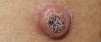
Actinic keratoma (senile keratosis)
It looks like rashes up to 1 cm in diameter, ranging in color from yellow to dark brown. As they develop, dry crusts and scales form on the spots, which when peeled off cause slight bleeding. The formation of compactions in the tumor indicates the transition of a borderline state into a malignant tumor.
Cutaneous (senile) horn
A cone-shaped formation resembling a horn of yellowish or brown color, which is why it got its name.
Characteristic of older people, it occurs mainly in open areas of the skin that are regularly subjected to friction or compression, and is formed from cells of the spinous layer of the skin.
It develops as an independent formation, a consequence of benign tumors (most often warts) or the initial stage of squamous cell carcinoma. It is removed surgically.
Rheumatic bumps
Painless bumps in the form of subcutaneous nodules can be a manifestation of erythema nodosum, characteristic not only of rheumatism, but also of some severe infectious diseases, for example, erysipelas or tick-borne borreliosis. Rheumatism is a connective tissue disease in which mainly the membranes of the heart become inflamed, as well as large joints, such as the joints of the legs, knees or hip joints. Rheumatism of the back is diagnosed quite rarely and accounts for no more than 8.4% of the total number of patients with this diagnosis.
Cones with rheumatic inflammation of the spine are painless, have a dark red, purple or burgundy color and clear contours. The structure of the formations is dense, motionless (less often, inactive). The size of the nodules can range from a few millimeters to a chicken egg, and their occurrence is preceded by high fever and other symptoms, which may include:
- intense aching pain in the back (mainly in the area where the nodules are located);
- progressive curvature of the spine;
- numbness, trembling in the limbs;
- pale pink rash;
- minor hemorrhages against the background of increased pallor of the skin;
- increased sweating.
The disease is caused by an infectious pathogen - group A β-hemolytic streptococcus - and often develops after illnesses of the oropharynx (sore throat, tonsillitis, scarlet fever, pharyngitis).
How to treat rheumatism of the back?
In the acute phase of the disease, with the development of acute rheumatic fever, treatment is carried out in a hospital. The patient undergoes sanitation of foci of chronic infection (washing the tonsils or paranasal sinuses, treatment of carious teeth, etc.), and is prescribed bed rest, which is necessary to relieve stress from the affected joints.
Drug therapy for cutaneous rheumatism is carried out according to the scheme presented in the table below.
Table. Drugs for the treatment of rheumatic fever.
| Pharmacological group | Drugs |
| Glucocorticosteroid hormones for relief of inflammatory processes and allergic reactions | "Hydrocortisone", "Dexamethasone", "Prednisolone". |
| Antihistamines | "Suprastin", "Claritin", "Loratadine", "Tavegil". |
| Antibiotics | "Amoxicillin", "Ciprolet", "Ciprofloxacin". |
| NSAIDs to reduce pain and inhibit the synthesis of prostaglandins (inflammatory mediators) | "Ibuprofen", "Ketorolac", "Diclofenac". |
| Immunosuppressants, antiprotozoals and antimalarials | "Delagil", "Chloroquine". |
Outside the exacerbation phase, the patient is shown restorative procedures and manipulations: massage, exercise therapy, paraffin therapy, physiotherapy (infrared irradiation, electrophoresis, UHF).
Drug treatment of papilloma on the back
There are many medications available for the treatment of papillomas over the counter in pharmacies. It is important to remember that the causes and treatment of pathology are inextricably linked. You should be careful when treating a wart with pharmaceutical drugs, as they harm healthy skin around the growth. Such medications are:
- Feresol,
- Cryopharma,
- Salicylic acid,
- Super Celandine.
The doctor may prescribe a course of antiviral and immunostimulating tablets:
- Anaferon,
- Amizon,
- Isoprinosine.
Taken together, such drug therapy helps relieve the symptoms of papillomavirus and improve the functioning of the immune system, getting rid of the problem without surgical intervention.
Benign neoplasms
New growths on the skin, classified as benign, as can be seen in the photo and their description, are not prone to rapid growth, metastasis and relapse after removal.
Differences between benign and malignant moles.
These tumors include:
- atheroma;
- hemangioma;
- lymphangioma;
- warts;
- moles (nevi);
- fibromas, etc.
The structure of a lipoma (benign tumor, popularly called a wen)
| View | Description |
| Atheroma | Damage to the sebaceous gland in the form of a cystic growth. Location: face. A raised formation, indistinguishable from normal skin in color, is tightly fused to it at one point. The contours are clear. Palpation of the atheroma is painless. |
| Hemangioma | Neoplasm of vascular origin, refers to childhood tumors. It can be either an independent disease or a manifestation of another pathology. The course depends on the patient’s age, location, size and depth to which the hemangioma has grown. Frequent locations are the head, face, neck, but other locations are also possible. The first manifestation is a red nodule (papule) up to 5 mm in diameter. Complaints include bleeding when accidentally touched, and sometimes dysfunction of the organ where the tumor is located. |
| Lymphangioma | There are two possible development options - a congenital defect or consequences of impaired lymph flow. The tumor can be located anywhere, but more often it is the oral cavity, neck, and upper limbs. Capillary lymphangiomas are multiple blisters with a yellowish transparent liquid inside. The same tumors that arose as a result of impaired lymph circulation look like plaques or spots, the diameter of which slowly increases. |
| Lipoma | Tumor of adipose tissue. More often they have a nodular shape. The course is asymptomatic. It is a painless pale pink neoplasm on a stalk. The consistency is doughy. The boundaries are unclear. |
| Seborrheic warts | Also called "senile". They arise as a result of impaired differentiation of cells in the basal layer of the epidermis. Appearance: Nodules or plaques raised above the skin. The surface is lumpy. The shape is often round or oval. The color of the wart varies from yellow-brown to black. |
| Moles | Or nevi. They are a developmental defect and consist of unchanged melanocytes. The characteristic color of the rash is from light brown to black, which is associated with different amounts of melanin (dark pigment) in the cells. More often, moles have a smooth surface and sometimes rise above the skin. |
| Fibroma | Fibroma is a tumor that forms from connective tissue. Dense to the touch. Reaches large sizes. Malignancy (transition to a malignant formation) of the tumor is possible. |
Neoplasms on the skin (photos and descriptions are presented above) of a benign nature, despite their relatively safe nature, can sometimes still develop into precancrosis and even cancer.
Medication self-treatment of papillomas
Reducing skin growths is the goal of everyone infected with the HPV virus. The fight against papillomas is not easy. In addition to the fact that neoplasms must be removed, care should be taken to ensure that they do not appear again. One of the treatment methods is medication. Preparations for removing papillomas can be purchased at any pharmacy. They are inexpensive and popular among people infected with the virus. These drugs include:
- Verrucacid,
- Solcoderm,
- Feresol,
- Cryopharma,
- Super clean,
- Oxolinic ointment,
- Salicylic ointment,
- Iodine.
Each of the remedies is called “time-tested”. For more than half a century, some of these drugs have been actively used to remove papillomas at home.
A simple way to remove papilloma is to treat it with iodine. If you coat the growth with this product 3-4 times a day, then after a week and a half the papilloma will dry out and fall off on its own. “Verrukatsid” has the same properties. This drug is produced in the form of an oil for external use. By treating the surface of the neoplasm 3-4 times a day, you can get a positive result already on the 5th day of the procedure. The exact amount of treatment time depends on the type, shape and size of the wart. Papilloma can be lubricated at home with oxolinic or salicylic ointment. Treatment with these is longer, but no less effective.
Oxolinic ointment, an inexpensive but effective drug
How to quickly get rid of papilloma
Neoplasms can be reduced in a quick way. For such cases, the modern drug “Cryopharma” is used. This product for removing warts is used in the form of freezing the tumor. After 2-3 uses, the growth disappears completely.
An old proven remedy is celandine. A balm based on it is sold in every pharmacy. The drug must be applied carefully. It is very poisonous due to the high content of sodium and potassium hydroxide in its composition. You can remove the growth using “Super Clean” in several sessions. This remedy leaves burns on the body of the growth, contributing to the necrosis of its tissues. At the 3-5th session, not a trace remains of the neoplasm.
"Cryopharma" freezes the growth
Why do they occur?
The study of skin tumors is still ongoing. The exact reasons for their occurrence have not been established, but scientists put forward several theories about this.
Provoking factors may be:
- family history (presence of neoplasms in relatives);
- individual characteristics of a person (light skin and hair, old age);
- exposure to ultraviolet, radiation and x-ray radiation;
- viral infection;
- long-term trauma to the skin;
- chronic exposure of the skin to chemical carcinogens (nitrosoamines, benzopyrene, aromatic amines, etc.);
- insect bites;
- metastatic processes in the presence of an oncological process in the body;
- violation of the trophism of the skin, hence chronic skin ulcers;
- weakened immune system (due to immunosuppressive therapy, HIV infection, etc.).
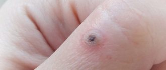
Type of infectious formations
Wart-shaped formation on the skin
that might be exactly what it is.
They can appear in children and adults under the influence of the human papillomavirus, which can appear several years after infection. Such warts on the skin
(
photo
) can differ in shape, structure and health hazard - everything will depend on the strain of the virus.
There are a type of warts on the skin
. They are:
- Ordinary or vulgar - often affects the surface of the fingers of the upper and lower extremities.
- Plantar - appear in the foot area and can greatly interfere with walking.
- Periungual - localized in the area of the nail plates, provoking the appearance of characteristic changes in it.
- Flat or youthful, which appear more often in children and adolescents. These growths are small, yellowish or flesh-colored.
- Thread-like, which can affect any part of the body - armpits, neck, face, chest, etc. When damaged, they tend to spread to healthy tissue areas.
- Pointed or genital - appear under the influence of certain strains of HPV, most of which have a high degree of oncogenicity. Affects the skin of the genital organs and anus.
Separately, we can highlight the appearance of senile or age-related keratomas, which appear in people of mature age and are not at all related to HPV. They are caused by physiological changes in skin cells.
Reasons why a lump appears and itches
What types of formations are there?
In medicine, many different types of cones have been recorded, which differ in some characteristics. Below are the most common external differences of neoplasms:
- cone size;
- location;
- the number of similar swellings;
- skin color on the lump;
- whether it itches or not;
- density of internal contents.
Education is not a problem
Possible causes of bumps
- allergic reaction;
- inflammation of the lymph nodes;
- folliculitis;
- hemangioma;
- infectious skin lesion;
- lipoma;
- bites of various insects;
- subcutaneous wen;
- cancerous tumors;
- atheroma.
Note! This is not a complete list that you can find in medical reference books. But, the above factors are the most common causes of all kinds of swelling that itch
Main features and differences
Diagnosis by a dermatologist
We also recommend that you consult a medical professional if you notice similar problems in your child. He will not be able to accurately answer whether the tumor itches and when exactly it appeared.
One of the most common causes is an allergic reaction of the body to some irritant. This is especially common in young children. When you notice small red spots on the body that cause itching (see photo), you should take an antihistamine in accordance with the age of the victim. Allergies can be caused by numerous external irritants, the most common being:
- household chemicals, detergents, washing and cleaning products;
- food products, most often sugar for a child;
- medicines;
- cosmetics and perfumes;
- pollen of flowering plants;
- dust and mold;
- animal fur.
Allergic rash in a child
When an infection enters the human body, the lymph nodes are most often the first to react to its appearance. They become inflamed and red, and quickly increase in size. If the lump on the head itches and is located behind the ears, then most likely it is a lymph node. When pressing on it, pain occurs. These are quite dense formations that can change their location, causing discomfort. These red bumps are a filter for the blood; they trap the infectious agent. They should be treated strictly according to the doctor’s recommendation. Lymph nodes can also be located on the arms, legs, neck, and head.
Painful lumps under the skin. Red bumps on the body itchy
Bumps on the human body can occur as a result of various skin diseases. The most common include: cysts, follicles, lipomas. Depending on the cause, the bumps may be hard or soft, painful or painless. Why do they appear and how to deal with them?
Instructions
The origin of lumps on the human body can be different, depending on the nature of the existing disease. Most often they form after injuries or bruises. Also, the reasons for the formation of bumps on the body include: inflamed lymph nodes - infections - insect bites - acne - tumor, folliculitis - allergic reaction of the body - skin cancer - warts.
Bumps can appear on various parts of the body. If such seals are detected, you should consult a doctor, since the true cause of their appearance can only be diagnosed by an experienced specialist.
For your first appointment, make an appointment with a therapist. If necessary, he will give a referral not only for tests, but also for examination by other specialists.
Once the diagnosis is made, you will be prescribed appropriate treatment.
Very often, bumps go away on their own without any outside intervention, but some diseases will require consultation with an experienced doctor and a competent course of treatment. First, a specialist will determine the reason for the appearance of these seals. There can be quite a lot of options.
If the bumps are the result of an infection, then most likely your doctor will prescribe antibiotics and anti-fungal medications, as it is very important to stop the spread of infection and prevent the formation of scars.
If the seals are the result of the formation of a cyst, then medical intervention may not be required. In most cases they go away on their own.
However, if an inflammatory process occurs, cortisone injections will come to the rescue.
If treatment does not produce results and the bumps do not go away, you should immediately consult a doctor, since if such symptoms occur, you may have to resort to surgical intervention.
Surgery is the only solution when the lumps are lipomas. They are removed if they cause real discomfort or if they are a cosmetic defect of the skin.
When the lump is an oncological pathology, it is removed along with the tissues surrounding it. Warts can also be called a lump, and they are removed only according to existing indications, either by medication (the result is not immediate, but gradual), or using a laser.
If bumps on the body appear as a result of an injury or bruise, then to avoid swelling, you first need to apply cold, then apply cream or ointment to speed up healing (there are many absorbable gels and ointments that are effective for bruises).
Another option for the appearance of seals is insect bites. In this case, you need to apply products that repel them to your skin. If you are nevertheless bitten, then the bite sites must be treated with special solutions: Fukortsin, Diamond Green solution, alcohol and others.
If lumps appear in the genital area, it is very important to get tested to detect sexually transmitted infections. When treating a herpes infection, acyclovir or valacyclovir is used as treatment. For condylomas, chemicals are used or they are removed surgically.
It should be taken into account that seals constantly appear under the influence of some factors (inflammatory processes, previous injuries, infectious and oncological diseases). The circumstance of the neoplasm determines the duration of its manifestation. Redness and swelling can be either single or multiple. Their sizes vary from 1 mm to 10 cm.
Do not forget that the appearance of red growths on the face and body of a person may indicate severe acne, at a time when a certain number of pimples turn into compactions. Quite often, such symptoms are observed with insect bites.
If the compaction resembles a smooth moving ball, a cyst may be suspected. In most cases, this pathology is not accompanied by additional symptoms.
Formations in the form of huge red pimples on the scalp, thighs and face indicate the development of folliculitis (inflammation of the hair follicle). They develop much more often in people with weakened immune systems, diabetes and obesity.
Please note that the appearance of impressive red bumps on the body and face may indicate infection or an allergic reaction of a person to any cosmetic products. When the lymph nodes are inflamed, the tumors are located above them. Along with this, a person experiences an increase in body temperature and severe intoxication.
It is fundamentally important to know that the red lump on the skin may be a hemangioma - a benign tumor of dense or soft consistency.
If not treated in a timely manner, it increases in size and destroys healthy neighboring tissues. With skin cancer, red lumps are fused with adjacent tissues and in most cases are painful.
In the later stages of the disease, they can transform into purulent bumps.
If red lumps appear on the skin of the back, abdomen, arms and legs, you should consult a therapist. In accordance with the expected diagnosis, he refers you to a specialist for consultation. Do not delay your visit to the doctor, because in some cases, neoplasms can be the result of important ailments, without causing any inconvenience.
Every neoplasm anywhere in the human body not only leads to frenzy, but also brings a person to panic. It is especially concerning if a lump appears under the skin on the back. Does it require removal? Definitely yes. It doesn’t matter if there is a feeling of discomfort or pain. Each growth can develop into a malignant formation at any time.
Bumps on the body under the skin of a person can appear at any time, regardless of age, in women, men, and children, including. They are distinguished by external data and divided into types.
Doctors to this day cannot confidently say and indicate the causes of the pathology. Of course, for the most part, the bumps are benign. But osteosarcoma is not uncommon in modern medical practice.
It is possible to say specifically what appeared on the back only after a thorough examination and diagnosis. An examination by a specialist is required, who will prescribe all the necessary types of tests.
The appearances of each species are different.
- Environmental factors, hormonal imbalances, and even some medications can provoke the formation of hemangiomas. There are cases when an illness suffered by the mother during pregnancy gives impetus to the growth of the growth already during the child’s adolescence.
- Lipomas can be caused by intoxication, improper metabolism, injuries in the spine, or an inactive lifestyle.
- Osteomas most often occur in males and are considered a hereditary disease.
A lump on the back can grow alone. Sometimes there are several of them and they merge into a cluster. The difficulty is that when examining with fingers, a specialist will not be able to say with accuracy how many there are in one place. The common causes of all neoplasms are:
- Disruption of sebaceous secretions, metabolism and hormonal changes.
- Minor injuries to the hair follicle or sebaceous gland.
- Congenital defects and hereditary factors.
- Seal rupture.
In cases where people discover a lump in any part of their body, there is a need to go to the hospital for examination and comprehensive diagnosis. The main thing is to determine the type of growth and its area of distribution. For this use:
- computer or magnetic resonance imaging;
- radioisotope scintigraphy;
- biopsy;
- histology and blood tests.
Once all the measures have been taken, you can judge the nature of the disease. After this, treatment is prescribed.
The appearance of bumps on the back, regardless of location (right or left), are internal or external in nature and are a fairly common occurrence in the modern world. There is only one solution to the problem - removal.
In cases of malignancy, chemotherapy or other types of cancer control are added.
How to determine that a tumor is malignant
Only a specialist can say more specifically after collecting all the tests. The distinctive features of such growths are:
- the appearance of a lump in the back area;
- nagging or aching pain in the lower abdomen on the right or left;
- back ache, significant pain at night.
With rapid metastasis. And also if the lower vertebra is damaged, lameness occurs.
What is the difference from inflammatory processes
Cases where lumps form under the skin of the back can also be of an inflammatory nature. For example, a boil or carbuncle has formed. They mainly form on the neck, forearms or shoulder blades.
It was noted that, in terms of frequency of occurrence, their favorite place is the right side. It is quite easy to recognize them, since the hair follicle or sebaceous sac is involved in the inflammation process.
The pus that forms in this area of the skin involves surrounding tissues in the inflammatory process. As a result, intense pain, redness and induration, and increased temperature appear.
If not treated promptly, the infection spreads further throughout the body, entering the blood.
If any lumps occur in the back area, do not self-medicate and consult a doctor immediately.
Swellings can be located on any part of the body and can be single or multiple, soft or hard. The size of the cones also varies from barely noticeable to impressive.
Some lumps are not accompanied by any other symptoms, while others are accompanied by redness of the skin, soreness and itching. Most often, lumps on the skin occur after injuries, but they can also appear for no apparent reason. In this case, you need to listen to your body so as not to trigger a serious illness, one of the first symptoms of which was a lump.
On the head. Folliculitis occurs as a result of inflammation of the hair follicles. The cause may be infection, mechanical or chemical exposure. The disease manifests itself in the form of red lumps on the scalp and itching. Folliculitis also occurs on the thighs.
If warty formations of brown or dirty yellow color appear on the skin of the face and scalp, raised above the surface, then this is most likely seborrheic keratosis. The bumps protrude above the skin and can be easily scraped off with a fingernail. This most often occurs in older women and men.
On the neck. In case of inflammatory processes of the lymph nodes, subcutaneous lumps are located strictly above them. As a rule, they are painful, hot and dense to the touch, and are not fused with the surrounding tissues. There are other signs of the disease: intoxication, increased body temperature.
Lipomas are harmless soft tissue tumors or nodules that develop from subcutaneous tissue. They are soft and elastic, most often found on the back of the head, back, shoulders or hips.
The average size of a lipoma ranges from 1-4 cm in diameter. It grows slowly and usually begins to bother you when it reaches a large size or when localized on an open area of skin.
As a rule, this cosmetic defect is removed surgically.
Lumps on the neck may be symptoms of leukemia or organ cancers in other parts of the body. In this case, they are located on the side areas of the neck and are very hard to the touch.
Under the arm. Hidradenitis is popularly called “bitch udder.” A lump grows due to a staphylococcal purulent infection entering the sebaceous glands. Inflammation engulfs the sweat glands and spreads to the subcutaneous tissue. More often than others, this disease occurs in those who sweat a lot, as well as in patients with diabetes or obesity.
On the body. An epidermal cyst occurs due to blockage of the sebaceous ducts, progresses slowly and without pain, resembling a ball with a smooth surface that can move under the skin.
The color of the skin over it is unchanged or slightly reddish, with a network of dilated capillaries. The diameter of the node can vary: from 0.5-5 cm to the size of a large apple. Cysts can be located throughout the body: on the face, ears, scalp, back and in the skin of the scrotum.
Periodic inflammation of the formation is possible, in which its contents can break out.
With oncological pathologies of the skin and internal organs, the bumps can be of different colors: both dark and normal. Such formations are usually painful and adherent to the tissues that surround them.
In the final stages of the disease, pus may be released from the lump. Bumps on the skin appear with basal cell carcinoma, soft tissue sarcoma, neurofibromatosis, etc.
A biopsy helps determine their exact nature.
On the genitals. Bumps under the skin of the penis and on the pubic area are most often formed due to viral or bacterial damage to the skin, blockage of the sebaceous glands and hair follicles, or an allergic reaction to contraceptives. In some cases, they may be a symptom of diseases that are sexually transmitted: for example, condylomas and herpes.
On the limbs. Painless bumps that form on the knuckles, wrists, elbows and knees can be signs of a rheumatic fever attack.
Hygroma is a herniated tendon.
A painless lump that appears on the dorsum of the hand, foot or wrist is an accumulation of serous fluid in the open cavity of the joint or tendon sheath.
The hygroma is slightly soft to the touch, with a diameter of 5 to 30 mm, and can be immobile or inactive. The reasons for its appearance are associated with physical activity, trauma and inflammation of the periarticular soft tissues.
Dermatofibroma is a benign connective tissue tumor localized on the lower extremities. It is not dangerous, but if necessary it can be removed under local anesthesia.
Sometimes, bumps on the legs - erythema nodosum - can appear in women during pregnancy. There are about 15 infectious pathogens that cause the appearance of these seals, which usually disappear after childbirth.
The appearance of a lump in the area of the big toe is most often associated with women wearing tight dress shoes.
If you find a lump on your body, contact a specialist: a dermatologist, an infectious disease specialist, a dermatovenerologist, an oncologist.
Source: https://elsaprof.ru/painful-bumps-under-the-skin-red-bumps-on-the-body-are-itching.html
Diagnostics
If lumps appear on your body, you can consult a general practitioner. After conducting an initial examination, he can redirect you for additional consultation with a dermatologist, surgeon or oncologist. To establish a medical history, the doctor will need answers to the following questions:
If you are worried about bumps on your body, you should consult a therapist, and then a surgeon or dermatologist.
- When did the patient first notice the lump?
- How many lumps are found on the body?
- Does the neoplasm cause discomfort?
- Are there any common additional symptoms?
After determining the overall picture, the doctor palpates the affected area to make a preliminary diagnosis. To confirm the diagnosis, tests and examinations are prescribed. The patient donates blood for biochemical and general analysis, undergoes ultrasound examination and computed tomography. If a malignant tumor is suspected, the doctor must prescribe a biopsy of tissue and internal contents from the affected area.
Why do bumps appear on the skin?
Dear ALIVE! For reference, I drink alcoholic beverages no more than 2 times a month, and now even less often... I don’t smoke and I don’t take drugs... Dear Anna Z! For any allergic manifestations, a person must completely eliminate alcohol in any form from your diet, and not experiment “after beer, wine and cognac.”
Those. You, having such obvious reactions of the body (the reason for which is clear to you as “white day”), intend to continue to study the chemical formula of alcohol (“allergy to alcohol? C2H5OH. I completely agree with the author of the answer Zh.I.V. very much in your issues related to alcohol, especially for an allergy sufferer. And it looks like you have a good understanding of medicine! After a week they go away, but as soon as you go to the bathhouse they appear again.
At first I thought it was a bite, but no, no one has such lumps at home. My brother has allergies, he would have had it all long ago. But his body is clean. Strange, but nothing like this has ever happened. Grandma said allergies. Can two people have an allergy at once? The same bump, it hurts like a bruise, itches a little, is warm, dense and red. I thought it was an allergy, I sat stupidly on chicken broth for days, no effect. As they appeared, they appear, both under clothes and on open areas of the skin. Someone tell me what this could be...
Sick and incurable: Allergy 100%! And its name is HURTIC, it can be unique every time. If the first one itches, and when you start scratching, blisters form in those places, then it’s an allergy! Gastritis. Do a gastroscopy. I also have the same symptoms...bumps on my head and elbows...what is this, please tell me guys.... 6 years of tsetrin and for that I feel good.
I also have a problem with these bumps, for about two months now. I went to an allergist and prescribed a strict diet and Suprastin injections. I’m generally an allergy sufferer with 25 years of experience (polinosis), maybe this is some kind of exacerbation. And that’s how spots appear. Fell asleep. maybe it’s true - nerves... Everything is as they said: subcutaneous bumps, like from a bite, are itchy and hot. Thanks, I'll try iodine. If it’s really an allergy, I’m shocked - it seems like it never happened. At first I thought I was leaning against the wall at night because I was cold.
A hard lump under red or blue skin
Skin abscess
If the skin around the lump is red or blue, it may be an abscess - a swollen area that contains a build-up of pus. A skin abscess is a lump usually surrounded by discolored skin.
The vast majority of abscesses are caused by infection. Inside they are full of pus, bacteria and dead skin cells. Abscesses are often painful and warm to the touch and can occur on any part of the body. However, the most common sites of occurrence include the skin of the armpits, around the anus, in the groin and around a tooth.
To effectively treat an abscess, it must be opened. Unlike other bacterial infections, antibiotics alone will not help. In rare cases, an abscess may open on its own, but most often it will need to be helped with a warm compress. If done incorrectly, it can lead to bruising.