Dermatofibroma is a benign formation that looks like a skin nodule. The formation consists of fibrous tissue; it can be very small, the size of a millet grain, but sometimes the tumor grows to large sizes.
Sclerosing hemangioma can appear on any part of the body, however, most often, nodules form on the shoulders, back, legs - feet or ankles.
Dermatofibroma most often occurs in adults, and men are affected much less often than women.
Causes and mechanism of development
The causes and mechanisms of its development are not clear. But it is known that the appearance of dermatofibroma can cause skin damage. In particular, this type of neoplasm is more common in those who suffer from chronic skin diseases. There is also a hereditary predisposition: familial forms of dermatofibroma are known, for example in Buschke-Ollendorff syndrome. Unfavorable environmental conditions also increase the likelihood of neoplasms, liver diseases. Dermatofibromas develop almost exclusively in adults, usually over thirty years of age. More often in women.
General information
This may explain the evolution of the tumor specifically in girls after thirty years of age and the frequent observation of fibroids in relatives. Certain viruses and concentrated injuries to the skin surface also have an effect. The development of dermatofibroma occurs at the site of a deep puncture or insect bite, with a predisposition to inflammation and after chickenpox.
Dermatofibroma is characterized by slow growth, without diffusion of healthy skin tissue and without dispersal through the blood and lymph systems. Its structure is the same; microcavities or daughter budding fragments are not found. After elimination, a connective tissue scar is formed at the site of the established pathology. If parts of the tumor are missed at the time of surgery, they can transform into several nodules of dermatofibroma near the first metastasis.
Symptoms of dermatofibroma:
- the presence of a round node deep in the skin surface or a large hemispherical papule on its edge, the average diameter of which reaches one centimeter;
- brownish, almost black, gray, purple or yellow-pink tint of the dermis in the area of the tumor, often with large anti-inflammatory pigmentation in the middle;
— there is no itching or burning, no feeling of pain;
— the dermis above the metastases is flat, defect-free;
— the skin sinks on the sides during compression of the fibroma;
— no diffusion and edema in the area of new carcioma, intact peripheral lymphatic vessels;
- slow increase without visible transformations in structure and appearance.
Types of nodules:
- single;
- lenticular - close concentration of several nodules that do not merge.
- fibrous - detection in tissues of multidirectional mature and young collagen fibers and fibrocytes, other elements are present in a small proportion;
- cellular;
- fibroxanthoma;
- sclerosing hemangioma.
- Delicate skin fibroma - to the touch it consists of lobules, a soft neoplasm that protrudes and is localized mainly on the face, in the shoulder girdle area and on the torso.
- Hard fibroma - compacted lobules, localization points - throughout the body.
Symptoms of dermatofibromas
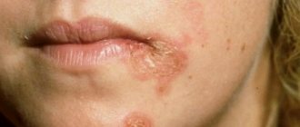
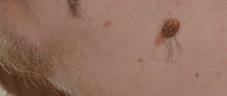
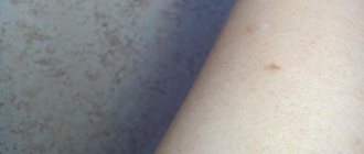
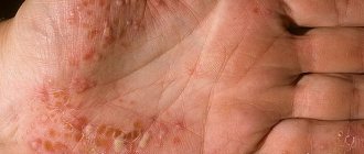
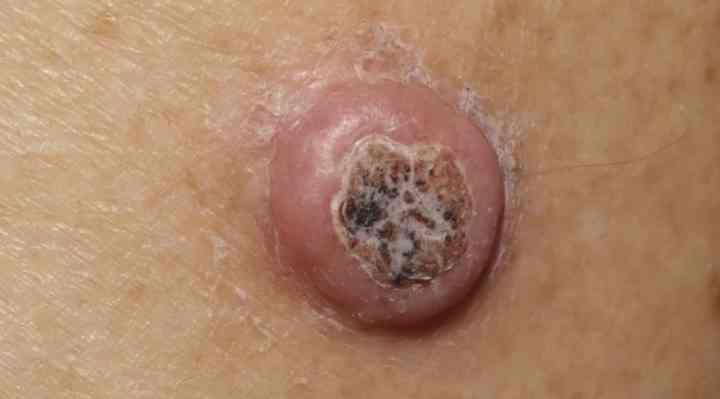
Externally, dermatofibromas look like round, smooth, slightly convex formations on the skin with a diameter of up to one centimeter, rarely more. Occasionally, the tumor may resemble a wart in appearance. Most often they occur singly; if more than one dermatofibroma appears in one patient, then they are usually located randomly on the body, not collected in groups and not tied to a specific area of the body. Dermatofibroma can appear anywhere on the body, but is usually found on the shoulders. lower back, upper back and lower legs. In some patients, cases of the appearance of multiple dermatofibromas on the palms and soles have also been described. The color of the formation can be different: red-brown, yellow-brown, gray, and sometimes does not differ in color from the surrounding skin. May be uneven: darker at the edges, slightly lighter in the center.
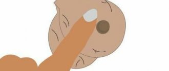
To the touch it has a very dense consistency, gives the impression of a foreign body in the thickness of the skin, and easily moves along with the skin fold. If you squeeze a dermatofibroma with your fingers, a dimple forms in its center.
Usually, dermatofibroma does not cause any discomfort to a person, other than being a cosmetic defect. But some complain of pain and itching in the area of the tumor. In addition, the tumor may be located in such a way that it will be regularly injured when shaving or rubbed against clothing or shoes.
Treatment of the disease
In most cases, dermatofibroma does not require treatment. This disease is not life-threatening and does not cause inconvenience to patients. However, if the neoplasm creates a cosmetic defect, or is located in a place where it is constantly injured, then it is better to remove the dermatofibroma.
The following methods will be used to remove the tumor.
Surgical excision. If the dermatofibroma is small, then the operation is performed on an outpatient basis. The operation consists of excision of a fragment of skin followed by exfoliation of the tumor from the surrounding tissue. Since the tumor is located in the deep layers of the skin, appropriate incisions will have to be made, so noticeable scars may remain after the operation. Therefore, it is necessary to ensure proper care of the wound and the resulting scar.
Removal of dermatofibroma using electrocoagulation. This method is based on thermal damage to tissue with simultaneous coagulation of blood vessels and capillaries. The operation is bloodless. At the site of the procedure, a dry scab first forms, and after it is rejected, a weakly pigmented spot or scar remains.
Laser removal of dermatofibroma is based on the destruction of the tumor using a laser beam. The operation is bloodless and infection is excluded.
An alternative treatment for dermatofibroma is to cut off the top of the tumor using one of the methods described above. The upper part of the tumor can also be removed by cryodestruction, that is, freezing by applying liquid nitrogen. In this case, there are no scars left. However, after such treatment, very often the tumor grows again in the same place.
Treatment with traditional methods
When treating dermatofibroma with folk remedies, patience will be required, since results can only be achieved after several months of regular procedures.
Treatment of dermatofibroma with camphor alcohol. It is necessary to lubricate the tumor with camphor alcohol three times a day. After several weeks of using the product, a burning sensation may appear. There is no need to be scared; you need to continue the procedures until the tumor completely disappears.
Treatment of dermatofibroma with tincture of aloe with iodine. You need to take the large (bottom) aloe leaf, wrap it in paper and put it in the refrigerator. Leave it there for three days, then grind it on a grater. Place the pulp in a dark glass jar and add 100 grams of medical alcohol. Leave for three weeks, shaking occasionally. Then, strain the tincture and add 10 drops of iodine tincture to it. Use the composition to lubricate the neoplasm with dermatofibroma.
Treatment with magnesium. Magnesia (magnesium hydroxide) is applied to the tumor for dermatofibroma for 10 minutes. Then the composition is thoroughly washed off the skin.
Diagnostics
Dermatofibroma can be recognized by visual examination. The final diagnosis is made after a biopsy and histological examination of the sample taken. In this case, it is necessary to differentiate dermatofibroma from glomus tumors, leiomyoma, xanthomas, nevi, dermatofibrosarcoma protuberans and melanoma. Typical histological signs of dermatofibroma are interweaving of collagen fibers, proliferation of fibroblasts, excessive pigmentation of the basal layer of the skin, large connective tissue cells that phagocytose large amounts of lipids and hemosiderin, and vascular sprouting. There are different types of tumor. The most common are dermatofibromas with a predominance of fibrous elements. Slightly less common - cell type of tumor: histiocytoma, endothelioma. Typically, dermatofibromas are heterogeneous in structure and include different morphological elements. But in all cases the tumor is penetrated by capillaries.
Dermatofibroma: causes
In the recent past, it was assumed that dermatofibromas arose as a response of the body to injury or bites of various insects. But they could not confirm this version, and to date there is no consensus on the causes of dermatofibroma.
This is partly due to the fact that the disease is persistent.
Experts suggest the following causes of dermatofibroma.
Perhaps the development of education is influenced by gender. Women are more susceptible to the disease. Causes such as a prick from a plant thorn, a mosquito bite, and other skin damage are also not discounted.
There have often been cases of family predisposition to the development of dermatofibromas, i.e. there is a hereditary factor. The patient's age also matters. As mentioned earlier, dermatofibroma in children is extremely rare.
Consequences
Dermatofibroma is considered a benign nodule and develops slowly. Degeneration into a malignant form occurs rarely. But to prevent an undesirable situation, you need to go to the clinic and undergo the necessary research. It is recommended to exclude the occurrence of cancer cells.
The scar after surgery must be corrected using plastic surgery. But sometimes there is a relapse - in about 20% of registered cases. If a relapse is detected, the problem area is re-excised, including all suspicious formations.
Dermatofibroma is a benign intradermal tumor that develops from undifferentiated reticular cells, which normally form endothelial cells, histiocytes and fibrocytes. Some experts believe that it should be considered more as a reactive hyperplasia of fibrous and vascular elements, rather than as a tumor.
Atlas of skin diseases
DERMATOPHIBROMA LENTICULAR (dermatofibroma lenticulare)Synonyms: cat nodule, simple fibroma, histiocytoma.
Etiology and pathogenesis
The etiology and pathogenesis are not clear. It is believed (Gertler, 1973) that dermatofibroma lenticular is a benign tumor that develops from undifferentiated perithelial reticular cells, which can differentiate into fibrocytes, histiocytes and endothelial cells. According to Beage and Cunliffe (1979), dermatofibroma is not a benign tumor, but probably represents a reactive hyperplasia of reticuloendothelial, vascular and fibrous cellular elements.
Development is promoted by injuries and insect bites. There are family cases.
Clinic
Single or, less commonly, multiple, intradermal tumors, on average, about 1 cm in size, round or oval in shape. They rise slightly above the skin level, which is more noticeable in the central part. Their tumor-like nature is clearly revealed only upon palpation. In this case, there is a feeling of a small dense plate embedded in the thickness of the skin.
The surface is smooth, but may be slightly hyperkeratotic and, in rare cases, verrucous. Dermatofibroma is mobile, easily displaced in relation to the underlying tissues. The color can be reddish, pale gray, yellowish, but more often reddish-brownish, more saturated in the area of old elements. It may be uneven, usually darker around the edges.
Dermatofibromas, as a rule, are not accompanied by subjective disorders, only occasionally some patients complain of itching. Lenticular dermatofibromas can be located on any part of the skin, but mainly on the legs, especially in the shins, and on the lower back. Bedi et al. described multiple dermatofibromas localized only on the palms and soles, ranging in size from 0.5 to 1.5 cm, round or oval in shape, and dense consistency.
Dermatofibromas predominantly occur at a young age, more often in women. Having reached a certain size, they exist without change for a long time. The general condition of the patients does not suffer.
The prognosis is favorable in most cases.
Occasionally, lenticular dermatofibromas may disappear spontaneously. After injury, ulceration and bleeding may occur.
The development of malignant fibrous histiocytoma in the lungs of a patient with multiple dermatofibromas on the skin has been described (Roberts et al., 1981). The possibility of metastases from a lesion in the skin removed 7 years before the onset of the process in the lungs was not excluded, although there was insufficient evidence for this assumption. Goette and Helwig (1975), Bryant (1977) observed the development of pigmented basal cell carcinoma in the dermatofibroma zone. According to Schoenfeld (1964), atypical proliferation of the epidermis is observed in 4.6% of cases of dermatofibromas. Histologically, as noted by Uanowitz and Goldstein (1964), they may be indistinguishable from basal cell carcinoma.
Halpryn and Allen (1959) found pronounced epidermal changes in 25% of cases of dermatofibromas, and in 8% it was basal cell carcinoma, in 11% it was seborrheic keratosis, and in 6% it was keratoacanthosis (hyperkeratosis, acanthosis, papillomatosis). Arnold and Tilden (1960) expressed doubt about such a high frequency of basal cell carcinomas given by the above authors. Although they often observed hyperplastic processes in the area of dermatofibromas, they did not note signs of basal cell carcinoma.
Korom and Simon (1983) found unchanged epidermis in only 15.2% of 255 cases of dermatofibroma, in 6.6% it was atrophic, in the rest hyperplasia was found (basalioma-like, follicular, pseudoepitheliomatous, similar to seborrheic keratosis, etc.), and in one - multicentric basal cell carcinoma. The reason for such frequent epidermal hyperplasia has not been precisely established. Among the factors that can stimulate the proliferation of the epidermis are the accumulation of PAS-positive and metachromasizing substances in the dermis, and the presence of dermatofibroma itself (Steigleder et al, 1962, etc.). Korom and Simon observed changes in the epidermis primarily where the dermatofibroma reached the dermo-epidermal junction. These data do not agree with the opinion of Halpryn and Allen, who did not find a relationship between the degree of epidermal changes and the depth of the tumor.
Multiple lenticular dermatofibromas can be combined with osteopoikilia (Buschke-Ollendorf syndrome). This syndrome is inherited in an autosomal dominant manner. There is a dissociation of characteristics in families. This may also apply to the nature of skin changes. Thus, Schorr et al. (1972) among 6 patients with Buschke-Ollendorff syndrome, skin changes were observed in only 3, and they were represented by different types of connective tissue nevus. Dermatofibromas in this case have a disseminated nature, a network-like or strip-like arrangement with a concentration on the thighs, buttocks, upper back, neck, and forearms. They are small, usually no more than 1 cm. The disease usually begins in childhood.
Piette-Brion et al. reported a variant of Buschke-Ollendorff syndrome with the presence, in addition to dermatofibromas, elastoma and bilateral deafness. Tansch et al. We observed a girl whose histological changes in the area of the rash were of a dual nature: corresponding to Nevuelasticus and lenticular dermatofibroma. Cole noted a predominant change in elastic fibers (elastomy) in his observation. A skin lesion appeared in a 63-year-old woman at the age of 58, initially regarded as pseudoxanthoma elasticus.
Raque and Wood consider disseminated lenticular dermatofibroma as a variant of connective tissue nevus. In all likelihood, the case described by S. A. Glauberzon and A. Ya. Lebedev as multiple flat dermatofibroma was Buschke-Ollendorff syndrome. Its peculiarity was the merging of individual rashes into rather large plaques.
Patients with this syndrome are sometimes noted to have a tendency to form keloids, band-like small focal atrophy with anetoderma, papillomas in the neck, ichthyosiform erythroderma, Rendu-Osler syndrome, palmoplantar keratoderma, blue sclera, dental dystrophy; growth may slow down. In addition to bone pathology, diabetes mellitus, decreased intelligence, infantilism, cataracts are found, Schorr et al. Precocious puberty was observed.
Histopathology
Acanthosis, hyperpigmentation of the basal layer, dense plexuses of newly formed collagen, fibroblast proliferation, vascular neoplasms, histiocytes containing hemosiderin, lipids. Gertler points out that histologically, various morphological structures can be detected, isolated or in different combinations within the same element - the structure of dense fibroma, neurofibroma, histiocytoma and endothelioma. In its pure form, the most common is histiocytoma, then fibroma, and rarely endothelioma. According to Lever (1983), on the contrary, the fibrous type of dermatofibroma is much more common than the cellular type (histiocytoma). In both cases, capillaries are detected, sometimes numerous, giving the tumor an angiomatous appearance (sclerosing hemangioma).
Differential diagnosis
It is necessary to differentiate dermatofibroma lenticular from xanthomas, leiomyomas, glomus tumors, nevi, melanoma, and dermatofibrosarcoma protuberans.
Treatment
Usually not carried out. In case of repeated injury - surgery.
Symptoms of cutaneous dermatofibroma
Basically, dermatofibromas have a single localization, but multiple formations on the surface of the skin are possible. The diameter of the formation rarely exceeds one centimeter.
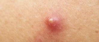
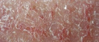
Dermatofibroma occurs in red, gray, brown or pink colors. Over time, the color of the formation may change. Dermatofibroma looks like a hard lump under the skin. The structure of the skin growth consists of fibrous fibrous tissue containing fibroblasts and histiocytes. When squeezed, a depression appears on the surface of the lump.
At first, dermatofibroma resembles a small dense pea or nodule. Gradually its size increases, density and color changes. And having reached about one centimeter, it stops growing. Dermatofibroma never degenerates into a malignant formation.
Typically, dermatofibromas do not cause any significant discomfort, because... their presence does not cause pain. And only sometimes, if dermatofibroma is localized in an uncomfortable area (wearing clothes, shoes, etc.), some patients experience slight itching and mild pain. Such cases are sometimes observed with dermatofibroma in children, who often become infected by scratching the skin.
The most common signs of education include:
- small lumps that rise above the skin, very rarely bleeding;
- nodules are usually localized on the legs below the knee, less often on the torso or arms;
- when pressure is applied to the nodule, a hole is formed;
- the color of the nodule is different from the main color of the skin, often darker;
- When touched, slight itching and soreness may occur.
It should be noted that dermatofibroma is similar in symptoms to the first stage of dermatofibrosarcoma, i.e. skin cancer Therefore, in order not to miss the development of such a dangerous disease at the first signs of formation, it is necessary to undergo appropriate diagnostics. Especially if there is rapid growth of the formation, severe pain appears, bleeding and the formation changes its shape.
Treatment of dermatofibroma
Therapy for fibrous formation consists of its surgical removal under local anesthesia. Methods of surgical intervention can be very different and depend on the level of technical equipment of the medical institution. Traditional excision of a benign tumor using a scalpel leaves a small scar because the surgeon has to make a deeper incision to completely remove fibrous tissue. Treatment using laser surgery allows the surgeon to excise dermatofibroma with minimal trauma to the epithelial tissue.
No special postoperative rehabilitation is required and the patient returns to his normal lifestyle the very next day. It is only necessary to periodically treat the surgical suture with a 3% solution of hydrogen peroxide. On days 5-7, the edges of the wound are completely healed and the stitches can be removed. For another 10 days after their removal, the surgical scar and the skin around it must be wiped with alcohol or a solution of salicylic acid. It is enough to do this once a day.
Diagnostics of education
A mole or wart may resemble a dermatofibroma. It can be distinguished by the presence of a characteristic pit when the edges of the nodule are compressed.
To clarify the diagnosis, you need to see a doctor and undergo additional examination. For dermatofibroma, dermatoscopy is performed - the affected tissues are examined with a special apparatus.
Biopsy and histology can determine the prognosis of tumor development. To do this, you need to take part of the biological material from the affected area. For a biopsy, a scraping is taken - scarification. Aspiration is carried out with a thin needle, incision - part of the nodule is pinched off, excision - the entire nodule is removed and sent to the laboratory.
Treatment
After confirming that the nodules are benign, the doctor often decides not to treat or remove them surgically. If there is no symptom of pain or itching, do not disturb the nodule. The decision to remove is made for cosmetic reasons. If the tumor has grown in a short period or is painful, the question arises of cutting out the diseased area with a scalpel or laser using the Surgitron device.
To prevent nodules from developing into cancer, the following measures are taken:
- Using a traditional scalpel with local anesthesia, the nodule is excised, capturing a healthy area.
- Laser treatment can cure a tumor with a small residual scar, but there is a risk of relapse.
- At the moment, the hospital often uses the cryodestruction method - freezing with liquid nitrogen.
Before using a laser or cryodestruction, a histological examination of the diseased area is carried out. After traditional removal, the biological sample is examined for cancer cells. If after some time the formation appears again, it becomes painful when pressed, you should urgently consult a doctor.
Diagnosis of fibrous formation
Only a specialized doctor can confirm or deny the presence of dermatofibroma on the patient’s body. Initially, diagnosis consists of a visual inspection of a foreign element. After receiving the biopsy results, conclusions can be drawn about a benign tumor of this type. Clinical signs of dermatofibroma are a large number of collagen fibers that are intertwined and do not continue to develop into surrounding tissues. Between them are the smallest vessels, capillaries, which provide nutrition to the bloodstream.
There is also always a large amount of fibrous type protein present. A distinctive feature of malignant tumors is that they do not have it in their structure.
Diseases by symptoms
Any symptom is a signal from the body that any organ, department or entire system is damaged. To find out why dermatofibroma occurs in newborns, you need to rule out certain diseases. Make sure that your baby undergoes timely diagnosis, find out why dermatofibroma appeared, and how to quickly and effectively improve the condition.
List of diseases in which dermatofibroma occurs:
- Fibromatosis and its varieties;
- Skin damage;
- Heredity;
- Liver diseases;
- Poor environmental conditions.
Because dermatofibromas usually develop in adults or children over five years of age, congenital dermatofibromas are common in newborns. In an infant, they can appear on the skin of the arms and legs, and sometimes dermatofibromas are found on the mucous membranes. Dermatofibromas, which increase in size, are especially dangerous - they can compress the thin vessels of an infant and disrupt the blood supply.