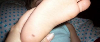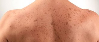
There is nothing unusual about moles on the body - such neoplasms appear in people at different years of life. But sometimes they appear on the head, and this is a little worse, because a mole on the head in the hair is not always noticeable, it can be accidentally damaged or it causes discomfort to its owner. Should I worry about a nevus located in such a place?
Why did a mole appear on my head in my hair?
The exact mechanism of the appearance of moles has not been established to date, but it is known that they appear due to the high content of melanin in the skin. Another factor is uncontrolled cell division, which the immune system does not have the strength to cope with.
In addition to the above, there are other reasons that can result in the formation of birthmarks on the head:
- genetic predisposition;
- the presence of skin diseases;
- weak general immunity;
- congenital skin defect;
- skin renewal;
- skin more susceptible to ultraviolet rays;
- radiation or x-ray exposure;
- hormonal disorders (pregnancy, adolescence);
- aging of the epidermis;
- pathological changes in the functioning of internal organs and body systems.
Over the course of a person’s life, moles can change shape and color, disappear or appear in other places.
Classification of nevi
Nevus on the head can be of two main types - pigmented and vascular, the latter is also called hemangioma.
Hemangioma or vascular nevus can appear as a result of abnormal development and growth of blood vessels.
Among the variety of types of nevi, the most common are two types - cavernous and capillary:
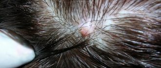
- Cavernous hemangioma is a nodular soft formation similar to a red mole on the head, its appearance is associated with pathological stretching of the blood vessels. As a result of an increase in the size of the choroid plexuses, this pathology can be complicated by bleeding. In some cases, after this, the hemangioma that appears spontaneously disappears.
- A capillary-type nevus of the scalp looks like a large, purple spot. The size of this benign formation on the head in the hair varies from 2 to 10 centimeters in diameter.
Pigmented nevi also come in different types. The most common are borderline, intradermal, complex.
Capillary nevus has a bright red color
Borderline pigmented
This is a flat, pigment-type formation that represents the very beginning of skin lesions. Thus, it is localized in the most superficial layer of the skin - the epidermis. For such a mole to appear, it is necessary to form a cluster of nevocytes in the epidermal layer almost at the border of the dermis. Borderline nevi can be exposed to various influences, turning into a malignant tumor - melanoma.
When they first appear, these moles are almost invisible, only a few millimeters in diameter. Since this type of pigmented neoplasm can lead to skin cancer, you need to monitor the size and structure of the nevus, and if it grows rapidly, consult a doctor.
Intradermal pigmented nevus
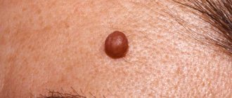
One of the most common forms of pigmented nevus is intradermal. It can be distinguished from other types by a number of characteristic features. For example, unlike a borderline nevus, such a mole has the shape of a plaque, not a spot, it has clear boundaries, is evenly colored, can be of various shades of brown, and has a dense texture. If the upper part of such a structure is not flat, but rather resembles a wart, then it can be classified as a papillomatous type.
The difference in structure is that nevocytes are not located at the border with the dermis, but in the depths of the dermis, usually forming clusters in the middle and lower layers.
These moles may appear in early childhood or be present on the skin at birth.
Intradermal pigmented nevus is located in the deep layers of the skin
Complex pigmented nevus
Another subtype of nevi is complex, located in both the epidermis and dermis. It protrudes above the surface of the skin, resembling a papilloma in appearance. Such nevi contain a large number of vessels. Nevocytes in these formations vary in degree of maturity and morphology. In the surface layer there are less mature cells, which have a cubic shape, are capable of synthesizing melanin and form clusters in the form of nests. Such cells are more likely to undergo malignant degeneration than more mature nevocytes. The latter are located in the deep layers of the skin, they are smaller and lighter in size, as they contain less melanin. However, the most mature nevus cells are localized in the middle of the formation and have a spindle-shaped shape. They are organized into bundles that resemble the tissue of peripheral nerve fibers.
Types of neoplasms on the head
Birthmarks on the scalp have different structures, sizes and shapes. There are several varieties:
KK Adapt. 5 paragraph
- Big ones. They are considered a congenital skin defect and are brown in color. Such a spot grows with the owner, so sometimes reaches significant sizes;
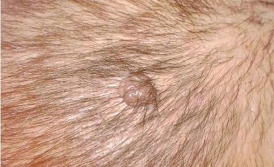
- Convex. Based on their color, nevi are classified into burgundy, red and brown. They appear in the inner layer of the epidermis and rise above it. Often moles are covered with hairs, and formations of this type are formed in mature people. They can be damaged accidentally;
- Flat. This type of formation is the most popular, and they are located at the same level with the skin, so that the person does not experience any discomfort;
- Hanging. They resemble warts and rarely form on the head. Since they are very easy to damage, it is better to remove such stains;
- Blue. Nevi of this type also rarely appear on the head, but they are no less dangerous than others. They have a cone-shaped shape, a bluish tint, and they protrude slightly above the skin level;
- Reds. These are vascular moles that form on a person’s head at birth. Over time, the stain may disappear on its own.
Having discovered any of these moles, it is advisable to monitor its condition in order to exclude signs of its degeneration into a malignant formation.
Moles and birthmarks on the head - parietal region
The meaning of a mole on the head, which is located in the crown area, lies in extraordinary intelligence. Holders of such marks are smart and resourceful. They may be interested in philosophy, esotericism, as well as other sciences that imply a tendency to think. As a rule, we are talking about thinkers deeply immersed in their own world.
At the same time, a person can be quite sociable; he often tries to convey a certain thought or idea to others. If he has oratorical talents, he succeeds. Success depends only on the person himself - if there is a desire to devote time to promoting ideas and spend effort on it, everything will work out.
Owners of such markings on the head usually do not go unnoticed. They almost always manage to bring something new into our world. The area of activity that turns out to be to your liking does not matter much. The main thing is whether you dare to put your innovative ideas on display. You will bring something to our world, but whether it will harm or benefit people is unknown.
Caring for a nevus on the head
Such stains must be handled with care. To minimize the risk of damage to them, it is enough to follow these recommendations:
- Do not use cosmetics that contain solid substances (peelings and scrubs), as they can damage the skin. Also, avoid using hair masks and shampoos containing strong chemicals;
- do not dry the area of the head where the mole is located with a hairdryer;
- do not scratch or itch the skin with sharp objects, use hairpins and hairpins with caution;
- if the formation was accidentally damaged, it should be treated with peroxide to stop the bleeding. If the mole is severely damaged, then it is recommended to seek qualified medical help.
Microtraumas of moles themselves do not pose a danger; the threat lies in infection and inflammation, so allowing both of these is extremely undesirable.
Prevention
No one is immune from the appearance of birthmarks. However, the development of malignancy can be prevented by taking certain precautions. The most common cause of melanoma is prolonged exposure to ultraviolet radiation. Therefore, it is necessary to avoid excessive exposure to sunlight and do not forget to wear hats.
You should know that a birthmark on the head must be monitored especially carefully, because anything located near the brain can lead to dangerous consequences. Therefore, if any changes are detected, you should immediately seek medical help.
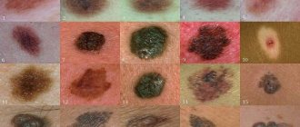
How to distinguish dangerous and non-dangerous moles on the human body
What is a mole
But benign moles are sometimes removed to prevent their degeneration if they are of rather large size (pedunculated) and are located in areas of the body where a person constantly injures them (during vigorous activity or parts of clothing).
Remember dangerous moles in the photo and be vigilant!
If nevi degenerate into cancerous moles, this is dangerous, since such neoplasms quickly metastasize to other organs.
1. The shape of the mole changes, it loses its symmetry and begins to grow in one direction.2. The edges of the nevus become uneven (“ragged”, “torn”).3. The color of the mole is uneven and contains yellow, red or black inclusions.4. The nevus grows or “shrinks”, its size changes rapidly.5.
The texture of the mole becomes different, smooth turns into rough, bumpy into flat, etc.6. Loss of hair growing from the nevus.7. Itching, peeling and burning in the area of the mole. There are several reasons why a nevus itches: - pathological cells multiply; - active processes of healthy tissue dying are underway; - the area around the formation becomes inflamed and swells.8. The appearance of microcracks and ulcerations.9. Bleeding and soreness of the mole.
Cancerous moles (melanomas): photo
Birthmark removal methods
The decision on the destruction of pigment formations is made by the doctor.
Indications for surgical intervention are:
- change in surface structure, shape;
- sudden and rapid growth;
- the spot began to itch, hurt, and was too open;
- has a large size;
- exposed to negative factors;
- the skin around has acquired an unhealthy appearance;
- causes inconvenience, interferes;
- hair does not grow on the birthmark, leaving voids on the head.
A mole is not an aesthetic problem; it can degenerate into a malignant tumor. If you have the described symptoms or suspicious changes, you must contact a dermatovenerologist.
Removal of birthmarks is carried out in one of the following ways:
- cryodestruction. Cold in the form of liquid nitrogen is used to remove elements. The patient does not require anesthesia, the process does not cause pain. The doctor directs a special nozzle or inserts a needle inside if the growth is convex and located deep in the dermis. Rejection occurs gradually over several days;
- laser. Painlessly and safely evaporates stains on visible areas of the body and head. The procedure takes on average 15 minutes. To prevent dangerous consequences, the doctor first scrapes the stain. The method is applicable to safe nevi that do not have signs of dysplasia. Before treatment begins, the patient is given an injection with an anesthetic drug. In the next 10-14 days, rest in the sauna, bathhouse, or on the beach is excluded. The wound requires special care for rapid healing;
- electrocoagulation. One of the common methods. Performed using electric current. Research of the mole is carried out after cutting. The current cuts out the growth body from healthy skin, which preserves its suitability for histology and cytology. Healing is fast. When removing large stains, a trace remains;
- radio waves. High frequency waves are sent to the problematic element. There is a neat, safe cutting from the surface of the cover. Suitable for working on open areas and hairy parts of the body.
- surgical excision. Traumatic procedure. It is prescribed in cases where there is active growth of the spot, the process of dysplasia has begun and the risk of metastasis is high. The surgeon cuts off the nevus with a scalpel, making an indentation onto healthy skin. The operation is performed under local anesthesia. The wound is closed with sutures, which leaves small scars after healing;
- phototherapy. Innovative methodology. Used in dermatology to solve problems with pigment formations. It is relevant when the spot has a light shade and is located on the head and face (left or right temple). Thanks to the effects of rays, pigment cells are reabsorbed without causing burns or injury to surrounding tissues. Today phototherapy is one of the safest and most effective methods.
After removal of birthmarks, the treatment site requires special care to speed up healing.
If you do not follow the doctor’s recommendations, inflammation will begin and a secondary infection will occur, which will lead to serious consequences and suppuration of the wound.