Formations for centuries
The eyelids and eyelash edges are covered with skin on the outside, so any skin formations can occur here.
such as papilloma, cysts, basal cell carcinoma, cholesterol plaques, etc. The carbon dioxide laser allows you to carefully remove any tumor with a good therapeutic and cosmetic effect and without deformation of the eyelid, which you will agree is very important for this small area of the face
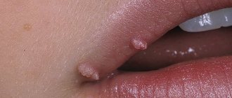
Papilloma
Papilloma is a benign tumor of the integumentary epithelium of the skin. It can be observed in childhood or adolescence, but more often after 45 years and in older people. Papilloma looks like limited or grouped warty growths with a villous or smooth surface, soft consistency, can have a different color (flesh, dirty gray, brown), rarely their sizes exceed 1-2 mm.
The reason for the appearance of papillomas is the same as for warts - viral. Infection occurs either through contact or in utero, that is, the child is endowed with viruses from birth. The favorite localization of papillomas is the face (eyelids, lips, nose), neck, armpits, under the mammary glands, groin area. In other words, the area of natural skin folds.
The greatest problems are caused by papillomas on the eyelids and even more on the eyelash area. As they grow, they can interfere with vision and often become inflamed. Self-medication with various cauterizing compounds in this delicate area is unacceptable and can lead to complications.
Laser vaporization of papillomas on the eyelids, including the eyelash edge, is absolutely harmless to vision and health. The laser allows you to quickly remove not one, but several papillomas. In one laser treatment session, it is possible to remove 10-50 or more papillomas.
Cyst
An epidermal cyst occurs as a result of retention of sebaceous gland secretions under the epidermis. Favorite localization is the eyelids, zygomatic and temporal areas. It can be of various sizes, but rarely exceeds 3-5 mm, has round or oval outlines, and is translucent in color. Cysts can be single or multiple, combined with fibroids. Prone to malignancy. Using a carbon dioxide laser, local vaporization of cysts is carried out with a good cosmetic effect.
Basalioma
Basal cell carcinoma (basal cell carcinoma) is the most common skin tumor. It manifests itself mainly in mature and old age, however, in recent years, cases of the disease have appeared in young people. Perhaps this is due to excessive tanning and environmental degradation. In 30% of patients, basal cell carcinomas are multiple (that is, from 2-3 tumors to a hundred or more). The disease begins with a small flesh-colored or pale pink nodule, usually on the face (forehead, nose, cheeks). Initially, it is perceived as an inflammatory pimple, which, as a rule, they try to squeeze out on their own. Gradually, the formation takes the form of a dense, flattened pea, the skin over which becomes thinner, acquires a bright pink tint, and a roll forms around it. The next stage is the appearance of a crusty ulcer in the central part. The ulcer slowly grows in depth and breadth. Gradually moves to other tissues: cartilage, bone, brain. Destroying them without mercy.
The most dangerous is basal cell carcinoma of the eyelids and especially the ciliary margin. Thus, harmless local redness of the skin, most often of the lower ciliary edge, ulcerates, and subsequently spreads to the tissue of the eyeball.
The sooner you start treatment for basal cell carcinoma, the better the prognosis for life. Surgical, electrosurgical excision, close-focus radiotherapy, less often cryotherapy, or a combination of several methods are used. The use of laser in recent years has shown its high effectiveness in this pathology. Using a carbon dioxide laser, the tumor is removed in one session within healthy tissue, followed by a good cosmetic effect. Treatment is carried out by dermato-oncologists or oncologists. The tumor material is necessarily sent for laboratory study of the cellular composition of the distant formation and the depth of penetration into the surrounding tissues.
WHAT IS CHOLESTEROL PLAQUE
(synonyms: XANTHOMA, XANTHELASMA)?
Cholesterol plaque can be localized or widespread. It is an accumulation of cholesterol and triglycerides. Limited xanthoma is most often located on the skin of the eyelids, mainly in elderly people against the background of lipid metabolism disorders. It is represented by a flat, soft, yellow oval or ribbon-shaped formation.
Effective removal of xanthomas on the eyelids with carbon dioxide gives a good cosmetic effect and is not dangerous for vision.
Postoperative care: lubricate the wound 2 times a day with Solcoseryl or Actovegin jelly for 10-14 days.
When to start worrying
Dry spots that appear on the skin and are accompanied by flaking and itching should be treated under the supervision of a specialist in the following cases:
- spots gradually occupy an increasing area of the skin;
- long-term treatment does not lead to positive results and the disappearance of spots, or the symptoms disappear, but reappear with the end of taking the medications;
- the appearance of spots is accompanied by pain or itching;
- wounds, ulcers or pimples appear around the pigments;
- pigmentation appeared in people living with the patient.
Important to remember! Identification of any of the described signs indicates the need to consult a qualified doctor to obtain an accurate diagnosis.

If you have a dry patch of skin that is flaky and itchy, do not put off visiting a dermatologist. It may turn out to be a disease.
Treatment methods
Treatment methods include only physical impact, since creams, drugs, sports and diets will not be able to destroy the plaque that has already formed in the vessel. There are three main directions for removing xanthelasma:
- cryodestruction;
- surgical removal;
- laser irradiation.
Only a doctor can prescribe treatment and removal of cholesterol plaques above the eyes, because before choosing a method, it is necessary to carefully examine the patient and make sure there are no contraindications, which include, for example, inflammatory processes, fatigue, damage to the vascular network.
Cryodestruction
The cryodestructive method will help you get rid of plaques on your eyelids quickly and without leaving a trace. The procedure involves a targeted effect of cold on neoplasms. During cryodestruction, the tissue surrounding the xanthelasma is destroyed, and cholesterol is released into the bloodstream.
Advantages of cryodestruction:
- in the case of a small plaque, one procedure is sufficient for complete removal;
- this method does not require physical damage to the integrity of the tissue, and therefore does not leave scars;
- removal does not require anesthesia or painkillers;
- the procedure takes up to half an hour.
Disadvantages of the cryodestructive method:
- danger of hypothermia of the tissues of the eye or eyelid;
- cannot be done for people under 20 years of age;
- multiple contraindications (inflammation, infection, glaucoma, cataracts and others).
Cryodestruction will help restore a beautiful appearance and will not leave scars or other marks. Remember that such an operation requires skills, because working with the eyes is always full of risk, so only a doctor can perform it.
Surgical removal
Surgical removal is an old and proven method to remove cholesterol plaques. Such removal requires anesthesia and subsequent care of the healing wound. The patient is given a small incision in the area of the xanthelasma, after which it is carefully separated from the vessels feeding the neoplasm and removed. The wound must be sutured; the threads can be self-absorbable or removable.
Today this method is used less frequently, because such removal of plaques leaves small scars on the eyelids, even if the operation was performed very well. The only advantage worth mentioning is that surgical treatment of xanthelasma guarantees complete removal of all parts of the tumor and almost completely eliminates its regrowth.
Treatment of xanthelasma can be carried out microsurgically; such an operation will cost more, but the scar from the section will be much smaller.
Laser irradiation
One of the methods for removing cholesterol plaques is laser irradiation, which allows you to safely and without a trace remove xanthelasma. The laser affects cholesterol plaques under the skin with high-frequency waves, due to which a resonance occurs, the tissues surrounding the xanthelasma are destroyed, and cholesterol is gradually released into the blood.
Advantages of laser irradiation:
- quick and painless procedure;
- the treatment leaves no marks on the skin of the eyelids;
- the likelihood of relapse is lower than with cryodestruction;
- the risk of surgical complications is lower.
Disadvantages of laser:
- cannot be done if there are metal implants in the facial skin;
- sensitive eyes may experience irritation;
- For some time after the procedure, eyelid skin care is necessary.
Getting rid of cholesterol plaques with a laser is safer than freezing them; this method does not leave cut marks on the skin and after the first procedure the eyelids acquire a normal appearance.
Causes of white dry spots on the skin
The appearance of white spots on the skin indicates the destruction of melanin.
There are several most common reasons:
- Lichen versicolor is a fungal disease characterized by the appearance of white spots covered with flaky skin and having clear edges. Over time, the size of the spots increases, but no burning, pain or itching is observed. Once the root cause is eradicated, the spots disappear within a few months;
- Excess of ultraviolet radiation. Burnt areas of the skin peel off, causing white spots to appear. When using special cosmetic preparations, the condition returns to normal after a few weeks;
- Vitiligo. To date, the disease has not been fully studied. The main factors that provoke it are liver diseases and problems with the endocrine system. A dry spot on the skin is flaky and itchy and has a light tint - the main symptom of the disease. Apart from its appearance, vitiligo does not cause any physical consequences. Treatment involves a comprehensive approach, including immunomodulators. Doesn't always give a positive result. In addition, the stains may disappear on their own.
There are other diseases that are characterized by these symptoms; a specialist will help make the correct diagnosis.
Products for external use
Local medications stop the progression of the disease, but the disease cannot be completely cured.
Local medications include:
- Ichthyol ointment. Apply to the formation up to two times. Therapy 2 weeks.
- Balsamic liniment (according to Vishnevsky). Apply once.
- Propolis. A piece of propolis must be applied to the affected area. To secure it we take a plaster. Keep it until the morning. Perform every day for 14 days.
- Cortisol. Removes inflammation and irritation. Use twice.
- Actovegin. With constant use, it eliminates postoperative scars. The course of treatment is 12 days.
Etiology and pathogenesis of plaque psoriasis
Today, experts adhere to two main theories that relate to the nature of this pathological process.
Psoriatic skin rashes appear as a result of increased division of keratinocytes (skin cells) and their accelerated growth. The exact cause of excessive cell growth and proliferation has not been established, but it is known that:
- Psoriasis vulgaris is associated with disorders of the immune and endocrine systems, as well as disorders of the autonomic nervous system.
- The prerequisite for the development of the disease are genes associated with psoriasis and influencing the functioning of the immune system, which control the cellular immune response and the interaction of cells with each other.
- Genetically determined disorders of cellular metabolism cause accelerated growth and proliferation of immature skin cells, which leads to the accumulation of lymphocytes in the epidermis. T-lymphocytes secrete inflammatory cytokines (peptide messenger molecules), which cause the accumulation of macrophages and neutrophil granulocytes in the skin, as well as the excessive proliferation of keratinocytes.
Surgical methods for removing plaque in blood vessels
In advanced cases, even if you take handfuls of tablets and wash them down with liters of water, you cannot do without radical methods. And here intravascular surgery and full-fledged angiosurgery come to the rescue. Through minimally invasive operations, it is possible to remove cholesterol plaques from blood vessels through minimal incisions, or to perform stenting (introduction of a special frame, a stent, into the lumen of a narrowed vessel) through a peripheral artery. Typically, stenting is a continuation of the diagnostic angiography procedure.
If necessary, angioplasty with the application of a vascular patch or bypass surgery is prescribed - transplantation of a section of the patient’s unchanged vessel, bypassing the blocked one. The graft is a corresponding segment of the thoracic artery, peripheral vessels or a synthetic shunt.
Surgery, even if it is performed in special clinics, is always associated with a certain risk to life. Conservative treatment lasts for years, accompanied by constant follow-up examinations. So isn't it better to prevent the disease? After all, for this you just need to eat right, move actively and not develop bad habits!
How to get rid of plaques on your face
Many patients are interested in how to remove annoying plaques on their face. General treatment directly depends on the root cause of the pathology.
You can find out how to get rid of cholesterol plaques on your face from any cosmetologist or doctor. Treatment in this case can be carried out according to the following types:
- Surgical removal.
- Cryodestruction.
- Laser cutting.
However, after physical elimination, it is necessary to correct lipid metabolism in the human body. If you do not ensure the normalization of the internal state, then pretty soon xanthelasmas will appear again.
It is virtually impossible to completely remove psoriatic plaques. You can only make them completely invisible. Pathology refers to chronic dermatoses that cannot be cured. Therapy is based on the use of hormonal ointments, creams and systemic therapy for severe forms of the disease.
Other types of plaques are treated according to a plan individualized by your doctor.
We hear about the risk of increased cholesterol levels at every step. This disease causes a dangerous disease - atherosclerosis. As a result of the formation of atherosclerotic plaques on the walls of blood vessels, blood pressure destabilizes, weakness appears, and memory decreases. Another unpleasant consequence of high cholesterol in the body is the appearance of plaques (xanthomas) on the skin. It is this manifestation that will be discussed in the article. We will tell you what the causes are, how to identify the disease, as well as methods of treatment and prevention.
Plaques that appear on the skin are called xanthomas. This is a clear external sign of an increase in low density in the body, with a reduced level of high-density lipoproteins.
What is the difference between these two types of cholesterol?
Low-density lipoproteins are a type of substance popularly nicknamed “bad.” This cholesterol is directly related to the appearance of atherosclerosis. It contains a high level of fat molecules and also has atherogenic properties.
It is important to control the content. Because it is low-density lipoproteins that are a component of the cell membrane and also affect hormonal levels
Therefore, if LDL is too low, a decrease in physical activity, mood, and even the occurrence of neuroses and depression may also occur. High density lipoproteins - the so-called. Its function is to regulate LDL levels. It captures cholesterol molecules and transports them to the liver for processing and removal from the body.
Folk remedies
How to clear blood vessels from plaques, avoiding surgical consequences and harmful side effects from taking medications?
First of all, you need to organize a dietary diet of vegetables and fruits. Fresh juices, cereals, cereals, nuts and fish are also beneficial. The listed products should replace fried, smoked and fatty foods.
Herbs and plant fruits contain substances that help the process of cleaning and preserving blood vessels. These are decoctions, infusions from:
They cleanse blood vessels throughout the body. Instead of regular stimulating tea, it is better to drink herbal tea with honey. The necessary funds can be grown in the garden and vegetable garden, found in the forest or meadows.
How to dissolve plaques in blood vessels using herbal decoctions and infusions? A collection of birch buds, chamomile, St. John's wort, and immortelle is used.
A powder is made from linden blossom and dandelion roots that helps when taken before meals daily. Such mixtures also reduce excess weight, which is also an important stage of treatment.
A very pleasant cleansing experience at home would be to eat walnut kernels. The green shell of the fruit is also used.
Garlic, which breaks down cholesterol formations, is considered an effective remedy to help solve the problem: how to remove plaques in blood vessels. By adding lemon and honey to it, the effect increases significantly, and the walls of the blood vessels become elastic.
Baths with the addition of nettle and a compress of wormwood with whey are also considered beneficial.
Note! If you discover signs of atherosclerosis, you should not self-medicate. Serious and effective measures can only be applied as prescribed by a doctor and under his supervision
But preventing disease through the right lifestyle is available to everyone
Serious and effective measures can only be taken as prescribed by a doctor and under his supervision. But preventing disease through the right lifestyle is available to everyone.
There are many recipes with garlic, including other herbal additives. When combined with lemon, the product becomes rich in antioxidants and vitamin C, which provide a preventive and therapeutic effect.
We will consider how to deal with bad cholesterol using natural resources at home in the form of the following recipes.
A forty-day cleanse is carried out using an infusion of lemon and garlic. Every week you need to prepare a three-liter container of the product, taking a total of 16 lemons and heads of garlic. This is enough for 4 containers.
You need to grind 4 lemons with peel and garlic in a meat grinder. Fill the mixture with warm boiled water and leave for three days without refrigeration. Then the product is filtered and consumed before meals, 100 ml.
Honey is added to the previous composition. You need to squeeze the juice out of 10 lemons, adding the peel. Grind 10 heads of garlic in a meat grinder, combining them with lemons and a liter of honey.
Let it brew for a week, protecting it from light. Once a day for 2 months, eat 4 teaspoons.
Mix a bottle of vodka with a slurry of lemon and 2 garlic cloves (crush with a meat grinder or blender). Add 5 bay leaves.
The tincture is aged for a month, after which the product must be strained. Take 2 teaspoons after meals daily.
350 grams of crushed garlic is poured with 200 ml of alcohol and infused in the dark. Take 11 days before meals, starting with 1 drop. Increase every day until the dose reaches 15 drops on days 5-6.
Then reduce it again so that on day 10 you take only 1 drop again. On the last day you need to use 25 drops 3 times.
When treating with the drug, the body will require a lot of water, and proper nutrition and diet are also necessary. Tibet recommends repeating the method after 6 years.
Atherosclerosis is a dangerous disease that creates a risk for the development of life-threatening conditions. In this regard, it is recommended to do prophylaxis that preserves blood vessels
It is especially important to create a healthy background predetermined by proper nutrition
Prevention measures
Statistics show that vegetarians suffer from clogged blood vessels very rarely. This fact does not require a complete abstinence from animal foods, but it is still worth thinking about creating the right diet.
To reduce the likelihood of excessive cholesterol plaque formation, doctors recommend the following preventive measures:
- limit consumption of fatty and smoked foods;
- switch to 5 meals a day;
- eat a lot of vegetables and fruits;
- during each meal, eat no more than 1 teaspoon of fat (required to remove bile into the intestines);
- engage in active sports at least 2 times a week.
Carrying out diagnostic procedures
The appearance of xanthelasmas is a sign of a severe disorder of lipid metabolism and also indicates the development of atherosclerosis. At the first signs of the formation of formations on the eyelids, you should consult a doctor. First of all, it is recommended to consult a dermatologist to rule out organic skin diseases. The next step is a consultation with a therapist, cardiologist and vascular surgeon.
In some cases, an objective examination of the patient with the collection of complaints is enough for an experienced doctor to make a diagnosis.
In order to clarify the diagnosis, it is necessary to carry out the following activities:
- Biochemical analysis from blood with an expanded complete lipid profile.
- Ultrasound examination of the heart and blood vessels.
- Dopplerography of heart vessels.
- Blood test for glucose, glucose tolerance test.
- Ankle-brachial index measurement;
- Calculation of body mass index.
- Electrocardiography.
Next, according to the principles of diagnosis, differential diagnosis is carried out.
Diseases with similar manifestations are:
- benign and malignant neoplasms;
- dropsy of the skin;
- syphilitic granulomas;
- tuberculous skin lesions;
- papillomas.
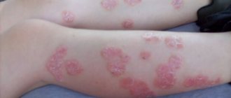
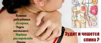
If the spots are red
The most common are red rashes, which indicate the presence of an irritant.
The presence of severe itching indicates an allergy. Stress or nervous overexcitement often leads to the appearance of shingles.
One of the most difficult diseases to treat, accompanied by the appearance of red spots, is psoriasis.
In this case, it is necessary to conduct a comprehensive examination, follow an individual diet and treatment under the supervision of a doctor.
Ways to slow down the formation of cholesterol plaques
If the goal is to at least partially get rid of cholesterol plaques, you need to work in two directions simultaneously: remove excess “bad” cholesterol and strengthen the vascular walls.
- The first is directly related to the normalization of lipid metabolism. But in order to know how to eliminate the imbalance, it is necessary to determine the concentration of its main indicators in the blood. The analysis is called a lipidogram, it is carried out both in public clinics and in private ones. Before the test, the patient must prepare properly, otherwise false results will be obtained. And since cholesterol concentration is not static and depends on age, gender, and area of residence, only a doctor can evaluate the lipid profile.
- The second direction is vascular training, eliminating harmful effects on the body, preventing infectious and allergic diseases, changes in blood pressure and body temperature.
Regular physical activity
Moderate physical activity will help partially remove atherosclerotic plaques from the arteries. No one is asking you to achieve world achievements in sports: regular exercise, morning exercises, swimming or outdoor games are enough. They not only increase the speed of blood flow, thereby creating good microcirculation in the tissues, but also train the muscular lining of blood vessels. And trained arteries adequately respond to changes in blood pressure, maintain it at the proper level, thereby protecting the internal lining from microtrauma.
Proper nutrition
Cleaning blood vessels from formed atherosclerotic plaques is impossible without regulated nutrition. First of all, it is necessary to remove trans fats from the diet, reduce simple carbohydrates and animal fats as much as possible. That is, give up unhealthy foods in favor of whole grain cereals and pasta, lean meat (poultry, rabbit, veal), high-quality fats (sea fish, olive and flaxseed oil, avocado, nuts and seeds). Be sure to consume plant fiber contained in vegetables, legumes, and bran.
To fully utilize “bad” cholesterol, it is recommended to speed up your metabolism. Vitamins, which are catalysts for chemical reactions, are primarily responsible for this. In any other case, spicy food would help, but not with atherosclerosis. The only allowed product is garlic in small quantities - 1 clove per day. Chicory, cinnamon, green tea, and spinach will help increase metabolism.
Separately, it is worth mentioning products that thin the blood and reduce the deposition of plaques. These are citrus fruits, berries (strawberries, raspberries, cranberries, blueberries, cherries), tomatoes, beets, cocoa and dark chocolate. The list of healthy foods is very long, and it is necessary to remember it not when the arteries are already clogged, but to remember it all your life. After all, rational nutrition is one of the main methods of preventing the disease.
Getting rid of bad habits
The same applies to bad habits: you need to quit smoking and drinking alcohol before starting treatment for atherosclerotic plaques, but much earlier. And not a single method of clearing cholesterol will help if you do not give up smoking and drinking alcohol. Bad habits include drinking strong coffee on an empty stomach. On an empty stomach, it is absorbed more intensively, and all the caffeine that comes in contributes to increased fluid excretion and thickening of the blood.
Recommended book: Allen Carr. An easy way to quit smoking
Medicines
Many pharmaceutical companies deliberately idealize their drugs in an effort to increase sales. The statins they extol cannot completely remove cholesterol plaques; they can only slow down their formation, and after long courses of treatment. Medicines are usually prescribed in combination with each other and in combination with other non-drug methods.
Help fight atherosclerosis:
- fibrates, blood thinners;
- cholesterol absorption inhibitors, reducing its intake from food;
- bile acid sequestrants, which inhibit the reabsorption of cholesterol residues after the digestion process;
- pharmacy vitamins and complexes;
- dietary supplements;
- homeopathic medicines.
Garbage - fight!
With regard to vessels, the statement is very true: “It is clean not where they clean, but where they do not litter.” Since atherosclerosis develops over decades, you need to maintain cleanliness throughout your life. And it's not just cholesterol. It is especially important to prevent calcium from depositing on the vessel wall. This “scale” is called calcinosis - and this is the most dangerous form of atherosclerosis. It's all about combining phosphorus with calcium in so-called phosphates. They are poorly soluble, and it is in this form that calcium is deposited in the blood vessels.
How to avoid this, says the famous biogerontologist, corresponding member of the Russian Academy of Sciences, professor, Doctor of Biological Sciences, head of a specialized laboratory at the Institute of Biology of the Komi Scientific Center of the Ural Branch of the Russian Academy of Sciences and the department at Syktyvkar State University Alexey Moskalev: “The simplest thing that can come to mind to reduce vascular calcification, is to reduce the intake of calcium and products containing it. But you don't need to do that. The body will not react very well to its lack in the blood - calcium will begin to be washed out of bones and teeth and will settle on the walls of blood vessels, in the tissues of the lungs, kidneys and liver. That is, it will only increase vascular calcification. It can be prevented by directing calcium, and with it the phosphorus contained in phosphates, into the bone tissue. What is needed for this?
Firstly, vitamin K₂ is important. It prevents calcium from being washed out of the bones and deposited on the walls of blood vessels. Most K₂ is found in fermentation products: cheese and fermented milk products. And the absolute record holder for its content is the traditional Japanese dish natto - it is made from fermented soybeans. In addition, vitamin K₂ is synthesized by some bacteria that live in our large intestine, which means that we need a diet rich in prebiotics - dietary fiber.
Secondly, you need to get enough magnesium. It can bind phosphates, preventing their deposition on the walls of blood vessels. In addition, magnesium can regulate the activity of vascular smooth muscle cells so that they prevent calcification. A lot of magnesium is found in buckwheat, oats, barley, legumes, and walnuts.
Features of the course and manifestations of the disease
This formation in the eyelid area has a medical name - xanthelasma. Such plaques look like yellow growths on the skin, small in size. Often such lesions occur in the area of the inner corner of the eye, mainly on the upper eyelid.
The number of these plaques can be different: both multiple formations and single ones occur. If there are many of them, then this means an advanced pathological process, and the affected area is not limited to the eyelids. Diagnosis of this disorder includes a blood lipid test to identify a cholesterol metabolism disorder. Treatment of this disease is based on the results of such an analysis.
Therapy cannot remove plaques from the eyelids; they are removed surgically, but drug treatment is still necessary to reduce cholesterol levels in the body. Women are susceptible to this pathology more often than men.
No one can say exactly what causes xanthelasma, but there is an opinion that plaques on the eyelids appear due to a malfunction in the fat metabolism of the human body, which is manifested by the accumulation of fat in the papillae of the skin.
What conditions contribute to the appearance of:
There are cases of hereditary transmission of xanthomatosis, then a person from the moment of birth is accompanied by a lipid metabolism disorder at the genetic level.
The appearance of xanthelasma resembles a yellow wart on the eyelid. The formation rises above the level of the skin, it is absolutely painless, even with pressure. Upon palpation, you can notice the soft structure of the plaque. Often the disease affects both eyelids. If formations cover the skin multiple times, they tend to merge together, creating conglomerates. Sometimes this pathological process goes very far, and the plaques form a single extensive yellow stripe on the eyelid, with uneven contours.
The appearance of cholesterol plaques on the skin remains for life; they will not disappear on their own. Their development is especially difficult for women, as it is an unpleasant cosmetic defect.
To clean VESSELS, prevent blood clots and get rid of CHOLESTEROL, our readers use a new natural drug recommended by Elena Malysheva. The preparation contains blueberry juice, clover flowers, native garlic concentrate, rock oil, and wild garlic juice.
Xanthelasma: symptoms and treatment
Xanthelasma is a benign growth that forms on the mobile eyelid and in the inner corner of the eye. It resembles a flat yellow plaque. You can get rid of it through surgical removal.
- Causes of xanthelasma formation
- Symptoms of the disease
- Types of xanthoma
- Diagnostic measures
- Features of drug therapy
- Folk recipes
- Surgical and laser removal
Causes of xanthelasma formation
When faced with the disease for the first time, patients are interested in what it is – xanthomas on the skin. The pathology is caused by a violation of fat metabolism. Modern diagnostic methods detect hyperlipidemia in the early stages - an increase in lipids and lipoproteins in the blood.
Experts identify a number of reasons that cause hyperlipidemia.
Pathologies of the biliary tract. The cause of increased lipids is gallstones, pancreatitis, and cancer of the pancreas.
Damage to liver tissue. The liver leads to hyperlipidemia in the case of hepatitis, Wilson-Konovalov disease. Cirrhosis can provoke pathology.
Diabetes. The risk category includes patients whose disease is difficult to control. Severe metabolic disorders provoke diabetic xanthomas.
Congenital lipoprotein lipase deficiency.
Hereditary factor. At risk are patients who have a family history of hypercholesterolemia and dysbetaproteinemia.
Alcohol addiction. Alcoholism provokes a malfunction of internal organs.
Thyroid dysfunction.
Hyperlipidemia leads to the accumulation of cholesterol and lipids in tissues. The cell, saturated with lipids, occupies the top layer of the skin. The accumulation of cells in the eyelid area and around the eyes leads to xanthelasma.
The reason for the appearance of xanthelomas is a violation of fat metabolism. The disease provokes histiocytosis with proliferation of macrophages in tissues. Experts identify an idiopathic form, in which a clear cause of the disease cannot be detected. It has been unequivocally proven that the occurrence of xanthelomas is influenced by the overweight of patients.
Symptoms of the disease
Xanthelasmas are typical for patients over 50 years of age. It has been statistically proven that the occurrence of pathology is more common among women than among men. At the same time, the symptoms of the disease are not divided by gender . In children and adolescents, the appearance of pathology is caused by hereditary hyperzolesterolemic xanthomatosis.
Xanthelasmas are not associated with:
restriction of eyelid mobility.
The pathology progresses slowly. The absence of acute symptoms leads to untimely consultation with a doctor. The main disadvantage of the pathology is a cosmetic defect. Therefore, experts recommend removing formations of medium and large sizes.
The localization of plaques affects the face; they are grouped on the moving and fixed eyelids, in the inner area of the eyes. Their formation is noted under the eyes, in the temporal lobe and on the bridge of the nose. In the case of general xanthelomatosis, plaques cover the patient's limbs and body.
Types of xanthoma
Depending on the structure, location and appearance of the formations, there are several types of xanthoma. Based on the location of lipid deposits, formations are divided into 2 groups.
1. Cutaneous neoplasms. The patient's skin is covered with xanthomas on top. The formations are easily amenable to therapeutic effects.
2. Internal xanthomas. The formations are localized on the meninges, tendons, and muscle surfaces. The pathology is difficult to diagnose and requires long-term therapeutic intervention. Tendon or Achilles xanthoma is especially dangerous.
Experts also classify xanthomas according to the nature of their formation.
1. Eruptive xanthoma. A distinctive feature is the acquired red color. Then the eruptive xanthoma changes color to a characteristic yellow. The tumor retains a burgundy rim for a long time. 2. The formations are round or spherical in shape, small and medium in size.
2. Tuberous xanthoma. A distinctive feature is symmetry in arrangement and a yellow or brown tint. The formations are large in size.
3. Flat xanthoma. The formations have a round, slightly convex shape. Their localization occurs on the hands and palms. Sizes range from small to large.
4. Xanthelasma of the eyelids. Medium-sized formations. They have a yellow or brown tint. Cover the upper eyelid and area around the eyes.
Separately, experts distinguish gastric xanthoma. Fatty growths are localized on the mucous tissues of the gastrointestinal tract. The formations are benign, but can develop into cancerous tumors.
Gastric xanthomas are asymptomatic. They are diagnosed by gastroscopy. They are typical for patients with diabetes, gastritis, and atherosclerosis. The antrum of the stomach is most often susceptible to xanthomas. Therapy involves taking lipid-lowering drugs and prescribing a diet. No surgery required.
Diagnostic measures
Diagnosis of the disease is carried out by a dermatologist and endocrinologist; patients can also seek advice from a dermatovenereal dispensary (DVD). The diagnosis is made based on external examination. The doctor pays attention to the size of the formations, their number, and location.
To study education, diascopy is performed. The method involves pressing on the formation with a glass slide. The plaque bleeds and its characteristic yellow color appears.
Laboratory tests are prescribed for patients.
1. Blood test. It shows the level of cholesterol in the blood serum. In a healthy person it does not exceed 5.2 mmol/l.
2. Lipid study. And analysis allows you to detect the presence of lipoproteins in the patient’s blood.
The patient's metabolism of fats in the body is also examined.
Features of drug therapy
Treatment of xanthelasma does not have a clear scheme. It involves the use of medications, surgery, and traditional medicine recipes. The prerequisites for the formation of xanthelasmas are obesity and metabolic failure. Consequently, treatment is aimed at normalizing metabolism and treating diseases of the digestive and choleretic system.
When treating xanthelasma, experts will highlight several areas.
Taking medications aimed at normalizing fat metabolism in the body. Products based on herbal preparations with lipotropic action are considered effective. Experts recommend taking the drug Livial.
Use of local products. Medicines act on skin growths. Promote its rapid resorption. Under the influence of ointments, the skin is smoothed after surgery, and the occurrence of postoperative scars is eliminated. Zinc-ichthyol, Mercury yellow, and hydrocortisone ointments are considered effective.
The use of medicinal decoctions. Traditional methods allow you to treat the stomach, esophagus and biliary system organs without the use of serious medications. Medicinal herbs improve metabolism, remove excess bile from the body, and have a beneficial effect on the body's protective functions.
Diet food. A diet is necessary to normalize the body’s fat metabolism, improve the functioning of the gastrointestinal tract, and eliminate obesity.
Surgical intervention. In cases where the xanthomatous growth causes inconvenience, it is surgically removed.
Therapy is selected based on the cause of xanthelasma. The main condition for a complete cure for the disease is strict adherence to the therapeutic regimen.
Folk recipes
Non-traditional methods have shown high effectiveness in the treatment of small xanthelasmas. Unconventional recipes are aimed at eliminating external signs of the disease and treating the original cause that caused it.
Yarrow, dill, mint, rose hips, immortelle, shiitake mushrooms, oregano, aloe, celandine and a number of other medicinal plants and products have been highly effective in the treatment of xanthelasma. Recipes include decoction, infusion, ointment, and lotions. The homemade recipe is easy to prepare and highly accessible.
Yarrow decoction. The recipe uses 2 teaspoons of dry leaves. The grass is poured with hot water (200 grams) and kept in a dark place. Use a quarter glass 3 times a day before meals.
Dill decoction . A spoonful of dill seeds is steamed with boiling water. After 30-40 minutes, filter the broth. The entire volume is drunk in equal parts per day.
Oregano decoction. For preparation you need 200 grams of water or milk and 1 spoon of oregano. The dry mixture is poured in and brought to a boil. The broth is kept on fire for 5 minutes, removed from the stove and placed in a warm place. The product is infused for 12 hours. Take a decoction of 1/3 cup 3 times a day. The course of treatment is up to 2-3 weeks.
Onion compress. The recipe requires an onion. It should be baked until soft and kneaded. The resulting slurry is mixed with a grated piece of laundry soap. The plaque is completely covered with the resulting mixture. A tight bandage or plaster is applied on top. The mixture should be prepared anew each time.
Aloe compresses. The leaf of a two-year-old plant should be cut in a wide part. The pulp is applied to the affected area and fixed with a plaster for 3 hours. 2 weeks of treatment and the tumor is opened. Compresses from plant juice are made in the same way.
When using traditional methods of treatment, you must first consult with a specialist and select suitable recipes. This is due to contraindications of medicinal plants. Priority is given to recipes that normalize metabolism, improve digestion, and facilitate the functioning of the liver and biliary tract.
Rules for cleansing cholesterol deposits under the skin of the eyelids
Cholesterol deposits on the eyelids can be removed surgically. The following types of surgical intervention are distinguished:
- laser surgery;
- electrocoagulation;
- exposure to cold;
- deletion.
Removal is carried out by dissecting the skin and excision of pathological tissues, followed by treatment with iron sesquichloride, which has a local coagulating property that stops bleeding. Allows the wound to heal by primary intention without the formation of a rough scar or suppuration.
Electrocoagulation involves the use of the coagulating ability of current. After removing the cholesterol deposits using scissors and tweezers, the base is cauterized with an electrode.
Exposure to cold involves treating by applying liquid nitrogen to the pathological area. The low temperature of nitrogen promotes the destruction of neoplasm cells and allows you to cleanse the skin.
Laser surgery is one of the effective ways to remove cholesterol plaques that have arisen under the eyes. The method ensures bloodlessness. Areas of the pathological process are affected by the laser. After the procedure, there is no tissue scarring or other consequences.
Surgery is a last resort. Before this, local and general treatment is carried out, aimed at correcting deviations in homeostasis that arose under the influence of the underlying disease. Of interest are diseases of the hepatobiliary system and hormonal disorders. They are the cause of plaques.
The diet aims to reduce blood cholesterol levels. Diet provisions:
- the diet should contain large quantities of fiber;
- In the daily diet, the consumption of protein foods should be increased;
- try to consume less animal fats and more vegetable fats; vegetable oils contain unsaturated fatty acids that help reduce cholesterol;
- do not eat yolks or red meat;
- Avoid eating foods with a high glycemic index (chocolate, sugar, cookies);
- consume low-fat dairy products;
- eat fresh fruits and vegetables rich in fiber.
To refuse from bad habits:
- Alcohol consumption.
- Smoking.
After treatment and removal of tumors, you need to focus on preventing the recurrence of the process. The patient must monitor nutrition, physical activity, and weight.
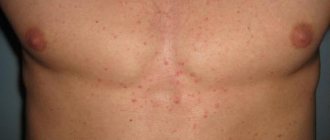
Cholesterol plaques on the face may indicate the development of an imbalance in the body. Yellow formations that appear on the face may indicate a lipid metabolism disorder (dyslipidemia). This phenomenon often occurs with high levels of cholesterol and blood sugar. However, not only people with pathologies of the cardiovascular system, obesity and diabetes mellitus can be susceptible to this disease.
Flat benign formations on the eyelids
Cholesterol plaques are classified as benign formations. According to topography, they are most often noted in the area of the inner corner of the eye of the upper eyelid. Can be either single or multiple. Moreover, localization is not limited only to the eye area.
Clinical manifestations
The typical location of plaques is the face, but the torso, feet, palms, and natural folds of the skin are also often affected. Symptoms are individual for each patient.
Clinical characteristics of formations:
- The color of the plaques varies from light yellow to dark brown. Dark xanthomas are often mistaken for melanomas.
- Xanthomas may be flat or raised above the surface of the skin.
- The dimensions usually do not exceed 10 millimeters. In rare cases, the dimensions reach several tens of centimeters.
- The surrounding tissue is dry and flaky. This indicates trophic disorders of the skin.
- Formations are characterized by slow but progressive development.
- Cholesterol plaques on the skin never degenerate into cancer.
Causes and signs of formation on the eyelids
Cholesterol plaques on the eyes are a form of xanthoma or xanthelasma. They appear as a result of lipid metabolism disorders and are classified as benign neoplasms of the skin. There are cases when pathology manifests itself at normal levels of lipids in the blood. The occurrence of xanthelasma is typical in older people, more often in women.
The etiology of the pathology could not be clearly established. There is an opinion that the deposition of cholesterol on the eyelids occurs as a result of the development of dyslipidemia and is caused by an increased content of low and very low density lipoproteins.
Formations occur in people with concomitant pathologies:
- diabetes;
- impaired fat metabolism;
- other disorders of endocrine regulation.
A number of factors contributing to the appearance of xanthelasma:
- metabolic dysfunction;
- excess weight;
- unhealthy diet (fast food, fatty, high-calorie foods);
- physical inactivity (low physical activity during the day);
- genetic predisposition;
- disturbance of lipid metabolism in the liver.
The scientific world believes that the main condition for the appearance of cholesterol deposits is obesity and genetic predisposition.
Pathology is characterized by the appearance of signs:
- cholesterol deposits on the eyelids are localized on the upper eyelid, at the medial angle, in the form of a spot;
- both eyes are affected;
- layers of cholesterol of soft consistency;
- the size of the tumors is no larger than a bean, there is no threat to vision;
- numerous formations gradually increase in size and tend to merge, forming extensive deformations;
- the disease occurs abruptly, progression can last for a long period;
- During the development of plaques, the patient does not experience pain or discomfort.
Xanthelasmas do not have a tendency to malignancy. There is no reliable evidence to the contrary.
Medications
Drug therapy involves the use of groups of drugs that effectively fight atherosclerosis:
Lipid-lowering drugs:
- Statins (Lovastatin);
- Fibrates (Clofibrate, Etofibrate);
- Bile acid sequesters (Colestipol);
- a nicotinic acid.
Hepatoprotectors: Essentiale, Choline chloride, Legalon, Lipoic acid.
Phytotherapy with galenic and new galenic preparations is used with drug therapy. Such therapy can correct metabolic disorders, restore liver function, and stop the progression of the disease. Cholesterol deposits will be removed by surgery.
External means
As an external agent, an ointment based on zinc and ichthyol, mercury ointment is used.
Yellow mercury ointment has a pronounced anti-inflammatory, antiseptic effect, helps restore damaged epidermis.
Zinc-ichitol ointment has the following effects:
- local anti-inflammatory effect;
- antibacterial;
- accelerates epithelization;
- antiseptic;
- disinfectants.
What information is missing from the article?
- Detailed review of medications
- More practical treatments
- Innovative developments in this area
- Qualified expert opinion
View results
Causes
The normal concentration of low molecular weight lipoproteins in an adult is considered to be 2.58-3.36 mmol/l. If values exceed 4.13 mmol/l, it is recommended to resort to treatment to reduce the likelihood of developing atherosclerosis. If the result of the disease is cholesterol plaques in the vessels of the heart or brain, myocardial infarction and stroke are quite possible, the mortality from which leads among other pathologies.
Can the process of cholesterol sedimentation be considered pathological? No. It occurs due to the inability of low molecular weight proteins to constantly be in a bound state, which is necessary for the normal performance of biological functions.
However, with a large amount of LDL and VLDL, cholesterol plaques precipitate excessively, gradually increase, provoke the proliferation of connective tissue in the vessels and the formation of blood clots. With a long process, blockage of arteries and capillaries cannot be avoided. Doctors identify several factors that contribute to increased concentrations of low-density lipoproteins and so-called bad cholesterol:
- metabolic disorders;
- obesity;
- sedentary lifestyle;
- poor nutrition;
- liver dysfunction;
- endocrine disruption;
- heredity.
Obesity, among other things, is considered the most likely cause of excessive formation of cholesterol plaques. Excess weight appears due to metabolic disorders and the predominance of fatty and simple carbohydrate-rich foods in the diet. If at the same time a person hardly eats plant foods and vitamins, then the body simply does not have time to process the incoming compounds, storing them in reserve.
And animal food, in turn, contains a large amount of cholesterol, the concentration of which at some point will exceed the permissible limit if there is an irrational approach to nutrition.
Excess weight often provokes liver dysfunction - this can impair the synthesis and excretion of bile into the intestines. Those. and cholesterol will also not leave the body. In addition, malfunction of the liver often occurs due to alcohol abuse or long-term use of medications - these factors always need to be taken into account during treatment.
In youth, clogged blood vessels are practically not diagnosed, since the person is in full bloom, and all serious illnesses are avoided (of course, except for congenital pathologies). Typically, problems appear closer to old age, when the intensity of physiological processes gradually fades.
Plaque psoriasis diagnosis
Plaque psoriasis is a multifactorial disease.
Experts still cannot say with certainty what factors cause widespread psoriasis vulgaris. In this regard, they only have assumptions, the correctness of which they are trying to verify by conducting all kinds of research and experiments.
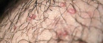
The main indicator of vulgar psoriasis is redness of certain areas of the skin. Moreover, in the affected area, the top layer of the skin is slightly raised and has a light gray or silver tint.
Vulgar psoriasis is also called plaque psoriasis. Most often, the disease gradually affects the skin in the form of a papular rash and the formation of red spots on the skin. Lack of treatment leads to rapid development of the disease.
Papules enlarge, merging into plaques. Peeling and a silvery tint of damaged areas may not be observed everywhere. But when scraped, the papules peel off easily.
Most often, the rash is observed in the area of large human joints or on the scalp. In case of acute disease, the papular rash quickly spreads throughout the body. This could be the back, buttocks, lower or upper limbs.
In medical practice, there are several main stages of vulgar psoriasis. These include:
- Progressive stage. In this case, the main symptoms of the progressive stage of vulgar psoriasis are an increase in papules, the formation of new spots on the skin and severe peeling of the damaged areas.
- Stationary. At this stage, the papules do not increase in size, and the total number of red spots remains the same. Moreover, it is at this stage that the formation of a shiny ring around the papule can be observed, which indicates the beginning of the transition of the disease to a regressive stage.
- Regressive. This stage is a period of decreasing number of papules and reducing the degree of desquamation.
Plaque (vulgar) psoriasis is a cyclical disease, it has three repeating stages:
- Progressive
- Stationary
- Regressive
- New rashes and papules constantly appear
- Already existing ones are growing in size
- Koebner's symptom - the appearance of new pathological elements in response to skin irritation (scratches, injections, aggressive ointments)
- Complete psoriatic triad
- Clearly noticeable peeling of the surface of papules, with the exception of newly formed psoriasis vulgaris
Stationary stage
- New papules do not appear, old ones do not stop growing
- Peeling is clearly visible on all papules, but its intensity decreases
- Around the papules, the so-called pseudoatrophic Voronov’s rim begins to appear - this is a shiny ring that is soft to the touch. Its full appearance indicates that the regressive stage of plaque psoriasis has begun.
- Koebner test is negative, psoriatic triad is still positive
- Peeling gradually disappears
- Psoriatic plaques disintegrate, papules disappear, leaving behind hypopigmented areas
- The Koebner reaction is negative, the psoriatic triad is positive
Psoriasis vulgaris can correlate its course with the seasons and worsen either in winter from cold, or in summer from solar radiation.
But there is also a non-seasonal form of plaque psoriasis, in which the onset of exacerbations does not depend on the season.
The acute onset of the disease is observed only in rare cases; psoriasis usually develops slowly and gradually.
It begins to appear in the form of localized rashes on the skin in the area of large joints or on the scalp.
Rashes in the form of reddish or pink nodules, some of which are covered with small gray-silver scales.
Psoriasis can occur in varying degrees of severity, which depends on the activity of development of the skin lesion process, the area of spread, and general symptoms of the disease: fatigue, drowsiness, increased levels of uric acid and ESR.
In mild forms of psoriasis, the area of skin damage does not exceed 3%.
Moderate severity of psoriasis is diagnosed when skin lesions range from 3% to 10% of the total area.
In this case, the papules grow in size, gradually merging with each other and forming psoriatic plaques. This is the so-called plaque psoriasis.
Plaques are mainly localized in the lower back, elbow and knee joints, in the bends of the limbs, on the scalp, palms and feet, but can (less often) form on other surface areas.
When exposed to external factors - poor nutrition, trauma to the surface of the plaques or scratching them, they tend to further spread.
In the case of psoriatic rashes affecting more than 10% of the skin surface, we are talking about a severe form of psoriasis vulgaris, which is also called vulgar or widespread psoriasis.
If plaque psoriasis in severe form has been going on for a long time, the rashes can swell, harden and cause significant discomfort with itching and pain.
If a patient with vulgar psoriasis does not receive adequate treatment, the disease becomes chronic.
In rare cases, the manifestations of ordinary psoriasis constantly progress over time or are in a stably active form.
Typically, plaque psoriasis occurs in the form of regular exacerbations with further remission.
Exacerbations can be caused by stress, infection or allergies, or they can occur for no apparent reason.
Vulgar psoriasis, if treated incorrectly or untimely, can develop into other severe forms of this disease: arthropathic, pustular or psoriatic erythroderma.
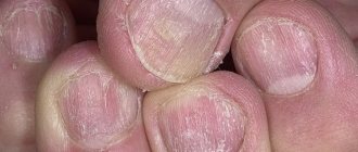
The appearance of psoriatic onychodystrophy, which is accompanied by damage to the nails, is also possible.
If a patient with vulgar psoriasis has metabolic disorders, obesity, or problems with the endocrine or immune system, psoriatic plaques may begin to become wet due to the release of a significant amount of exudate from the plaques - an inflammatory fluid that permeates the scales of the plaques.
Because of this, they stick together and form yellow crusts on the surface of the skin. This is the so-called exudative form of plaque psoriasis, which most often manifests itself in children, the elderly, and those who work outdoors.
Plaque psoriasis, as well as its other forms, is characterized by the presence of three main signs that can be easily detected by scraping the top layer of the papule.
- “Bloody dew” - very small pinpoint bleeding due to damage to the capillary network
- “Stearin stain” - the surface of the papule is removed in one piece with a soft layer, similar to a layer of stearin.
- “Terminal film” - after removing the scales from the papule, a wet layer is formed on its surface, resembling cellophane film in appearance. This is another protective layer of the papule, if violated, bleeding will begin.
Vulgar psoriasis, the manifestation of which can be seen in photographs depicting sick people, develops in stages. The main symptoms of the disease do not make themselves known immediately. Its severity increases gradually. Only in rare cases does the pathological process manifest itself with acute symptoms that adversely affect the patient’s quality of life.
The plaque type of psoriasis is recognized by the following symptoms:
- The appearance of papules on different parts of the body;
- Severe itching in the affected area;
- Redness of certain areas of the skin;
- Dry skin and flaking;
- Severe irritation.
The common plaque type of psoriasis, which occurs in a mild form, affects no more than 3% of the total human skin. Plaques cover individual areas, after which they begin to lose their rich hue. Eventually they just disappear.
Psoriasis is well diagnosed based on its external manifestations.
A blood test allows you to find out the presence of inflammatory, autoimmune, biochemical and other disorders in the body, which allows you to adjust the treatment plan.
The chronic form of plaque psoriasis is very easy to diagnose. This is because the disease is characterized by characteristic rashes on the body. So doctors do not have to carry out specific procedures and tests to determine the patient’s diagnosis.
Psoriasis vulgaris is recognized by plaques accumulated on the skin, which can cause bleeding.
If necessary, the doctor may prescribe the patient to undergo a series of tests that help detect psoriasis at an advanced stage. Their results will indicate the development of autoimmune, inflammatory and rheumatic processes in the body.
Vulgar psoriasis can develop either gradually or quite quickly, affecting large areas of the skin. Therefore, lack of timely treatment can cause serious problems.
Diagnostics in this case does not require any specific procedures. To do this, it is enough to examine the patient’s skin. But if it is necessary to exclude other skin diseases, specialists may prescribe a skin biopsy.