What is angioma
Venous angiomas are localized in organs and tissues and can be multiple or single.
They are located in most cases on the upper half of the body. Their morphological basis is highly dilated lymphatic/blood vessels. The shape and size of pathological areas can vary, color - from colorless to red-blue. The lesions tend to progress rapidly. There are angiomas of the brain, liver, external genitalia, and bones. Angiomas are classified according to their structure:
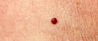
- Simple (hypertrophic, capillary). Small venous and arterial vessels grow. If you study a photo of such an angioma, it will become clear that it is a bright red or bluish-purple spot. Its sizes can be different, even gigantic. If you press on the lesion, it will immediately turn pale due to the outflow of blood. Capillary angiomas of the skin extremely rarely transform into malignant formations.
- Branched (racellose). They are represented by plexuses of tortuous dilated vascular trunks. They are characterized by the presence of constant pulsation. The limbs and face are most often affected. Traumatization of venous branched angiomas is fraught with severe bleeding, which cannot always be stopped on its own.
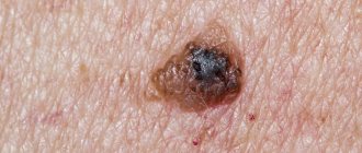
- Cavernous (cavernous). They are formed by wide spongy cavities that are filled with blood. Cavernous angioma looks like a purple-bluish bumpy spot. Most often located under the skin. It feels hotter than the surrounding tissue. When stressed, it increases in size. A complex form is cavernous angioma of the brain, the treatment of which is very long and involves restoration of impaired blood flow.
- Combined. Combine subcutaneous and superficial location. Formed by overgrown vessels and other tissues (depending on location on the body).
The shape of angiomas is:
- stellate (point angiomatous formations, from which dilated small blood vessels extend in all directions);
- flat (vascular spots of pink-blue color located in the upper layers of the skin);
- nodular (seals protruding above the surface of the skin);
- serpiginous (rashes on the skin in the form of small burgundy nodules).
Ways to eliminate red dots
In ninety percent of cases, the presence of an angioma causes feelings of a psychological nature. However, the growth of the tumor, the appearance of itching and pain requires immediate examination. Only after the nature of the pathology has been identified can we talk about its removal.
- Surgery. This type of surgery is performed in a hospital setting. After the operation and examinations, the patient can go home. Excision of the growth can lead to the formation of postoperative scars. Considering this factor, the surgical method of removal is used only to eliminate tumors that are small in size. It should also be noted that this type of surgery is not performed on the face.
- Laser removal. One of the modern and popular methods. Carrying out several sessions allows you to remove even those angiomas that have grown deeply into the tissue. Before starting the procedure, the surface to be treated is numbed using special means. The laser treatment itself lasts less than a minute. After the growth is removed, a small crust forms in its place; it disappears within three weeks after the session. After the damaged tissues are restored, shallow scars remain at the site of the neoplasm.
- Cauterization. One of the most effective and safe methods. Cauterization using a special mixture based on carbon dioxide and nitrogen allows you to remove the angioma without leaving any traces. This method shows high efficiency even when removing particularly large growths. Before the procedure begins, the patient is given an intramuscular injection of an anesthetic.
- Coagulation. You can remove a red mole using electrical impulses, photocoagulation, radio wave therapy and infrared exposure. Each of these methods has its own characteristics, contraindications and side effects.
MORE ABOUT: Heparin ointment. Composition, instructions for use in medicine and cosmetology
Before carrying out the removal procedure, you will need to undergo certain types of tests and undergo a complete diagnosis of the body. Such measures are necessary in order to eliminate the risks associated with the presence of cancer. Based on your medical history, the dermatologist will select the most appropriate method of performing the procedure.
Causes of angioma formation
Angioma in newborns is a standard clinical picture, since this disease in most cases is congenital. The source of the development of neoplasms in adulthood are persistent fetal anastomoses located between the veins and arteries. The proliferation of blood vessels in a benign tumor leads to the growth of the tumor itself.
The causes of angioma are as follows:
- hereditary predisposition;
- changes in hormonal levels;
- diseases of the gastrointestinal tract;
- dysfunction of lipid metabolism;
- skin pigmentation disorders.
In rare cases, venous angiomas of the skin occur after trauma to the skin area (bruises, cuts). They can also accompany malignant neoplasms of internal organs and cirrhosis of the liver.
Red warts on the body: causes of appearance
If such formations appear immediately after birth, this is not a sign that there are any abnormalities in the baby’s body. The reason may be that during pregnancy the mother suffered a viral disease or an exacerbation of a chronic infection. Such moles disappear from the body of children up to the age of seven and do not require any treatment.
On the skin of adults, red moles are somewhat less common than in children, and their appearance indicates existing changes in the body . If an angioma has formed on the body of an adult, the reasons for its appearance may be as follows:
- hormonal disbalance;
- lack of essential vitamins in the body, which leads to thinning of blood vessels;
- inflammation in the gastrointestinal tract;
- cardiovascular diseases;
- oncological tumors present in the body;
- hemophilia;
- pyelonephritis;
- diabetes;
- heredity;
- pregnancy or menopause;
- pathologies in the endocrine system.
If the cause of angioma is liver pathology , then it has a more saturated cherry color.
Symptoms of angioma
Symptoms of angioma depend on the type of vascular tumor, its size and location. In newborns, it is diagnosed immediately or several months after birth, and can grow very quickly, covering large areas of the skin.
Vascular angiomas can affect:
- Integumentary tissues - skin, mucous membranes of the genitals, subcutaneous tissue. Angiomas are also found in the mouth.
- Musculoskeletal system - muscles, bones. A severe form of the disease includes hemangioma of the vertebral body.
- Internal organs.
If integumentary hemangiomas cause cosmetic defects, then hemangiomas of internal organs provoke the occurrence of disturbances of important functions - urination, breathing, vision, defecation.
Angiomas of muscles and bones are accompanied by:
- skeletal deformation;
- severe pain in the joints;
- frequent fractures;
- radicular syndrome (compression of the spinal nerves).
Venous angiomas of the brain (cerebellum, left/right frontal lobe, etc.) are very dangerous. They lead to subarachnoid hemorrhage and epilepsy. Also, due to compression of the brain vessels by the tumor, the following symptoms may occur:
- convulsions;
- headache;
- nausea, vomiting;
- dizziness;
- noise in the head;
- taste disturbances;
- speech problems.
During the growth process, angiomas can become inflamed, causing phlebitis and thrombosis. A common complication is bleeding due to trauma.
If you notice similar symptoms, consult a doctor immediately. It is easier to prevent a disease than to deal with the consequences.
Diagnostics
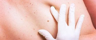
Angiomas located on the surface of the skin are diagnosed during examination and palpation. The characteristic coloring, ability to contract and increase in size when pressed and strained allow the doctor to easily make the correct diagnosis.
If the localization of the lesion is more complex (internal organs, bones, brain), a number of diagnostic measures are required:
Skeletal hemangiomas are identified using x-rays of the spine, ribs, bones, skull and pelvis.
To identify angiomas of internal organs, antiography (contrast x-ray examination of blood vessels) of the kidneys, brain, lungs, etc. is performed.
Pharyngeal angiomas are examined by an otolaryngologist.
Ultrasound diagnostics allows you to determine the exact depth of growth of the angioma, its structure and location features. If a vascular angioma is suspected, the doctor may additionally prescribe a puncture. The resulting yellowish liquid is examined in the laboratory. This makes it possible to differentiate angioma from lymphadenitis, lipoma, cyst, hernia.
Treatment of angioma
Treatment of angioma is aimed at stopping its growth, eliminating the pathological process and normalizing the functioning of the vascular network. Indications for urgent intervention are:
- extensive tissue damage;
- rapid growth of the tumor;
- localization in the neck, head;
- frequent bleeding;
- dysfunction of the organ in which the angioma is located.
Watchful waiting is justified only if regression of a vascular benign tumor is observed.
Among the most common treatments for angiomas:
- Diathermoelectrocoagulation. Cauterization with electric current is indicated if it is necessary to remove hard-to-reach punctate angiomas and angiofibromas. The method cannot be used if the angioma is deep and occupies a large area.
- Laser treatment. Using a laser, the doctor removes pathological tissue layer by layer. The advantage of laser treatment of angiomas is minimal bleeding.
- Radiation treatment. Allows you to achieve good results when removing angiomas of complex anatomical localization.
- Surgery. It is used if the vascular tumor is deep and it is not possible to remove it without affecting the surrounding healthy tissue.
- Cryotherapy. Makes it possible to remove simple small angiomas of any location. During the procedure, liquid nitrogen is applied to the tumor. The method is painless and does not cause bleeding.
- Sclerosing therapy. Seventy percent alcohol is injected into the tumor. The treatment is painful and is suitable if the angioma is located in the deep layers of the skin.
- Hormonal therapy. Relevant if the lesion is extensive, angiomas grow quickly.
- Excision of angiomatous areas followed by reconstruction of the vessel.
- Ligation of the arteries supplying the angioma. A ligature is applied to the end of the artery, as a result of which the neoplasm gradually dies.
It is possible to treat angioma with folk remedies:
- Apply the kombucha to the tumor site and fix it. After a day, replace the compress. The duration of such treatment is 2-3 weeks.
- Dilute a tablespoon of copper sulfate with 100 ml of clean water. Wipe the neoplasm with the resulting liquid 4-5 times a day for 10 days.
- Apply a compress of onion pulp to the affected area of skin for 10 days. Change the bandage every 12 hours.
- Cover the angioma with fresh grated carrots and tie gauze on top. Change 3 times a day.
- Mix fresh celandine juice in a ratio of 1:4 with petroleum jelly and add a drop of 0.25% carbolic acid. Use the ointment daily in the morning and evening.
- If the angioma has affected the internal organs, you can use the following recipe: pour 3 tablespoons of potato flowers into 300 ml of boiling water. Leave in a thermos. Drink half a glass 3 times a day half an hour before meals. The treatment course is 2 weeks.
It should be remembered that any folk recipe can be used only after consultation with your doctor. Some medicinal plants can provoke the growth of angiomas, so preparing decoctions and infusions from them yourself is not recommended.
Removal methods
Removal of hemangioma is the only method of completely getting rid of the tumor. The actual procedure is selected by the child’s attending physician. Common ways to remove growth are:
- Surgical excision. It is performed in cases of deep tumor placement. The most invasive method. It is resorted to when other procedures are not possible.
- Laser removal. Used for small formations. The advantages of this method are the targeted effect on the problem area, while healthy tissue remains untouched. Sometimes, to completely eliminate the entire growth with laser, several laser therapy sessions are required.
- Cryodestruction or cauterization of the formation with liquid nitrogen. Under the influence of low temperature, tumor cells are destroyed. Within a few days, the treated tissues die. There may be a small scar or scar left at the surgery site.
- Sclerosis. A loading dose of Prednisolone is injected into the growth tissue. It is assumed that after the blood supply is blocked, the formation dies.
Whatever method of removal is chosen for the child, it is necessary to understand that delaying the operation is dangerous. The sooner the removal occurs, the faster the rehabilitation period begins, the lower the risk of complications.
In children, vascular tumors on the lip appear in the first week of life. Experts advise parents not to panic in such a situation: by the age of one year, the hemangioma may disappear on its own without the use of any treatment. You need to see a specialist if the nodules filled with blood protrude strongly above the surface of the lip.
MORE ABOUT: Chlamydia in women - causes, symptoms, diagnosis and treatment
Various methods are used to treat vascular tumors in adults:
- injections,
- hormonal drugs,
- radical treatment.
Sclerotherapy is the introduction of drugs into the body, the action of which stops the growth of hemangioma - a painful method of treatment. As a result of this procedure, the nutrition of the tumor is stopped, so it turns pale and disappears over time.
When choosing drug treatment, the patient is prescribed hormonal drugs and beta blockers for oral administration. They affect pathological vessels, reduce or block cell division processes. Thanks to this, the tumor gradually dies.
When the formation is located deep in the tissues from which the lip is formed, radical treatment is resorted to.
| Name | Description |
| Laser therapy | When it is used, the tumor is affected using a beam of directed light. It generates thermal radiation, which evaporates the fluid in the pathologically altered cells. They die from dehydration. The surgeon has the ability to control the depth of penetration, so healthy tissue is not harmed |
| Surgery | During the manipulation process, the hemangioma is removed using a scalpel. The operation is performed under local anesthesia |
| Cryodestruction | Used for superficial tumor localization. It is exposed to liquid nitrogen. Low temperatures cause instant necrosis of pathological tissues |
| Cauterization | This treatment method is used only for the treatment of small capillary hemangioma. She is burned with electric shock |
Independent removal of the formation using traditional medicine is unacceptable. This is fraught with the appearance of severe bleeding or transformation into a cancerous tumor. It is also impossible to leave a hemangioma unattended, as there are risks of developing unwanted complications.
The method of eliminating hemangioma is selected depending on its type, size and depth of germination.
Basic methods:
- Laser removal is a method that minimizes damage to surrounding healthy cells. This treatment method is ideal for small hemangiomas.
- Exposure to cold (cryodestruction) - using liquid nitrogen. The method helps eliminate the problem without damaging the facial nerve, which may be located nearby.
- Surgical excision of hemangioma is used in cases where the tumor is located deep and cannot be eliminated by other means.
The most promising method of treating lip hemangioma in adults and children is laser. This method allows you to act as precisely and delicately as possible on the problem area.
- Speed of the procedure;
- No seams or scars;
- Prevention of blood loss due to the cauterizing effect of the laser;
- The process is almost completely painless;
- Accuracy and accuracy of hemangioma removal on the lip in adults and children;
- Minimal risk of reappearance (relapse);
- The result meets the high requirements for a cosmetic surgical procedure performed on the face, in the area of the delicate and sensitive skin of the lips.
In our clinic, hemangioma removal on the lip is carried out using innovative equipment, the FOTONA laser, and is completely painless.
Large tumors, especially those that are convex and deform the surrounding soft tissue, require preliminary ultrasound examination. Using ultrasound, you can determine the boundaries and depth of penetration of the node or monitor the dynamics of its growth rate, comparing the data obtained with previous examinations. This is a fairly common method of preliminary examination.
If there are large vessels in the tumor, preparatory drug treatment may be necessary before surgery to reduce and close their lumens.
If it is necessary to remove a hemangioma on the lip, treatment may have some limitations in use.
So, it is contraindicated for:
- Acute diseases and exacerbations of chronic health problems (diabetes, etc.);
- The presence of purulent processes in the area where the tumor is located;
- Problems with blood clotting;
- Autoimmune diseases;
- Oncological diseases.
Most of the contraindications are not permanent and after recovery or improvement of the condition, you can again think about eliminating the tumor.
- Complete correction of a cosmetic defect, restoration of the correct proportions and shape of the lips;
- Elimination of the potential danger of developing a malignant process;
- Relief from difficulties with eating, drinking, breathing (in case of large sizes or deep germination of the node into the thickness of tissues).
Contact the Neo Vita clinic and sign up for a free consultation with our cosmetologists and we will quickly and painlessly solve all your problems related to the elimination of hemangiomas on the lips.
You can get an online consultation with a doctor on this problem from anywhere in the world. The cost of consultation is 5,000 rubles.
| Removal of hemangioma up to 2 mm | 800 |
| Removal of hemangioma from 2 mm to 5 mm | 1 500 |
| Removal of hemangioma from 5 mm to 10 mm | 2 000 |
| Removal of hemangioma from 1 cm to 2 cm | 2 500 |
| Removal of hemangioma more than 2 cm | 3 000 |
- injections;
- hormonal drugs;
- radical treatment.
In some cases, one type of hemangioma appears on the lip of adults or children. Hemangioma on the lip is removed in various ways, depending on the extent of the formation, its type and other features.
A hemangioma on the lip in a child or adult is a benign formation that appears on the surface of this part of the body. This localization is due to the fact that this is where a large number of blood vessels and capillaries are located. The cause of such capillary formation is vascular disorders.
Hemangioma of the lip in adults or children is shaped like a spot. This benign neoplasm consists of a fine network of blood vessels and capillaries. It is painted pink, red, bluish or purple.
The main features of this education include:
- These are congenital capillary benign tumors, predominantly red in color. They can develop as they grow older under the influence of certain causes or provoking factors.
- Dense formations can become larger in size over time, trapping and deforming neighboring tissues.
- Sometimes they tend to grow excessively quickly. In such a situation, surgical intervention is indicated as quickly as possible to prevent malignancy of the pathological process.
- The small red lesion may disappear on its own.
According to statistics, more than 40% of all benign formations are hemangiomas. The appearance of hemangioma in children and adults is due to various reasons. There are congenital and acquired formations.
Doctors say that congenital ones appear as a result of disturbances in the functioning of the circulatory system, when it is not formed correctly in the fetus during the gestational period.
Also, the reasons for the appearance of congenital hemangioma may lie in diseases suffered by the expectant mother during pregnancy.
Acquired pathology refers to vascular disorders and may be caused by genetic predisposition and frequent exposure to ultraviolet radiation.
Danger
A small spot formed on the skin can go unnoticed or be ignored by the patient for a long time. Since it can transform into a malignant formation, the lack of necessary treatment can lead to dire consequences.
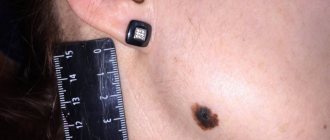
Particular attention should be paid to angiomas located in places of increased friction with clothing (neck, chest, abdomen, shoulders), on the scalp. Frequent traumatization can lead to inflammation of a benign tumor and its rapid growth and bleeding.
The greatest danger to life is posed by angiomas of the brain and internal organs. As practice shows, if you do not treat a cerebral angioma, the prognosis is unfavorable - the area of accumulation of blood vessels will increase, which will lead to their rupture, hemorrhage in the brain and death.
Preventive measures
The expectant mother can prevent the onset of the disease. It is necessary to approach the process of conception and pregnancy competently. Having decided to become parents, a couple should undergo a medical examination, identify and get rid of existing diseases, and eliminate cigarettes and alcohol-containing drinks from their lives.
Particular attention is paid to diseases of the cardiovascular system. Angioma is on the border between a benign tumor and a disorder in the formation of vascular tissue. A woman should identify vascular problems and begin treatment before conception.
MORE ABOUT: Mammography: on what day of the cycle should it be done to ensure the reliability of the result |
Expectant parents need to maintain healthy reproductive organs and normal hormonal levels. A woman should stop taking contraceptives for a long time.
During pregnancy, it is not recommended to stay in the sun for a long time. Daily walks are required, but direct sunlight should be avoided.
If the disease is present, the patient must regularly measure blood pressure, stop smoking and alcohol, carefully take medications that affect the blood, follow a sleep and rest schedule, and not force the body through physical and mental labor.
Angiomas are neoplasms on the skin that look like red moles. These growths are considered to be benign. The appearance of moles is usually associated with disturbances in the circulatory or lymphatic system. Angioma can form on the body throughout a person’s life, however, having appeared in childhood, such moles can subsequently completely disappear. Let's find out why red moles appear on the body and how to deal with them.
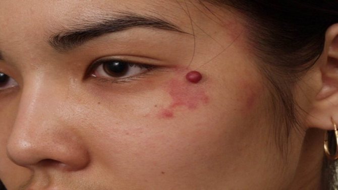
Angioma refers to benign tumors, the structure of which is represented by lymphatic or blood vessels