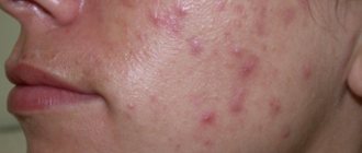
A mole, or, in medical terms, a nevus, does not pose a danger to human health until the neoplasm begins to degenerate or mutate. If problematic symptoms occur, you should consult a doctor. Sometimes the only treatment is to remove the mole.
Electrocoagulation, a modern method of getting rid of a mole
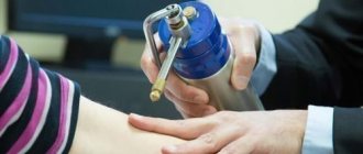
Electrocoagulation: what is it?
There are several options for removing moles that may be harmful or cause discomfort. Most often, when doctors tell their patients that they need to eliminate a problematic formation, people become afraid of surgery, pain and scars for life. But today various techniques are used that allow you to get rid of tumors without pain and scars.
Electrocoagulation is a method of removing various tumors on the skin with electric current. Using this method, moles, viral papillomas, warts and other unwanted formations are eliminated.
How can I remove it?
People faced with the question of how to remove a tumor have to choose a method for doing it. Each method has its negative and positive sides, and is suitable for a certain type of growth.
Papillomas, moles and other neoplasms can be removed using several methods:
- Electrocoagulation method. When carrying out this procedure, high frequency current is used.
- Laser excision. The tumor is eliminated using a laser, with minimal blood loss, which prevents possible metastasis.
- Cryodestruction. Ultra-low temperatures are used for removal.
- Surgical removal. This excision produces maximum damage to the skin with noticeable scarring.
- Radio knife. The elimination process occurs without damaging healthy tissue. This method is one of the most promising.
Cryodestruction
A widely used method for removing tumors for several decades is cryodestruction. The procedure is carried out with liquid nitrogen or another refrigerant. The essence of the technique is to expose the skin to extremely low temperatures (down to –196 C). Flash freezing destroys unwanted tissue and turns it white. Complete rejection of pathological elements occurs within 5–6 weeks.
The removal itself is not accompanied by any unpleasant sensations (only slight tingling, numbness), however, after “defrosting” the skin, pain is possible. In case of superficial formation, a cotton wool with a reagent is applied. In case of deep tissue damage, a cryodestructor is used. A thin needle with a sensor is immersed to the desired depth and immediately removed. One such session lasts 2–3 minutes.
Advantages of cryodestruction:
- There is no need for pain relief.
- Well tolerated by patients.
- Fast wound healing.
- No scars.
- Impossibility of infection and spread.
- Low cost.
Disadvantages of cryodestruction:
- Inability to accurately control the depth and area of exposure to the refrigerant.
- In some cases, repeat sessions are needed.
- Temporary appearance of spots and swelling at the site of exposure.
- Contraindicated for cold intolerance.
During cryodestruction, there is a possibility of removal of healthy tissue or incomplete elimination of the formation.
Radio wave coagulation
The essence of the radio wave coagulation method is the exposure of tissue to high frequency radio waves (3.8-4 MHz). This leads to their overheating and a kind of “evaporation”. For the procedure, special devices are used, the SURGITRON TM device (American made) is especially widely used. Today, the radio knife is the most modern “tool” for removing tumors.
During coagulation, radio waves are concentrated at the end of the electrode, which allows the affected tissue to be removed with pinpoint precision. The doctor has complete control over the depth and area of exposure.
The advantages of radio wave coagulation are:
- Painless procedure.
- Fast healing.
- It is possible to conduct subsequent histological analysis.
- Minimal likelihood of scarring.
- No subsequent swelling or infection.
There are some restrictions on radio wave coagulation. The procedure is contraindicated in the following cases:
- Presence of a pacemaker.
- ORZ.
- Fevers.
- Epilepsy.
- Diabetes mellitus.
- Suspicions of cancer.
Electrocoagulation
The electrocoagulation method has been used since the mid-twentieth century. Its use in modern medicine speaks of reliability and effectiveness. The device for carrying out the procedure is similar to a soldering iron, and the principle consists of point cauterization of tissue. The specialist performs layer-by-layer cauterization, trying not to touch healthy tissue. At the site of “welding” of the neoplasm cells, a crust remains, which disappears over time. You can't tear it off. Complete healing of the wound occurs in 10–14 days.
Advantages of electrocoagulation:
- Complete removal of unwanted growth.
- Minimum material costs.
- Possibility of collecting biomaterial for analysis.
The disadvantages of electrocoagulation are:
- Need for pain relief.
- The likelihood of a scar or pit appearing at the site of formation.
- Possibility of damage to adjacent tissues.
- The need for subsequent care of the affected area (until the crust falls off).
Electrocoagulation is suitable for removing only small lesions.
Laser
For laser removal of tumors, a light beam is used to destroy the affected cells, evaporating them. The beam parameters are selected individually. The procedure is carried out in layers without affecting healthy tissue. A crust forms at the site of the treated area, which disappears on its own after 2–3 days. Local anesthesia will eliminate discomfort.
Advantages of using laser:
- Complete removal of the pathological area.
- Impossibility of traumatizing healthy tissue.
- Bloodlessness of the procedure.
Disadvantages of laser removal:
- The procedure is expensive.
- The need for local anesthesia.
- Possible scarring.
- The likelihood of a burn on dark skin.
- Existence of contraindications: diabetes, acute phase of infectious processes.
- Lack of ability to analyze affected tissue.
Surgically
The most reliable and proven way to remove any, especially large, deep, tumors. It is preferable for excision of dangerous and hanging moles. The procedure is performed under local anesthesia. A scar remains at the incision site, and the postoperative area requires care for some time.
Disadvantages of surgical removal:
- High price.
- The appearance of a scar.
- The need for postoperative measures.
- Need for anesthesia.
Diathermocoagulation
The method of cauterization and excision of tissue using high-frequency alternating electric current is called diathermocoagulation. The principle of the method is based on heating to high temperatures, which leads to irreversible folding of cell proteins. The diathermy apparatus produces a current of up to 3–5 Amps, with the help of which specialists achieve coagulation. It is important that this cauterization eliminates bleeding due to thrombosis of blood vessels. This fact prevents the spread of tumor cells throughout the blood system.
Advantages of diathermocoagulation:
- Bloodlessness.
- Simplicity and affordability.
- Sufficient penetration depth.
Disadvantages of this method of treatment:
- Formation of scar tissue.
- There is a rehabilitation period after the procedure.
With diathermocoagulation, there is a possibility of wound infection.
Reasons for removing nevi
It often happens that a new mole on the body suddenly appears in an area where it constantly clings or causes inconvenience.
As a rule, nevi are removed if they are located in the following places:
- on the neck, where the frequency of friction and snags by a collar or chain is increased,
- in areas where moles are often injured by an elastic band, strap or other parts of clothing,
- in the armpits, which makes the process of hair removal inconvenient, nevi on the scalp when they are often touched by a comb.
In such cases, many turn to a dermatologist to examine the formation and recommend the necessary method of removal.
Moles under the armpit are usually recommended to be removed
Recovery period
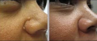
After removing a mole using electrocoagulation, a dry crust forms on the skin. This is a scab under which new healthy tissue is forming.
Rules for patient behavior during the recovery period:
- Do not wet the crust with water or try to tear it off yourself.
- In the first days after electrocoagulation of a mole, minor painful sensations may appear. In this case, it is permissible to stop them with anesthetic drugs.
- It is forbidden to apply decorative cosmetics to the scab, especially for patients who have had a nevus removed from their face. If the wound is on the torso or limbs, it is not advisable to even use shower gel. Recommendations regarding skin care are provided by a dermatologist. Typically, your doctor will recommend showering with baby soap.
- Until the crust falls off, it is prohibited to visit swimming pools, saunas and steam baths. Swimming in open water is also prohibited.
- Reduce the intensity of physical activity to a minimum. This is due to the need to reduce the amount of sweat produced.
- Avoid prolonged exposure to sunlight for two months. This can cause hyperpigmentation.
The duration of the recovery period is, on average, 1 month. During this time, the scab disappears, and in its place remains smooth skin of a slightly pinkish tint. Over time, the fabrics acquire a natural color.
Advantages of removal using electrocoagulation
When compared with other possible options that allow you to get rid of pigmented formations, the electrocoagulation method has a set of characteristic advantages.
Among the advantages of this type of mole removal are the following:
- the operation does not take much time,
- no cases of relapse,
- scars and cicatrices are excluded, which is suitable for removing moles on the face and other prominent places,
- no bleeding,
- This method can remove even small formations,
- the possibility of further examination of the epidermis for the risk of future complications.
Most often, this method is used by people who want to get rid of a mole for aesthetic reasons or when it causes physical discomfort.
It is advisable for representatives of the fair sex to take into account that the most favorable period for the operation is the first half of the cycle.
The electrocoagulator does not leave bleeding wounds or scars
When electrocoagulation is not suitable
As a rule, this method is excellent in cases where nevi have an insignificant base or are small in size.
Despite the fact that removing moles using this method has a number of advantages, there are also situations when you need to refrain from electrocoagulation:
- during pregnancy and breastfeeding,
- patients with malignant tumors on the face,
- if the blood does not clot well,
- when a person finds it difficult to tolerate operations using electric current,
- the body is susceptible to chronic infection.
If you find yourself with at least one of the listed symptoms, then this option for excision of a mole is not suitable for you.
Electrocoagulation is not performed when there are signs of degeneration of a mole into a malignant tumor. Since there is a risk of starting the process of developing metastases. In cases where the likelihood of further transformation of the nevus into skin cancer is detected, a surgical removal method is used.
It should also be noted that large safe moles, with a diameter of 10 mm or more, are best removed through surgery. And the wounds formed in the areas should be stitched with threads to speed up healing and also to avoid the accumulation of pus in the cells.
Poor blood clotting, contraindication to electrocoagulation
What to do after?
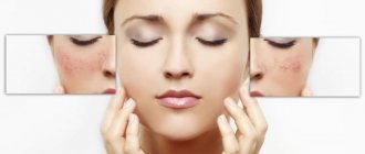
During the recovery period, you should avoid ultraviolet rays, do not wet the crust and do not touch it under any circumstances. Over time, it disappears on its own. Do not be alarmed if a pink spot remains in place of the crust; soon the skin of the face will acquire a normal and natural color, all skin defects will disappear. If you notice inflammatory processes, you should immediately consult a doctor.
You can often hear from patients: “I removed a mole, and a scar remained.” Such situations also happen, and there is no need to be afraid; if the work is done in the best possible way, then there should be no scars. It would not be superfluous to mention that you need to seek help from a highly qualified specialist who will do the job efficiently. Elena Vladimirovna Salyamkina has been working in the field of plastic surgery for many years, and the increase in clients every year speaks of the quality of the work performed.
Features of the procedure
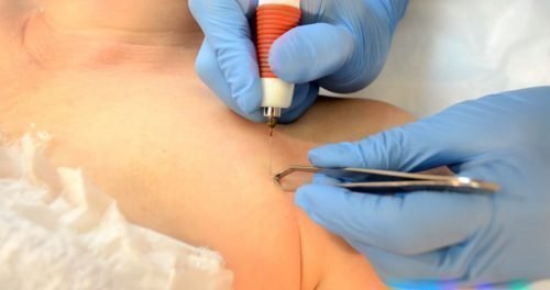
In this way, tumors can be eliminated on any part of the body. The mole is completely removed in one session. As a rule, the entire removal procedure, which does not require special preparation, takes about 20-30 minutes. For the operation, a medical device is used - an electrocoagulator, which is equipped with an electrode at one end.
First of all, the specialist administers local anesthesia. This may be a pain-relieving injection or application of a special cream to the area. Then, with a single movement, cut the mole. At the time of removal of the nevus, the adjacent layers of skin are cauterized, which allows the procedure to be carried out without the appearance of blood, swelling or infection.
It is necessary to say separately about the nature of pain during the operation. Many people worry that cauterizing moles is incredibly painful. The maximum that the patient can feel is a slight tingling in the area where the removal takes place.
Requirements for the postoperative recovery period
The precautions that must be taken after a mole has been removed by electrocoagulation are in many ways similar to other methods of eliminating nevi.
It is recommended to follow a few simple rules:
- you can't sunbathe in the sun,
- you need to refrain from going to the sauna or bathhouse,
- use creams or other cosmetics to apply to the healing area to dry it out,
- To protect against infection, the wound should be treated with dissolved brilliant green.
If you strictly follow these rules and the doctor’s instructions, the rehabilitation period will end quickly (up to 5 days) and there will be no complications such as pigmentation or suppuration.
Zelenka will protect the wound from infection
Advantages
According to medical reviews, removal of moles by electrocoagulation is the most modern and safest method of excision of nevi.
Advantages of the method:
- Efficiency. Once removed, the tumor will never appear again.
- Aesthetics. During the procedure, no excision of healthy surrounding tissue occurs. After the recovery period is complete, smooth skin without scars remains at the site of the mole.
- Safety. The risk of complications is minimized.
- Availability. Electrocoagulation of moles is cheaper than excision, for example, with a scalpel.
- The duration of the procedure is only a few minutes.
- Possibility of adjusting the depth of electrode exposure.
- Hemorrhage is excluded.
- Minimal risk of infection.
In addition, the procedure is not associated with any unpleasant sensations. Immediately before removing a mole, the doctor injects an anesthetic into the required area.
Possible complications after surgery
When removal is performed on the face, it is likely that a person will experience mild soreness at the excision site, as well as swelling. These small changes disappear within a few days on their own and do not require additional medical measures. In extreme cases, a person may be prescribed painkillers.
Usually, at the end of 14 days, the resulting protective crust is rejected naturally. And in this area, the growth of a new layer of pink epithelium begins, which later becomes colorless.
Sometimes, due to violations in the technology of the removal process or improper care during recovery time, some complications may arise.
- Infection. If the protective crust falls off before healing is complete, there is a risk of infection entering the wound, which can cause the formation of pus.
- Scarring. When an infection enters the wound immediately at the moment of cauterization, pus collects under the protective crust, which provokes peeling. As a result, a scar may form at the site where the mole was removed.
Therefore, careful care of the area where the removal was performed will help avoid the formation of an unsightly scar.
A scar after mole removal may occur if it suppurates
Removal of papillomas, grasses and moles using electrocoagulation
Once upon a time, I had a mole in my head, just behind my ear. The most common one is just a brown spot on the skin. But then it began to grow, as if on top of the skin. in short, it turned into a nevus.
Every time I went to the hairdresser or when someone touched my hair there, I felt shame and embarrassment. Because the mole was big, I was embarrassed by it. And hairdressers generally almost ripped them off.
Since I don’t have a lot of hair, it was often visible with smoothly combed hair.
In short, she poisoned my existence, and I decided to get rid of her.
Somehow it didn’t work out all the time, and then a month and a half ago I made up my mind.
Surprisingly, I was not afraid of pain, and as it turned out, in vain! Because I won’t forget the shock that I experienced, probably until I die!
So, I didn’t go to a private clinic, as most people do, but to oncology. Since I was, as they say, an acquaintance, I was not taken through the registration desk, which I now very much regret. And I’ll explain why below.
So, I was told to bring insulin syringes and lidocaine.
In total, I removed three moles. Two in the head, including the big one, and one on the back of the neck. The doctor didn’t send me home, he said that everything would be fine, since the summer is cool now, and in the winter I can’t wear a hat either.
On the neck and one on the head - it didn’t hurt at all. It smelled like burnt skin, but there was no sensation. But the second one was something special! I haven't felt such horror in a long time! I was in terrible pain, blood was flowing down my ears and face, and there was no way to stop it.
But everything comes to an end, and this test too.
So I went home in complete shock, and at home my mother (the health worker) asked me in surprise: why didn’t they sew up these holes? How will it all heal now?
The doctor had only one prescription: apply brilliant green twice a day. Do not wash your hair for three days.
Okay, said and done. A couple of days pass, and I begin to feel that I cannot sleep at night on the side where the mole once was, and now there are holes. After these couple of days the wounds looked like this:
There is that same black crust. which should have departed safely. So, the morning after an unpleasant night, I ask my mother to check what is happening there, and the trouble turns out: the wounds are rotting.
In short, I had to tear off these crusts several times and fill them with brilliant green again and again, because pus accumulated endlessly under the crusts! After several such days, the wound on the neck looked like this:
Time passed. I washed my hair for the first time a week later, although I was afraid of causing harm. It was painful to sleep, and the edges of the wounds were very itchy.
I couldn’t go and present it to the doctor, since I was going, as they say, unofficially.
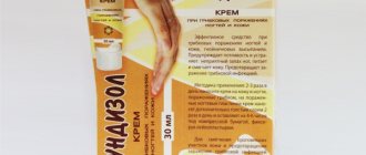
I went to the oncologists’ website, read people who had faced the same problem, and found out that in such cases everyone is prescribed Baneocin.
In general, I engaged in what is called self-medication.
A month and a half after the procedure, one wound was completely healed, but the second two were not.
The one on the neck also could not be tightened, and only now things have improved a little. View after a month (white is Baneocin, powder like this):
Today (a little over a month and a half after the procedure) this wound looks like this:
It is clear that there will be a terrible scar, and it is still too early for complete healing.
But the wound in the head itself looks much more terrible. Today I discovered that pus had accumulated under a thick crust. Here's what it looks like:
I continue to rinse and sprinkle Baneocin.
This is the conclusion I made after scouring the Internet. Many people had similar complications after this method. Who is to blame: I picked up an infection somewhere myself (which is no wonder, since it’s summer, it’s hot, plus it’s my hair, more dust and rubbish cling to it), or the doctors brought something in. We'll never find out. But! If it had been a standard excision followed by suturing the wound, this would definitely not have happened, since the wound would have been sutured and not open, like mine was.
If the wound after the mole is not large, then I agree. that there is no need to sew it. But, it seems to me that we need to look at the place of removal, at the depth... In short, at various associated factors.
And after such a scar, which many are afraid of, will naturally remain.
I also pray to God that these troubles will end only in this way, and not somewhere deep down they begin to degenerate, these moles, and then I wouldn’t have to run to all the oncologies in Kyiv!
I do not recommend this method to anyone! And take care of yourself!