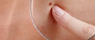
Consultation with a doctor
It is imperative to go to see a doctor if the birthmarks have increased in diameter so much that they only cause pain and inconvenience to the person when they come into contact with clothing.
Sometimes the pain and redness is only temporary, but don't let your body confuse you. After some time, all the unpleasant symptoms will return again, but at this time they will become much stronger and more unpleasant. Light skin, which has a large number of birthmarks, requires a more attentive and serious attitude. Deaths often occur when melanoma is detected late. Its main danger lies in the fact that metastases occur almost immediately, affecting the internal organs of a person.
Currently, modern diagnostics includes methods for identifying dangerous moles that are completely safe and painless for humans (good procedures are: x-rays, computed tomography, laboratory tests, biopsy). Large moles must be removed without fail to prevent any oncology from occurring. At the moment, their elimination occurs surgically with the use of radiation therapy.
Many of us think that melanoma is something terrible and scary, it is necessarily noticeable. However, in fact, even a small speck can develop into a dangerous tumor. However, it does not have to be huge. Very often its size does not exceed 5 mm and resembles a dark spot on the surface of the skin. In this case, there may not be any pain in this area.
Of course, most often melanoma begins to bleed and hurt. But usually this happens in the later stages, when treatment is not particularly effective, and the likelihood of a complete cure is minimized. Thus, only regular examinations, as well as constant monitoring of one’s own health and timely consultation with a doctor, contribute to recovery.
Melanoma
Thus, a specialist will be able to almost immediately identify a malignant neoplasm or a common nevus. The fact is that under a microscope, as well as a magnifying glass, all the bends and fractures, as well as the relief of the mole, are much better visible. Thus, melanoma can be detected quite quickly and easily at an early stage, which guarantees recovery.
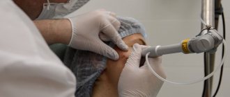
Diagnostic options
Why do moles become prominent?
Moles that settled on our skin after birth are conventionally divided into three groups:
- Superficial, located in the upper layers of the skin.
- Borderline, located on the border between the dermis and epidermis.
- Deep, lying deep in the dermis.
The depth of the melanocyte group mainly affects the shape of the mole itself. Flat moles are mostly safe neoplasms, because... contain a minimal amount of pigments due to their location on the surface of the skin. While raised moles are located inside the dermis.
This suggests a conclusion: if a mole protrudes above the skin, this may mean an increase in the number of atypical cells and the growth of a neoplasm deep into the dermis. Such changes in nevi require an immediate visit to a dermatologist, regardless of whether the mole has become bulging on the face or another part of the body.
A raised mole requires mandatory examination if:
- It is located on those parts of the body that are most often exposed to ultraviolet radiation: the face, neck from the back, shoulders and the back of the hand.
- Often subject to injury due to friction with clothing and accessories: on the neck, waist, wrists, palms, feet.
- An increase of more than 5 mm is observed.
- Vegetation spontaneously fell from its surface.
- It became asymmetrical and uneven at the edges.
- Any changes in color and shape are observed. For example, a previously brown and flat mole has become red and raised.
- The skin on its surface began to itch, peel, and became crusty.
- The skin around it is inflamed and discharge is observed.
- Blotches of a different color appeared on its surface, as well as nodules.
- The characteristic skin pattern has disappeared from its surface. In all these cases, it is necessary to seek advice from a dermatologist or therapist as soon as possible. After examining your mole, even with the slightest hint of a threat, he will write a referral to an oncologist or oncodermatologist. There is no need to postpone your visit to the doctor for a week or two, because if the tumor is confirmed to be malignant, time may not be on your side. Treatment provided at the earliest stage is most effective.
The color of moles is affected by the accumulation of melanin: the more of it accumulated in a mole, the darker it will be. Mega-doses of melanin can turn nevi dark brown and even black.
Such a mole is a rather dangerous formation when compared with other facts: it is the only one on your body, significantly different from other moles, and appeared relatively recently. If a black spot appeared on the skin in adolescence, then you don’t have to worry about it - the color could become intensely black due to hormonal changes in the young body.
A double danger is warned by the fact that a once flat mole has become convex and acquired an intense black tint. Such a sign, although not always, can be a signal of a sharp increase in the number of cancer cells. If, along with a change in color, the mole begins to quickly increase in size, begins to itch and peel, you should urgently seek advice from a dermatologist. And even if it spontaneously dried up and fell off, the risk of developing cancer may remain at a fairly high level.
In the end, I would like to add: so that one day flat moles do not become convex, protect them from the harmful rays of the sun, constant injuries and regularly check for suspicious changes.
If there are many large moles on the body, then they should be carefully monitored. Rapid growth and bulge should be a reason to contact a specialist. Early diagnosis will help avoid serious consequences.
Such nevi may not cause much discomfort to the patient, but they are easy to injure. Then dermatologists advise removing the nevus in order to avoid infection and its degeneration into a malignant neoplasm.
A flat mole becomes convex for a number of reasons:
- Excessive exposure to ultraviolet radiation.
- Hormonal changes in the body. This could be pregnancy or hormonal treatment.
- Disturbances in the functioning of internal organs.
Often, patients turn to a dermatologist with complaints about changes in the structure of the nevus (the mole is swollen, swollen). Such transformations are not always dangerous, but require increased attention from a specialist. If a mole has increased in size in just a few days, it is possible that the cause is a hormonal imbalance. Many may remember that a flat mole became convex during puberty.
Excessive exposure to the sun can cause nevus growth
In adulthood, hormonal imbalance can lead to the active production of melanin. As a result, new brown spots appear on the body, and old growths increase in size. If, in addition to this, other problems appear, such as increased hair growth, enlarged mammary glands in men or menstrual irregularities in women, a visit to the doctor cannot be postponed.
A flat mole can also swell during pregnancy. The reason is also the increased production of hormones. Again, melanin begins to be actively produced, which acts as a “foundation” for nevi.
We suggest you familiarize yourself with Brown flat spots on the body
Ultraviolet rays can also aggravate the problem. It is no coincidence that people with a large number of nevi on the body are not recommended to be in the open sun. Ultraviolet light will not only cause the mole to swell, but can also lead to irreversible malignant changes in some neoplasms.
How to determine the oncogenicity of a tumor
Let's look at the main signs of malignant moles. First of all, various changes on the surface of the growth indicate the degeneration of birthmarks. Peeling, increase in diameter and change in structure are the main symptoms of nevus transformation. Symptoms such as itching, burning and tingling sensation in the area of moles are also important. Less commonly, various allergic reactions occur in those parts of the body that are adjacent to the growths.
Hair loss, color changes and the formation of papillomas are one of the clear signs of metamorphosis. Subsequently, new small-sized moles are added to the above-described symptoms, located close to the main growth. The appearance of small wounds and hemorrhages is also a negative symptom.
Skin cancer is a deadly disease!
Above are photos of moles that need to be removed in order to prevent the development of cancer. Each of the symptoms indicated in the list may be the beginning of pathological processes that have a destructive effect on the body
That is why it is very important to seek qualified help in a timely manner.
Modern methods
If a specialist notices any negative changes in the area of the mole (the presence of bleeding or a significant increase in size), he decides to remove the nevus.
There are a large number of medical and cosmetic techniques that allow you to remove a raised mole painlessly and without visible marks.
- Mole removal using laser surgery is the most popular but expensive method. With this application, there is no bleeding, since the outer skin is not damaged, and the person does not feel pain.
- During electrocoagulation , a specialist uses a high-frequency current; it has a destructive effect on pigmented formations, gradually leading to their reduction and discoloration. In such a procedure, currents with a temperature of over 800°C are used, after their influence the protein coagulates, and the area where the mole was located, being the site of the burn, is treated to stop bleeding. Experts often prefer this type of surgical intervention because of the ability to completely control the depth of penetration of the current, because nevi can be close to the surface or deeply located.
- The use of radiosurgery is similar to a laser; high-frequency radio waves are used here; if a convex mole is located in the deepest layers of the skin, then it is necessary to make a similar surgical dissection.
- Often, to get rid of moles and warts the cryodestruction method ; it is based on the use of low temperatures. The problem area freezes, the tissue becomes numb, and its complete destruction occurs. The whole procedure takes from a few seconds to 2-3 minutes.
This is interesting: Mole above the lip on the left
What is a mole?
A mole is a skin growth filled with cells capable of forming the pigment melanin. These elementary units that produce melanin, located in the lower layer of the epidermis, are called melanocytes. The creation of melanin by melanocytes occurs under the influence of natural or artificial ultraviolet rays.
People have approximately the same number of melanocyte cells, but each person produces melanin individually. Based on the amount of suspended polymer compounds produced that color the fabric, humanity is conventionally divided into four classes.
Representatives of the first class have snow-white skin, light-colored eyes (blue or green) and red hair. Often their face is covered with freckles. Little melanin is produced, so the skin almost never tans.
The third class is represented by dark-skinned people with gray or almond eyes, coffee-colored or dark-brown hair. Due to the high amount of melanin produced, their skin quickly darkens in the sun, and damage to the skin during prolonged exposure to sunlight is extremely rare.
And the participants in the fourth grade are not sensitive to ultraviolet radiation, they are dark-skinned, brown-eyed, with dark hair.
Methods for removing flat moles
Removal is carried out by a dermatologist or oncologist surgeon. The course of the operation is determined strictly according to the indications and examination results. Pigment formations should not be injured to avoid dangerous consequences.
Serious reasons for surgical intervention are:
- degeneration into a malignant tumor;
- begins to enlarge, become convex, causes discomfort, increases the risk of injury;
- The mole is located on an open area of the body and does not look aesthetically pleasing.
Removal cannot be carried out during viral diseases, acute viral infections, during inflammatory processes, or in people with mental disorders.
Modern techniques:
| Name | Advantages and disadvantages | Contraindications |
| Cryodestruction. The birthmark is treated with liquid nitrogen. The result is gradual destruction. |
|
|
| Laser. The high temperature beam excises the tissue layer by layer. |
|
|
| Radio wave therapy. The doctor uses a special instrument - a radio knife. The tissue is cut by thermal energy without contact, which preserves the health of the surrounding skin. |
|
|
| Diathermo-electrocoagulation. The fabric is burned away by electric current. |
|
|
| Surgical excision. A flat nevus and a healthy area around it are cut out with a scalpel to avoid dangerous processes. |
| There are no contraindications; it is used when identifying prohibitions on other techniques and in complex cases of precancerous and cancerous conditions. |
The choice of method is made by a dermatologist, oncologist or surgeon, based on the location (head, lower back, face, other areas), shape, size, type of formation
It is important to choose an effective and safe option for the patient.
Treatment
Before starting treatment, a detailed diagnosis of the tumor is carried out. The doctor examines the patient and talks with him. During the survey, the following facts are established:
- amount of time spent in the sun,
- chronic diseases,
- violation of the body's immune function,
- hormonal imbalances,
- hereditary factor.
Then the surface of the mole is examined using special equipment under magnification. This way you can notice the smallest details. Afterwards, the temperature of the mole and healthy skin is measured, and blood and urine tests are taken. A nevus biopsy is also taken, i.e. scraping from its surface. Only after studying the results is treatment prescribed.
It is impossible to get rid of a raised mole with the help of medications, as a relapse may occur. The most effective way is to remove the tumor.
Modern treatment methods suggest the following options for removing moles:
- Using a laser, the procedure is safe and does not require incisions. Usually there is no scar left, except in cases where the nevus has a wide base.
- Cryodestruction is the removal of tumors using liquid nitrogen. During the procedure, nevus tissue is frozen. The method is used for small size moles.
- Electrocoagulation - a method that involves exposing the birthmark to high-frequency waves. It is used to excise filamentous nevi. When large moles are removed, a scar may remain.
- Radio waves are an effective method for removing nevi of any type.
- Surgical removal - this method is used if there is a risk of nevus degeneration, as well as with poor biopsy results. During the operation, nearby tissues are also removed.
Type of malignant neoplasms
At the moment the baby is born, there are no pigment spots on his body. They actively appear on the skin of children by the age of four.
The types of moles in children and adults differ.
Intradermal, Various in size and color. Some species resemble blackberries, others grow on a stalk.
Congenital. The most common pigment formations among babies. They develop as a result of a failure in the process of converting the skin of the embryo into melanin.
To prevent an ordinary mole from causing trouble, you must follow some simple rules:
- sunbathe at the right time - before 10 am and after 6 pm
- use high-quality creams and lotions that protect from bright sun rays
- do not visit the solarium
- do not injure the mole with belts, straps, chains and bracelets
- visit a skin doctor
- if necessary, visit a medical institution specializing in the removal of skin tumors and perform surgery
Carcinoma has two distinct forms, called basal cell and squamous cell. The reason for the appearance of this pathology is prolonged exposure to sunlight on the skin. According to experts, this disease is most often observed in patients whose age exceeds forty years. Carcinoma is considered to be a curable disease, provided that the pathology is detected in a timely manner.
Basal cell carcinoma is a disease better known by its abbreviation basal cell carcinoma. This type of cancer is considered one of the most common. Tumor formation occurs under the influence of atypical dermal cells. Basalioma has the appearance of nodules with a dense structure.
Over time, benign neoplasms can change, grow, change color, thereby becoming substandard
Most often, neoplasms appear at the site of accumulation of individual branches of the vascular system. These areas of the body include the upper torso, neck and face. Basal cell carcinoma does not metastasize to healthy tissue. The development of the disease is accompanied by inflammatory processes and the formation of ulcers on the affected tissues.
Squamous cell carcinoma spreads deep under the skin, attacking healthy cells. This type of disease is formed against the background of the activity of certain cells that are located deep under the epidermis.
Melanoma is the most aggressive type of cancer. Let's find out what a malignant mole of this type looks like. This type of neoplasm looks like a small nodule that rises above the surface of the skin. Less commonly, melanoma is flat in appearance and literally pressed into the skin.
The degeneration of education is accompanied by certain modifications, which are easy to determine visually
The development of a tumor leads to the fact that cancer affects the lymph nodes and individual elements of the vascular system. The disease is formed under the influence of pathological processes occurring in the cells responsible for the pigment. If pathology is detected early, melanoma can be cured. However, this type of cancer is considered to be the most dangerous, since cancer metastases spread at high speed. In order to make an accurate diagnosis, a specialist will need data obtained through laboratory testing.
Wine stains
A child's mole is a plaque-like, spherical formation, delimited from adjacent tissues, diameter: 0.5-0.7 cm. A characteristic sign is angiomatosis (severity of capillaries). The skin on the surface of the formation is thinned
Requires extremely careful handling, because trauma can activate the mechanisms of malignant degeneration
Types of raised moles
photo of a hanging mole on the neck
A convex mole is often localized on the face, in the eyelid area, on the skin of the neck and décolleté, and back. They are diagnosed less frequently in other areas of the body. Such formations cause severe cosmetic discomfort. Removal of nevi is usually carried out to correct defects, but in some cases treatment and disposal of them is necessary to minimize health risks. Various types of growths can be dangerous.
Types of raised moles:
- Fibroepithelial formations. Such an element has a round shape, rises sharply above the skin, the outlines and boundaries are clear, the color is light brown or slightly pinkish. The surface is smooth and covered with hair. The consistency is dense. The diameter is 3-10 mm. The danger of growth lies in the fact that a large number of them pose a high cancer risk. If the number of fibroepithelial nevi is more than 10, it is worth visiting an oncologist. These elements may also indicate the presence of liver disease.
- Intraepidermal and mixed nevi. Such formations have a dark brown color, the surface is smooth, and the outlines are sharp. They rise slightly above the surface of the skin and look like spots. As a rule, intraepidermal moles are flat, but their danger lies in the fact that growth above the skin may indicate their degeneration. Normally they are not convex. If the early spot was flat, but has become convex, you should consult a dermatologist or oncologist.
- Papillomatous growths. The appearance of such formations is filamentous if they are single, and warty if they are multiple. Often such growths can be found on the skin of the eyelids of mature women, as well as on the skin of the chin. The color of the elements is flesh or light brown, the surface is covered with hair. Sometimes large warty-type elements acquire a dark brown color. They are often found on the neck and head.
- Hemangiomas are benign growths of blood vessels. They are bright red, burgundy or purple. The surface may be bumpy. Their danger lies in the fact that the risk of permanent trauma can lead to bleeding. Hemangiomas, as a rule, appear in childhood, have a pronounced tendency to grow and stop developing by 4-6 years.
- A red, raised mole usually appears in adults. Such elements often have multiple lesions of the skin of the body and are associated with pathology of the gastrointestinal tract.
The vast majority of such benign neoplasms are melanoma-free. A large number of growths on the skin and the constant addition of new ones are a sign of diseases of the gastrointestinal tract and endocrine system.
Is it possible to remove moles?
Often the removal of flat spots is associated with a cosmetic defect. For example, a rash of moles on the face will not look piquant, unlike one small spot near the mouth a la Marilyn Monroe. In this case, it is recommended to carry out cosmetic procedures for lightening. Moles need to be removed only when they have reached the stage of malignancy.
How to get rid of it at home?
It is difficult to independently distinguish a “good” mole from a “bad” one. A specialist will help with this problem, because doctors know the criteria by which they recognize danger and give recommendations for removal. Removal at home is dangerous: if an injury occurs, irreversible cellular changes may occur, not for the better. After treating the wound, we urgently make an appointment with an oncodermatologist.
Removing flat nevi at home should not involve mechanical methods.
True, you can “disguise” small scattering with folk recipes. Some natural ingredients have a “lightening” effect. Among the safe ones are:
- celandine juice - lubricate the tumor 3 times a day;
- apple cider vinegar - apply one drop for 5 days;
- vinegar essence - a drop per day is enough, use for a month;
- garlic and lemon juice - use in turn, half a minute every day (period - 2 weeks);
- honey and flaxseed oil - compresses are made from this mixture for 3 minutes;
- castor oil - has a whitening effect, 2 times a day is enough.
Removal by a specialist
The doctor will prescribe correct removal based on the reason:
- surgery is the only way to remove malignant melanoma, which leaves scars and scars on the body;
- removal with liquid nitrogen (cryodestruction) is used to remove nevi located on the skin;
- exposure to radio waves (electrocoagulation) provides a good result with minimally noticeable scars, but can cause burns;
- Laser removal of flat moles is considered a popular method because it is safe and leaves almost no marks.
Removing a house
It is necessary to get rid of moles that cause inconvenience in everyday life:
- while putting on clothes, bras or underwear;
- if it is on the head and interferes with combing the hair, it is constantly injured;
- with noticeable changes in the appearance of the mole.
It is better not to eliminate a nevus at home without prior approval from a doctor.
The most frequently used and most dangerous methods of getting rid of moles on your own are:
- apple cider vinegar, applied undiluted to the mole, it should dissolve after a certain time if a similar procedure is carried out periodically;
- garlic, ground to a mushy state, which must be applied to the problem area, leaving overnight; similar manipulations must be carried out at least 5 times in a row, then you can achieve the desired result;
- if a person experiences discomfort when using vinegar or garlic, it is better to use iodine; it must be applied several times a day to the intended removal site and wait for the result;
- apple juice is a less fast and effective method, but it acts a little softer; to do this, you need to apply the juice of sour apples to the mole, and it will disappear within a month;
- a scrub based on pineapple pulp and sea salt can only lighten and exfoliate the top layer of the skin, but it will not be able to remove the mole forever;
- castor oil, honey and soda are methods of additional action and will not give a quick and desired result.
The use of such means to eliminate such a formation as a raised mole can be a key factor influencing its possible degeneration into melanoma - a malignant neoplasm, the untimely treatment of which leads to death.
Medical classification
Nevi are diverse and changeable - the variety of their shapes, structures and degree of danger requires different approaches to classification. Let us describe the types of moles that are identified by practicing dermatologists when making a diagnosis.
Lentigo
Somewhat reminiscent of freckles. The main difference is a more saturated color. In addition, under the influence of ultraviolet radiation, the number of such spots and the intensity of their color do not change. Border formations located in the basal layer between the epidermis and dermis.
Epidermo-dermal
They are non-convex nevi of small size up to 1 cm. Like lentigo, they are based in the border area. Color from flesh to black. Localized on the skin of the feet, palms and genital area.
Intradermal nevi
The melanocytes that form this type of nevus are located in the thickness of the dermis. The deeper the melanocytes lie, the more convex the nevus. In this case, the plaques necessarily protrude above the skin. Color - from beige to dark brown.
Complex nevi
Melanocytes of such a nevus are located in both the dermis and epidermis. These moles always protrude above the skin and are very dark in color.
Sutton's nevi
A characteristic feature of this type is the ability to spontaneously disappear and appear. They are easily distinguished from others by the presence of a ring of unpigmented skin around the nevus.
Dysplastic nevi
They have a number of characteristic features:
- They first appear in people over 35 years of age.
- Diameter - up to 12 mm.
- They are located in areas hidden from sunlight (buttocks, chest, scalp).
- Often these are numerous clusters.
- Passed on by inheritance.
- Paradoxically, these irregularly shaped plaques with blurred edges and uneven coloring, although they contain signs of cancer, extremely rarely turn into malignant forms.
Blue nevi
The color range of such nevi is varied - from gray-blue to blue and dark blue. A distinctive feature is color variations within the blue palette. These are towering formations with a smooth surface and a clear boundary. The size does not exceed 2 cm, hair does not grow in their area. Located on the face, limbs and buttocks.
Cellular blue nevus
Visually, this type of nevus is indistinguishable from a simple blue nevus. Histological examination shows that this type of mole is distinguished by its ability to rapidly divide melanocytes. This is an unfavorable sign indicating the possibility of developing melanoma.
Giant pigmented nevus
It is a flat spot, the color of which can be either brown or dark gray. It can reach impressive sizes, since it is a congenital formation that tends to grow with the child.
Degeneration of a mole
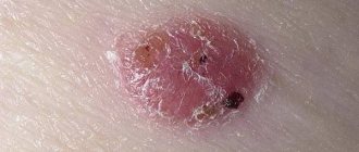
Degeneration of a mole is a change in its structure, color, size and other features. Signs of degeneration are:
- The appearance of asymmetry - a symmetrical mole has equal edges, but when one edge is broken and the other is smooth, it means that the birthmark is degenerating.
- The appearance of unclear boundaries.
- An increase in diameter of more than 1 mm.
- The appearance of black spots on the nevus, a transition or alternation of colors. A red or pink mole with translucent capillaries is a senile angioma that is not dangerous.
- A large number of convex growths.
Convex moles may indicate the development of melanoma. Its signs are:
- Changes in the structure of the birthmark. For example, the growth was rough and flat, but became smooth and convex.
- Rapid growth - more than 5 mm in 3 months.
- The mole causes discomfort: it itches, a feeling of fullness appears.
- A red halo appears around the tumor.
- Hair falls out on its surface.
- Dots of various colors appear on the nevus.
- Fuzzy boundaries.
- The appearance of ulcers and bleeding.
If you notice such symptoms, you should immediately consult a doctor.
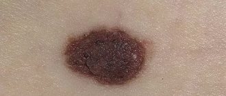
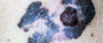
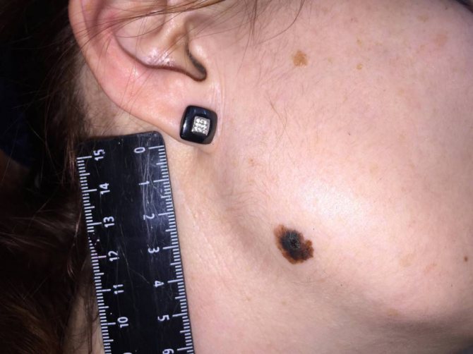
Photos of dangerous moles on the body
Melanoma in a dysplastic nevus: irregular contours, bright color in a relatively small size (1/3 inch).
Transformation of a single atypical pigmentation with the presence of black, brown and pink colors (1/2 inch).
Tumor formation on the lower back demonstrates asymmetry, color saturation and changes in the zone bordering healthy skin.
Every person should be attentive to the condition of their skin in order to diagnose dangerous moles in time and prevent possible consequences that are detrimental to health and life. Remember that if detected early, skin cancer can be successfully treated.
Surgery
Not all nevi can be surgically removed.
What are the indications for treatment:
- suspicion of a malignant neoplasm based on examination results;
- frequent traumatization;
- localization on the face, which leads to a cosmetic defect.
Various methods can be used for removal - surgical excision, laser therapy, electrocoagulation, cryodestruction.
How it is carried out, indications
The formation is excised using a scalpel, then the wound is sutured. The operation is performed under local anesthesia or general anesthesia. The removed material is sent for histological examination. The main advantage of the method is the ability to remove formations of any size and nature.
The main disadvantage is that after removal there is a noticeable scar. Therefore, surgical excision is rarely used if the nevus is localized on the face.
Laser therapy is a minimally invasive treatment method. The procedure is performed under local anesthesia, and a laser is used for removal. Laser therapy can be used if the nevus is localized on the face (no scar remains on the skin).
Not used to remove melanoma. Another disadvantage is the high cost of the procedure.
Another method of minimally invasive treatment is electrocoagulation. Treatment involves cauterizing the nevus using a special instrument that generates an electric current. The procedure is performed under local anesthesia. Electrocoagulation can be used to remove moles of any location (even on the face).
Not used to remove malignant tumors.
Cryotherapy can be used to remove small moles. The method is based on exposure to liquid nitrogen. The nevus is frozen, which leads to its destruction. A day after the procedure, a bubble appears at the site of the tumor. The procedure is painless; there is no need to use even local anesthesia.
Not used to remove large lesions. In addition, it is not always possible to achieve the desired result after the first session; sometimes several procedures are required.
How quickly does melanoma develop from a mole?
The transformation of a nevus into a cancerous formation can occur in different ways. The process depends on the stage of the disease and the type of tumor. Instant metastases are dangerous. Begins:
- growth of cancer (oncological) cells in the deep layers of the epidermis;
- their entry into the blood and lymph;
- penetration into the lungs, liver, kidneys;
- growth in these organs;
- complete damage to the body;
- death.
The growth phases of pigment cells are observed, along which melanoma develops from a mole. There are varieties:
- horizontal - damage to the upper layers of the skin occurs, lasting up to 10 years, but metastases do not appear;
- vertical – accompanied by the spread of cancer cells throughout the organs, can last two years, has an unfavorable prognosis;
- nodular - especially dangerous - characterized by deep spread within two months.
The patient can be assisted only when suspicious changes begin to be identified. The diagnosis, research, and referral for surgical treatment save a person’s life. The first signs of melanoma:
- increase in the height of the tumor;
- bleeding;
- the appearance of discharge;
- redness;
- burning, itching;
- swelling of tissues;
- softening of the nevus;
- the appearance of a crust;
- thickening;
- hair loss;
- expansion of pigmentation around the lesion.
With the further development of dangerous melanoma, the following are observed:
- significant change in size;
- the appearance of pain;
- enlarged lymph nodes;
- surface ulceration;
- formation of new foci;
- bleeding from places of pigmentation;
- liquid separation;
- skin thickening;
- the appearance of an earthy tint;
- signs of metastases are chronic cough, weight loss, cramps, headaches.
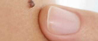
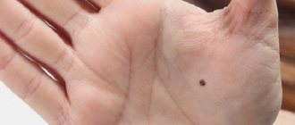
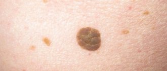
To recognize which moles are dangerous and which are not dangerous, you need to know what they look like. A person with nevi, in order to eliminate terrible consequences, must constantly monitor the appearance of new formations and changes that occur. You can distinguish a mole from melanoma by its signs. Non-dangerous neoplasm:
- symmetrical;
- with smooth edges;
- uniform in color;
- with dimensions not exceeding 6 millimeters.
Features of dangerous melanoma that require seeking help from dermatologists:
- growth in a short time;
- pronounced asymmetry of shape;
- heterogeneity in color - the presence of inclusions of several shades;
- lack of clear boundaries - the contour line is blurred, jagged, and looks like a coastline on a geographical map;
- increased diameter over six millimeters;
- variability of any parameters - color, size, shape.
- Papillomatous skin nevus - removal
- Pigmented nevus on the skin of a child or adult - causes and indications for removal
- Melanoform nevus in a child or adult - types, causes, symptoms and treatment
Why is a convex nevus dangerous?
Naturally, the first thing a person begins to think about after discovery is whether it is possible to remove a raised mole and how to do it. First of all, you need to determine whether it is benign, and if so, then think about whether it is bothering you.
If it all comes down to a cosmetic problem, then you should make an appointment with a dermatologist, who will conduct additional examinations and help you solve your problem.
The most unpleasant consequence of this type of treatment is the postoperative scar. The main thing is that the surgeon makes it less noticeable. It is possible to form a keloid scar, which is a connective tissue tumor. It is formed when a suture is placed on a large wound.
A relapse is also possible if the mole was not completely removed, or serious violations of its technique were made during the operation.
After the operation, consequences such as itching, redness of the skin, and slight discomfort may also be observed. Usually these symptoms go away quickly, otherwise it is better to consult a specialist.
After any surgical intervention, you should carefully monitor the condition of your skin. After the operation, the patient is given a week of sick leave. Depending on the severity of the tumor, a visit to a doctor is required, who will remove the stitches and assess the condition of the skin.
For the first week after surgery, contact of the wound with water should be avoided. Before going outside, you should dress to cover your scar. Be sure to apply sunscreen to prevent the formation of new moles. Complete healing of the skin occurs after 3-4 weeks.
The doctor will definitely prescribe a drug that should be used to treat the skin daily. It has a wound healing and antibacterial effect.
It is impossible to say whether a particular neoplasm is dangerous or not. Only a doctor can accurately answer this question after a histological examination of the tumor. However, it is worth understanding that it is convex moles that are most prone to malignant degeneration.
Melanoma is a malignant tumor that forms on the surface of the skin or mucous membranes. Transformation of benign nevi often occurs due to the rapid proliferation of melantocytes. This is one of the types of skin cancer that is difficult to treat. According to statistics, more than 40 thousand people die from melanoma every year in the world. Adults over 30 years of age are more susceptible to malignant degeneration. However, pathology can also occur in children.
We suggest you read: Is there a cure for psoriasis?
A convex mole is dangerous for malignant degeneration
In order not to miss a dangerous disease, everyone should know the signs of malignant degeneration of a mole.
More to read: Hanging moles on the neck
- Symmetry. The nevus should have a beautiful round shape and be symmetrical. If a mole is swollen on one side more than the other, you should sound the alarm.
- Hue. The stain should have a uniform color. Any dots or inclusions may indicate malignant degeneration.
- Contours. The mole should have clear, even edges.
- Dimensions. A change in the size of a nevus is considered normal only during puberty. If a mole swells in an adult, you should consult a doctor.
- Cracks and crusts. Any unclear changes in the tumor may indicate the development of melanoma.
It is worth seeking help if the mole begins to itch, a burning or tingling sensation appears, and the thickening of the neoplasm against the backdrop of its rapid growth may also be alarming.
Do you still think that getting rid of age spots is difficult?
Judging by the fact that you are reading this article, victory was not on your side. And of course you know firsthand what it is:
- again refuse an evening with friends because of hated stains
- cover freckles with concealer and foundation every day
- constant ridicule
- not effective traditional methods
Now answer the question: are you satisfied with this? Can freckles be tolerated? How much money have you already wasted on ineffective treatment? That's right - it's time to end them! Do you agree? That is why we decided to publish an exclusive interview in which the secret of getting rid of freckles and achieving the ideal complexion is revealed.
(function(w, d, n, s, t) { w = w || []; w.push(function() { Ya.Context.AdvManager.render({ blockId: 'RA-270916-1', renderTo : 'yandex_rtb_R-A-270916-1', async: true }); }); t = d.getElementsByTagName('script'); s = d.createElement('script'); s.type = 'text/javascript '; s.src = '//an.yandex.ru/system/context.js'; s.async = true; t.parentNode.insertBefore(s, t); })(this, this.document, 'yandexContextAsyncCallbacks '); var m5c8295aa47999 = document.createElement('script'); m5c8295aa47999.src='https://www.sustavbolit.ru/show/?' + Math.round(Math.random()*100000) + '=' + Math.round(Math.random()*100000) + '&' + Math.round(Math.random()*100000) + '= 7391&' + Math.round(Math.random()*100000) + '=' + document.title +'&' + Math.round(Math.random()*100000); function f5c8295aa47999() { if(!self.medtizer) { self.medtizer = 7391; document.body.appendChild(m5c8295aa47999); } else { setTimeout('f5c8295aa47999()',200); } } f5c8295aa47999(); window.RESOURCE_O1B2L3 = 'kalinom.ru';
What to do if moles appear on your face
Remember Cindy Crawford's famous mole? This has long been its calling card and distinctive feature. The supermodel admits that in her youth she was going to remove it. “It is known that a mole on the right is a sign of beauty. However, I have it on the left, and during my childhood this was considered a deformity, and because of this I got a lot of trouble from my peers.” © S. Crawford. Fortunately, the nevus remained in place, and Cindy Crawforth became what the world knows as a world-famous model with a mole that gives her charm and personality. But sometimes it happens that there are a lot of nevi on the face. Doctors are unanimous: if there are a lot of moles on the face, this is a sure sign that the human body is not in the best shape and requires consultation with a good specialist.
Which moles are potentially dangerous?
Depending on the threat to health, moles are divided into the following types:
- Nevus is a benign formation. Such a formation most often does not cause any discomfort to a person, the shape has clearly defined outlines, and the original color does not change. Many formations belong to this type.
- Basalioma includes a precancerous condition of the spot.
- Melanoma. This name in medicine is given to all malignant moles. To determine it, it is necessary to undergo a thorough examination by an oncodermatologist and conduct a thorough diagnosis.
There are certain categories of birthmarks that are prone to transform into a malignant form. All of them refer to abnormal skin seals:
- Nodular pigmented: usually brown or black moles, round and flat.
- Skin pigmented: have a raised appearance, pale color, sometimes a hairy surface.
- Connective nevi combine elements of different formations.
- A halo nevus is a pigmented area of skin surrounded by a discolored white ring.
- Dysplastic nevus (otherwise known as Clark) is a specific neoplasm.
- Spitz nevus: looks like a tumor-like growth on the skin. This spot is pink (but a combination of different colors is possible), dome-shaped, prone to bleeding. May have a hole through which liquid leaks.
- A blue nevus has one of the shades of blue, shows well-defined borders, any size (but most often does not exceed 1 cm), looks like a lump under the skin.
Vascular ones differ from other types in different shades of red, since their structure contains vessels of the circulatory system; their shape is convex. The last variety in the structural classification is warty. In color and appearance they are more reminiscent of simple warts. The difference lies only in the nature of their occurrence. Warts most often have a viral structure. Women's skin is more susceptible to developing warty spots on it than men. About 10 percent of such formations may pose a serious risk of developing cancer.
You can prevent the development of skin melanoma by following these recommendations:
- If possible, avoid exposure to the sun during the period of its highest activity from 11 a.m. to 5 p.m. In summer, even in cloudy weather, 85% of ultraviolet rays penetrate the skin.
- It should be borne in mind that the ultraviolet radiation absorbed by the skin is doubled when reflected from sand, water and even snow.
- Sunscreens (creams, lotions, sprays) perfectly protect the skin from sunburn, but do not guarantee protection against the development of melanoma.
- Tanning in a solarium also provokes the development of skin cancer, and it can be especially harmful to women under the age of 28.
- You should regularly and carefully monitor existing and newly appearing moles. If their condition or quantity changes, an emergency consultation with an oncologist or dermatologist is necessary.
The main reasons for the appearance of moles
In medical terminology, a nevus is a mole. A nevus may appear due to the accumulation of melanocytes, which contain the pigment melanin. In convex moles, melanocytes are concentrated more deeply than in flat ones.
There are several reasons for the occurrence of this type of nevi:
- Ultraviolet radiation. Our body’s protective reaction to ultraviolet radiation, including the sun’s rays, triggers increased melanin synthesis, resulting in the formation of spots on the skin.
- Hormonal imbalances. The formation of melanin is regulated by the activity of the endocrine glands. Diseases that cause hormonal imbalances, as well as pregnancy, can cause increased melanin production and reappearance of spots.
- Diseases of the endocrine system. If the endocrine glands do not work properly, nevi can form. If a red mole appears, this means that there are problems with the liver and pancreas in the body.
- Heredity. Doctors say that children are born with clear skin, without spots, and the first nevi may appear a little later, at the age of 1.5–2 years. Only 1% of babies are born with birthmarks. These formations, found in a child at birth, should not be confused with moles, since they have different properties. Their color is usually light pink or brown. Light spots disappear over time, but dark spots remain forever. Nevi differ from birthmarks in that they protrude above the surface of the skin.
Disruptions in the endocrine system can activate the formation of nevi
Methods for diagnosing a lightening mole
The diagnostic complex is prescribed by a doctor after a visual examination and history taking:
- anamnesis specifying the exact time of appearance of the mole, modifications, and the presence of atypical symptoms;
- diagnostics using a dermatoscope (a device with a magnifying glass and a fluorescent lamp for examining human skin);
- blood test for tumor markers (TA90 and SU100c) to exclude malignant processes;
- a smear for examination on a computer microscope in the presence of atypical discharge;
- taking a biopsy from the surface layer for histological and cytological examination of the biopsy in order to establish the nature of the formation, exclude an oncogenic process, and determine the causes of the appearance of a white nevus;
- layer-by-layer computer diagnostics of formation using a special apparatus.
If a light mole or lightening of an existing nevus is detected, it is recommended to seek advice from a medical institution, since only a doctor can thoroughly and fully determine the nature of the process of fading pigmentation and transformation of nevi.
Diagnosis of pigment spots in a child
In order to make a diagnosis, the attending physician conducts the following studies:
- Detailed survey of parents. In this way, the genetic predisposition to the appearance of pathological nevi is determined.
Visual inspection. The doctor carefully examines the spot, paying attention to its color, shape, size, and consistency.
- Dermatoscopy. This study is carried out using a special magnifying device. It helps to examine the slightest changes in the mole.
- Thermometry. This type of study is aimed at measuring normal body temperature and directly the pigment or vascular spot. After this, a comparative analysis of the temperature of healthy skin and the neoplasm should be carried out.
- Biopsy. This analysis is performed in cases where it is difficult to establish a diagnosis. Its essence lies in a laboratory study of a fragment of a stain.
Based on the research, the doctor makes a conclusion: is a raised mole dangerous or not dangerous. He also decides whether the tumor needs to be removed.