A mole has crusted over: the reason, what does it mean and is it dangerous?
› Moles ›
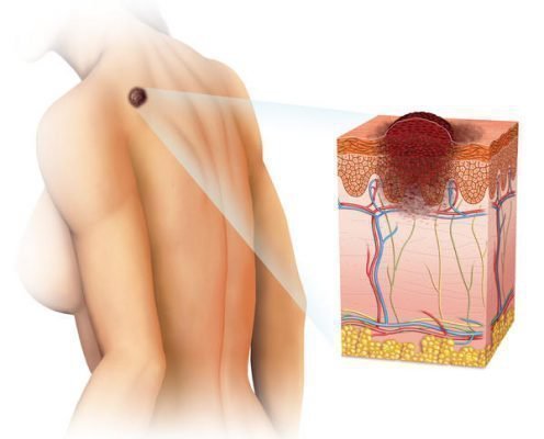
Despite the initially benign nature of moles, they are capable of undergoing a variety of adverse changes. One of these changes is inflammation of formations, which is fraught with transformation into a malignant process. To trigger it, the element must turn into melanoma - a real malignant focus.
https://www.youtube.com/watch?v=4q_FgHF7-II
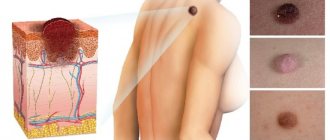
The pathology is recognized as the initial stage of skin cancer, and a sore on a mole can behave as follows:
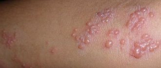
- the pigment spot of the nevus is surrounded by noticeable redness;
- the mole changes its tone up or down, that is, it becomes either very bright, or whitish and even colorless;
- the contours of the formation become distorted and unclear, but some people notice completely opposite changes;
- growth of the neoplasm in size (the severity and severity of the process are judged by speed).
What triggers the mechanism of inflammation of a mole? There may be several predisposing factors, but they are all classified into categories such as exposure to sunlight and household injuries.
Pale-skinned and freckled people should avoid prolonged exposure to the scorching rays of the sun. Excessive exposure to ultraviolet radiation is harmful to any organism, but this category of people is designated by experts as the most vulnerable.
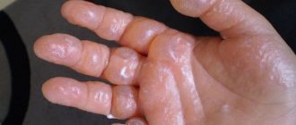
In the process of household activities, human skin is subject to friction and microcracks. And even the small size of the lesion already becomes a gateway for infection, which will subsequently affect the birthmark. Therefore, if a mole becomes inflamed, begins to itch, turns red and sheds its top layer, the reason for its behavior should be clarified in consultation with a dermatologist or oncologist.
Moles can form on any part of the human body. Medicine knows cases of turning to specialists regarding neoplasms on the eyeball, mucous membrane of the mouth or nasal passages. Nevi develop regardless of whether a given anatomical area is hairy or not.
When a mole festers, increases in size and changes in every possible way, you need to hurry to the doctor.
The specialist will give an objective assessment of the condition of the tumor and prescribe medications that will help solve the problem. In certain cases, doctors take a biopsy, that is, they pinch off cells from the changed tissue. The purpose of the manipulation is to determine the nature of the nevus - suddenly it has become malignant.
At the same time, some scientists in the field of dermatology consider taking a biopsy to be an unjustified and even risky measure, since damage to an already inflamed formation can aggravate the situation. They suggest resorting to more gentle diagnostics - computer examination or dermatoscopy.
If the fact that the mole is at an intermediate stage of degeneration is confirmed, the doctor will recommend getting rid of it.
What to do if a mole becomes inflamed, but there is no way to see a doctor? Is it possible to calm down the process on your own? Yes, but for this you need to have calendula tincture, alcohol, antibiotic ointment and other medicines with you.
Self-medication can be carried out in the form of activities such as:
- treating the inflamed element with medical alcohol. The action is repeated until the redness disappears completely. Alcohol can be replaced/alternated with tincture of calendula raw materials.
- Antibiotic ointment provides long-awaited relief. It will be good if its components include zinc or salicylic acid.
- The nevus can be sprinkled with powder obtained from Streptocide tablets and lubricated with linseed oil.
- A cotton pad is sprinkled with celandine tincture and a lotion is made. After 10 minutes, the application is removed. The manipulation is performed 3 times. in a day.
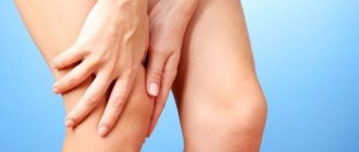
In order not to accidentally injure a mole with your own hands, you need to monitor the condition of your nails and give preference to comfortable clothes and shoes. Poor-quality and tight wardrobe items can put pressure on new growths, and unkempt nails can scratch them while dressing or performing hygiene procedures.
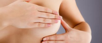
A woman can injure a nevus with unkempt or too long nails.
How to help yourself, what to do if you hurt a mole? First you need to stop the bleeding. Since the formation is enriched with small capillaries, even a minor injury can result in prolonged bleeding. A 3% solution of hydrogen peroxide will help cope with the task. An irrigated cotton-gauze swab is applied to the affected area for 10 minutes.
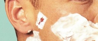
The second stage is treating the damaged element with brilliant green or medical alcohol. This is a very important point, because any wound on the body facilitates the penetration of pathogenic bacteria and fungi into the body.
Frequent injuries leading to cutting of moles provoke the replacement of healthy cells with mutated ones. Their danger lies in the possible transformation of nevi into malignant tumors.
After providing first aid, it is a good idea to see a doctor. A dermatologist or surgeon will assess the condition of the affected area and make a prognosis regarding the future behavior of the mole. Based on the results of a histological examination of the cells taken, it is judged whether the tumor should be removed. Doctors advise getting rid of accidentally torn moles due to the high probability of re-injury.
If the mole has festered, you should not take risks and self-medicate. It is necessary to urgently contact a surgeon and choose the appropriate method for removing the diseased lesion. Modern types of surgical intervention are carried out very quickly and painlessly and do not require long-term hospitalization.
Spots on the skin can be vascular (hemangiomas) or pigmented. Among the vascular ones there are large hemangiomas, which degenerate under the influence of various factors. Pigmented nevi are completely harmless and are slightly darkened small areas of the dermis.
The reasons for the development of nevi range from genetic predisposition to diseases and external factors.
It is not dangerous when there are no symptoms or signs of inflammation, growth, pain, or itching in a specific area.
From the point of view of malignancy, the formation of a crust on a healthy red mole can be considered safe. They rarely lead to skin cancer.
Damage to the nevus can lead to trouble if a foreign infection has been introduced. If there is inflammation on the spot, peeling begins, bruising or bloody discharge appears, there is a possibility of cancer. To eliminate the possibility of developing cancer, you need to undergo examinations by a dermatologist and oncologist.
Crust on a mole: when it’s not dangerous (self-diagnosis)
› Moles ›
Moles are congenital or acquired spots on the body of different sizes, structures and colors. They don't bother you until they start behaving dynamically. Sometimes they change shape or increase in size so that clothes begin to touch them. If a crust appears on a mole, this may signal a modification of hormones in the body that adversely affect health.
Reasons for the appearance of a crust on a mole
All birthmarks appear under the influence of changes in pigmentation and vascular system. Deformation serves as a signal to visit a doctor. The appearance of a crust on a mole is influenced by several reasons:
- mechanical injury;
- constant touching by clothes;
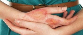
- getting burned;
- hormonal disorders leading to pathology.
A crust on a mole is not always a precursor to cancer. If it appears during an injury or burn, there is no health risk. Here it is necessary to eliminate the entry of pathogenic microorganisms into the wound so as not to aggravate the course of the disease.
The following symptoms indicate the possibility of developing cancer:
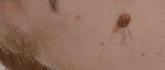
- hair on the formation began to fall out;
- redness of the area around the birthmark;
- secretion of lymphatic fluid;
- burning, itching, tingling;
- change in density.
If a mole has crusted over or enlarged, what does this mean, a dermatologist or oncologist can answer. Therefore, if you experience any of the above symptoms, you should immediately visit a specialist.
Can the crust fall off on its own?
When a mole becomes covered with a dry crust, you need to find out the reasons for this process and consult a doctor. This may be a sign of melanoma development.
The crust along with the mole may fall off in some cases:
- damage has occurred from regular contact with synthetics;
- temperature effect;
- lack of the required amount of nutrients;
- prolonged exposure to ultraviolet rays.
A hanging mole can easily be touched, twisted, thereby cutting off the blood supply and causing it to fall off. New growths occur due to hormonal imbalances. When the hormones return to normal, the mole with the top layer disappears.
The most dangerous case is when the birthmark peels off, causes itching, and a dry crust. Then, over time, the top layer falls off, which may indicate the beginning of melanoma formation.
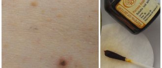
After an injury, a scab often appears that falls off on its own. This process cannot be accelerated. If you accidentally hit someone and start bleeding, you need to urgently perform the following manipulations:
- Take a piece of cotton wool or gauze, soak it in hydrogen peroxide and place it on the wound. The injured area will not become inflamed and bleeding will stop;
- After the bleeding has stopped, you need to remove the cotton wool and cover the wound with a bandage, and then seal it with a band-aid;
- The detached layer with the growth should be placed in a sterile vial or wrapped in a bandage and taken to the laboratory for examination. By conducting a histological analysis, specialists will be able to verify its benign nature.
Sometimes the crust can be touched and it will be partially torn off. In this incident, it is best to visit a dermatologist and listen to his recommendations for further actions.
What to do if a mole has crusted over
Normal moles look like small, dark, round spots. They can be on the back, arms, stomach, neck and other parts of the body. Flat nevi are considered the most harmless. All others require a thorough examination.
The following growths require medical attention:
- Rapidly increasing in size over 6 mm.
- They have an asymmetrical shape.
- Ragged edges.
- If there is pain.
- The appearance of dryness.
- If you start bleeding.
A birthmark that has become crusted over should absolutely not be touched. Do not apply medicated ointments or lotions with medications to soften the top layer. You cannot use traditional recipes for removal. This will not only help, but will also make the problem worse.
The color of the top layer can tell a specialist a lot:
- brown color indicates that the mole was injured and began to heal;
- black color remains on the area of the removed growth;
- a gray, yellow tint indicates the beginning of rebirth.
The crusts must not be torn off or scratched. They must be carefully hidden from bright ultraviolet rays. It is not advisable to keep all birthmarks in direct sun for a long time. If the skin begins to bleed, you should immediately visit a doctor.
Diagnostic and treatment methods
To prevent skin cancer, you need to closely monitor your birthmarks and conduct regular examinations.
Therapy is prescribed only after diagnosis. Diagnostic methods are as follows:
- visual study of shape, color, size;
- taking a smear in the area of the growth;
- The discharge under the crust is taken for examination;
- computer modeling is carried out;
- fluorescence microscopy;
- biopsy of a piece of tissue for microscopic examination;
- a blood test is taken for tumor markers, which determines whether the growth is benign or malignant;
- prescribing adequate treatment.
You can carry out self-monitoring using the American Melanoma Accord system. Thanks to this system, everyone can recognize dangerous changes at an early stage. The procedure is carried out in several steps:
- “A” is the presence of asymmetry in birthmarks;
- “K” – state of the ends of the build-up;
- “K” – blurred edges, characteristic of a malignant form of oncology;
- “O” – study of color distribution, color change in relation to the original state;
- “P” – dynamic increase in size, diameter over 5–6 mm indicates the development of skin cancer;
- “D” - intensive growth dynamics, changes in structure, the presence of a crust and other changes are included in this parameter.
Therapy directly depends on the transformation of the mole into melanoma.
MethodologyWays of treatment
| Radiosurgery | Radiation therapy is carried out using a radioknife. This is a non-traumatic method in which the resulting crust falls off after a week. |
| Laser removal | A safe and painless method that leaves no traces. After removing a mole with a powerful light beam, the top layer disappears after 14–20 days. |
| Surgical intervention | The method is ideal for large, hanging birthmarks that are dangerous. There may be a scar that takes a month to heal. |
| Electrocoagulation | Removal using current. A medical coagulator is used. After such an operation, it is possible to study the cut material. |
| Cryodestruction | A thin needle with a temperature sensor is inserted into the growth, then cooled to the desired temperature and abruptly removed. Freezing occurs with liquid nitrogen. With this method, a crust is formed that lasts about 30 days. |
If there is a crust on a birthmark, you should not immediately panic, tear it off or treat it yourself. Be sure to visit a doctor to make a diagnosis and prescribe treatment. Not all cases require mole removal, and timely examination can save lives.
A crust on a mole: when it is not dangerous (self-diagnosis) Link to main publication
Source: https://beznarostov.ru/rodinki/korochka
Inflammation of a mole
Any changes in the human body can lead to an increase in the number of moles, changes in the shape and size of existing formations. Thus, decreased immunity can also lead to inflammation of nevi. If a mole is covered with a crust, this does not mean at all that a terrible diagnosis will be made. However, it is recommended to consult a doctor as soon as possible.
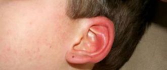
A common seasonal cold can lead to inflammation of the mole. But convex nevi, which are located on the neck or folds, are especially common. Such formations are more susceptible to injury than others. And if such a mole becomes covered with a crust, it is possible that it was damaged.
In addition, nevi can burn in the sun. Therefore, in the summer from 12:00 to 16:00 it is better not to sunbathe at all.
Not only a crust will indicate inflammation of a mole. The area around the growth may turn red. The inflamed area usually becomes denser. And if a bacterial infection gets into the mole area, pus may appear.
If the mole is located in the wrong place and is often injured, the doctor will recommend removing it. Frequent damage to nevi is one of the reasons for their malignant degeneration. Popular methods for removing moles will be described below.
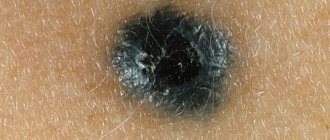
If a mole is covered with a white crust, this is a dangerous sign. Melanoma is an aggressive malignant tumor of the skin that can develop rapidly. Therefore, everyone should know when to postpone a visit to a dermatologist.
There is a technique that includes a number of signs suggesting that it was a malignant tumor that we had to deal with:
- Asymmetry is the first thing you should pay attention to. A healthy mole always has the correct shape. If the round formation suddenly begins to blur, you should worry. The edges of the mole must be smooth.
- Bleeding is another dangerous sign indicating that something is wrong with the mole. If the nevus does not cause any discomfort, but blood periodically bleeds from it, you need to make an appointment with an oncologist.
- The color of the formation also matters. If a mole has darkened and crusted over, this is one of the signs of melanoma.
- You will also have to pay attention to the size. Healthy nevi can also increase in size, but if the size of the mole changes rapidly, you should sound the alarm.
MORE ABOUT: 5 nootropics with proven effectiveness [Pumping up the Brain]
An accidental injury, a scratched mole, leads to the formation of a dry crust. A small child can accidentally tear off a piece. When a crust forms on a mole, a number of additional factors should be taken into account:
- the appearance of asymmetry;
- the edge of the nevus blurs;
- changing ends;
- formation of bruises from under the crust;
- color change;
- reduction or enlargement of the spot;
- growth intensity.
When dealing with symptoms, diagnosis, smear testing and treatment will be prescribed. It is important to carry out tumor measurements; they will help to draw a conclusion about the nature of the formation (malignant or benign).
It is advisable to get rid of a malignant mole as soon as possible. Metastases can reach the lymphatic system.
If symptoms are detected, the following types of procedures are prescribed:
- laser therapy;
- freezing;
- elimination using a radio knife;
- cauterization;
- surgery.
The removal procedure is carried out in accordance with established medical norms and rules, after which you should not wet the skin for at least a week. Doctors insist on covering the damaged area with a band-aid. It would be advisable to avoid cosmetics. If a crust begins to form at the removal site, do not touch it; over time, it will fall off on its own. The rehabilitation period must take place under strict control. The treating specialist should observe and advise.
Damage to a mole
Many people hear warnings throughout their lives that they should never injure, scratch or rip off such a skin growth. And it is true.
Nevi are very sensitive and unpredictable skin formations. A small impact on them is enough for them to begin to change and grow. The presence of a crack in a mole may be an alarm bell. Sometimes, in the event of an unfortunate fall or other injuries, they can simply burst. No one is immune from this, but if you have large raised moles, you should make sure that everything is fine with them.
Sometimes they can crack for reasons beyond a person’s control, which means that some process has started in the cells of the mole and it has begun to change on its own.
Sometimes, after a mole has cracked, bleeding begins, which you need to try to stop and then contact a specialist. The places where such skin tumors are located are penetrated by capillaries, so even a small mole can bleed for quite a long time.
Capillaries approach the mole and it can bleed for quite a long time
First aid
It is best to stop the bleeding with hydrogen peroxide, after which you can additionally treat the crack with an antiseptic (chlorhexidine), then carefully seal the injured nevus with a bactericidal adhesive plaster so that the wound does not get infected.
In other cases, the mole sometimes becomes covered with small cracks and begins to dry out; gradually, pieces may even fall off from it.
Under no circumstances should you remove a cracked nevus yourself, cauterize it with iodine, or try to “cure” it using traditional methods. After self-removal of the nevus, in the best case, it will grow again in the same place, and in the worst case, the incompletely removed melanocyte cells will degenerate into a tumor. Wrong actions can lead to even greater complications that will be difficult for specialists to cope with.
Once the bleeding has stopped, you should seek help from a dermatologist or dermato-oncologist. If it is not possible to visit a doctor in the near future, it is necessary to carefully monitor the nevus to see if any pathological changes occur. This information will help the doctor in the future. If a piece comes off from a cracked mole, you must put it in a sterile container and show it to the doctor during examination.
After any damage, the doctor will generally recommend removing the mole and examining it. Sometimes, upon examination, it turns out that a person’s mole is not cracked at all, but a completely different, harmless skin formation that only looks like a nevus. Such growths on the skin may consist of a different type of cell, so a crack in them does not pose a threat and can calmly heal with the use of special ointments.
If a mole nevertheless cracks, then the melanocytes, which secrete the coloring pigment, can degenerate into cancer cells. These cells begin to divide extremely quickly, forming a skin tumor called melanoma. If the cells of such a tumor enter the blood, they begin to spread throughout the body and then treatment becomes almost impossible, because they settle in any tissue and develop there, forming new tumors and metastases.
A cracked mole should be seen by a dermatologist.
Mole removal methods
Removing a nevus for no reason is dangerous. If, after consultation with a dermatologist and oncologist, there is no other option, you need to think about a method for removing the skin lesion. You can get rid of skin lesions using:
- laser skin correction;
- cryodestruction (freezing with liquid nitrogen);
- radio knife;
- electrocoagulation (cauterization);
- complete removal by the surgeon during surgery.
Regardless of the method of mole removal, it is recommended to follow certain rules after the procedure. You cannot wet the area where the nevus was for a week. It is recommended to limit sun exposure, even when using sunscreens with a filter of 60-100. It is important to stop using cosmetics for a while.
As an independent phenomenon, moles are a benign formation, which in some cases can transform into a small ticking bomb, ready to turn into a tumor.
Reasons for this behavior include:
- A large number of sunburns at a young age can cause tumors
a large number of sunburns at a young age;
- frequent exposure to the sun - for example, from eleven o'clock in the morning to four o'clock in the evening. Those who like to get a good tan are at risk. Ultraviolet radiation produced by the sun causes many skin diseases;
- with a love for the solarium. This is also a very serious risk, since a few minutes in it can be equal to a whole day in the sun;
- nevus injury. Even a simply accidentally touched mole can turn into a tumor if damaged;
- disturbances in the functioning of organs responsible for the body’s hormonal levels.
People of Aryan appearance, with fair skin and red hair are most at risk. In pregnant women, the skin is susceptible to the development of new nevi. In general, anyone who has a large number of freckles and moles should be careful going out into the sun. Those whose relatives were sick with a similar disease, as well as those who have suffered many severe sunburns or who have moles that are darker in the center than the edges, should be constantly examined by specialists.
A crust on a mole shows that any changes are being recorded in the body, the nature of which needs to be established
A mole is a completely safe spot on the skin that does not cause discomfort. Over time, it may become covered with a thick crust or change color. In this case, any changes are recorded in the body, the nature of which must be established. Quite often, a crust on a mole forms in the event of hormonal changes that negatively affect the condition of the tissue.
The appearance of a tumor on the skin is not always harmless. A person should know in what cases it is necessary to be extremely attentive to changes of this type on the skin.
Among the symptoms that should alert a person when a nevus is detected are:
- the appearance of a crust on the mole, peeling around it;
- the appearance of red pigmentation in this area;
- there is discomfort at the location of the mole;
- if hair grows in a mole, it may fall out;
- increase in size, distortion of shape;
- mole falling off;
- when touching a nevus, painful sensations occur and lymph oozes;
- has a shiny appearance.
This list is not complete. If something worries you, it is better to consult a doctor, especially in the cases listed above or if the mole has changed in any way.
Crust on a mole
A mole or nevus is a benign growth on human skin. It is dangerous when a mole becomes crusty, changes color, falls off, or causes discomfort. The main reasons why a crust appears are trauma to the nevus, hormonal disorders, or irreversible changes begin in its tissues.
New growths on a mole can be either harmless or a signal of complications.
Mole: simple or dangerous?
Spots on the skin can be vascular (hemangiomas) or pigmented. Among the vascular ones there are large hemangiomas, which degenerate under the influence of various factors.
Pigmented nevi are completely harmless and are slightly darkened small areas of the dermis.
There are epidermal-dermal (flat neoplasms up to a centimeter, different shades), intradermal (rise above the dermis), lentigo, dysplastic and blue nevi, a large pigmented nevus.
In terms of size, the formations are small (up to 1.5 cm), medium (up to 10 cm), large (more than 10 cm), giant (covering part of the body almost completely).
The reasons for the development of nevi range from genetic predisposition to diseases and external factors.
Reasons for appearance
Nevi appear under the influence of sunlight and are hereditary. It is possible that they appeared due to hormonal changes in the body (for example, during puberty or during pregnancy).
Most often, the reason for the appearance of moles on the body is a kind of genetic code. A newborn has virtually no pigmentation, but teenagers have many times more pigmentation.
By the age of 30, a person has about 30 different spots; by old age they disappear.
Risk group:
- people with hereditary nevi;
- persons exposed to radiation and electromagnetic irradiation;
- those who like to abuse ultraviolet radiation (especially between 11 a.m. and 4 p.m.) and tanning in solariums;
- people with very light skin;
- patients already suffering from cancer;
- those who have crossed the 50-year mark;
- those with a total number of nevi of more than fifty;
- people with injured skin at or around the mole;
- those who eat carcinogens and GMOs, and those who abuse household chemicals.
Symptoms of mole degeneration
Degeneration, namely the transformation into a malignant neoplasm on the dermis, is melanoma or aggressive type cancer. The prognosis for recovery from this disease is 50/50. According to statistics, this type of oncology accounts for 10% of all cancers.
Around the world, about 40 thousand people die from melanoma every year, and every 3rd person suffers from melanoma. The size of moles affects their degeneration. In 50% of cases, it was the giant formations that turned into cancer. If there is any change (even a crust on a mole), you should immediately contact a doctor.
Symptoms of malignancy (degeneration):
- The mole has changed its usual color and density. Different shades with the addition of red or black dots are alarming. The formation has become hard to the touch or, conversely, soft - also a negative condition of the nevus.
- The edges of the nevus begin to spread, for example, a flat one becomes convex and noticeably rises above the skin. Normally, the halves of the mole are symmetrical.
- Crusts on the mole or cracks appear on it with the release of clear liquid and blood.
- The site of pigmentation brings discomfort (pain, burning, colitis, itching).
- A new mole has formed and the old one has fallen off.
- There was a loss of hair that grew from a mole.
The appearance of crusts on moles may be preceded by accidental injury or exposure to irritants.
If a crust appears
A crust has formed on the top of the mole - perhaps there was trauma or scratching. A black, dry crust appears at the site of removal - this situation is not always bad.
Most likely, the wound will heal and new pink skin will grow underneath. Black crusts can be the result of a burn that a person received, for example, in a sauna (bath).
Overgrowth with a crust is not dangerous, but only if the wound does not become infected.
Diagnosis of a crusty mole
Before starting treatment, a thorough examination is necessary.
Diagnosis of the condition of a nevus involves a smear from the surface of the mole (if there is discharge), dermoscopy (hardware examination), laboratory tests for tumor markers, biopsy, removal (the removed material is sent to determine malignancy). Melanoma can be confused with a wart, papilloma or varicose veins (the neoplasm is mistaken for spider veins or an enlarged vessel).
To control moles, use the melanoma primer - ACCORD. The method is popular in America, where a reminder with a self-examination algorithm hangs in every public place. ACCORD is a home self-examination, and stands for indicators for determining the nature of a pigmented spot:
- A - asymmetry;
- K - ends;
- K - blood formation;
- O - color;
- P—size;
- D - dynamism.
Determination of the degree of danger
Any spot on the skin is formed through the influence of the vascular system or pigment changes. However, during the development process, such a spot can be reborn at any time. This process is negatively affected by external and internal factors.
Nevus, as a rule, changes color only in certain areas of the dermis. Depending on the degree of color change and protrusion above the surface, all moles are divided into groups. In the process of analyzing their good quality, size plays an important role.
Any nevus is formed under the influence of genetic factors and past diseases. There is also a group of internal factors, the nature of which cannot be fully established.
For what reasons is blood released from moles?
There are several factors under the influence of which blood can be released from a nevus, namely:
- trauma to the birthmark;
- degeneration of moles into malignant neoplasms;
- the development of a strong inflammatory process in the body.
Many people know why moles bleed, but not everyone knows that such a phenomenon can be a sign of the development of pathology in the human body.
Features of the degeneration of moles into a malignant formation
Melanoma or cancer pose a serious threat to human life and health
Melanoma or cancer poses a serious threat to human life and health. In medical practice, recovery is recorded only in 50 cases out of 100. Today, this type of cancer accounts for approximately 10% of the total statistics. Unfortunately, according to worldwide data, 40,000 people die from this disease every year. To prevent the development of a dangerous pathology, it is recommended to be attentive to the condition of the skin and, if abnormalities are detected, seek advice from a doctor.
A person should know the following symptoms that indicate the start of the rebirth process:
- The structure of the nevus begins to slowly change. One formation may contain several shades and colors at once. When palpated, compaction or excessive softness is noticeable. In this case, the condition is negative and requires immediate consultation with a doctor.
- The outlines lose clarity, and the formation begins to protrude above the surface of the skin. Additionally, it should be noted that a mole in its normal state should have symmetrical edges.
- The mole is covered with a crust, on which cracks appear over time. The formation may also ooze fluid and blood.
- Pigmentation changes the character and causes discomfort to a person.
- The nevus disappeared, and after that a new formation appeared in its place.
- Hair began to fall out around the mole.
A crust can also form on a mole if it has been injured. The situation arises in case of regular scratching. In this case, the formation of a crust is considered completely normal.
After a certain period of time, it will heal or become covered with pink skin. If the patient had a burn before, the crust becomes black. A person can get such a burn in a bathhouse or sauna. It is important to prevent the development of infection, because due to it the nature of the disease can be aggravated.
In what cases could this be a sign of cancer?
Skin cancer can be treated quite successfully in the first stages if it is noticed in time, because melanoma is a very aggressive skin cancer that quickly metastasizes and leads to death.
Direct signs of a malignant neoplasm are:
- Change in color - the mole may turn black, or vice versa - become lighter.
- Violation of the contour around the pigmented formation.
- Inflammation (redness) in the area of the nevus.
- The appearance of loose edges.
- Unpleasant sensations (itching, burning) and constant sensitivity appear in the mole area.
- The appearance of ulcers and cracks, nodules or other neoplasms on the formation (near it).
- Bloody or other discharge.
- Complete or partial drying of the mole.
- Hair loss occurs in the pigmentation zone.
You need to carefully monitor the condition of moles on your body, and to prevent skin cancer, promptly seek advice from a dermatologist - oncologist.
When to see a doctor
Adequate treatment can be selected only after a correct diagnosis
Doctors do not advise removing nevi without a good reason, even if they have changed color. Hanging moles are subject to excision if they are located in an unfavorable location and permanent damage occurs. The appearance of even black moles on the body is normal. Especially if a person has to constantly be under the sun's rays. But if the nevus, which is located under clothing, has darkened, then you need to have it checked by a specialist.
MORE ABOUT: Antispasmodics (drugs): list with names
It is also not recommended to get rid of black formations with home remedies, such as acetic acid lotions. Traditional recipes will allow you to remove only the upper part of the nevus, which is located on the surface of the skin. Its base will remain in the deeper layers of the epidermis and can transform into a dangerous form - melanoma. If a malignant tumor is suspected, tests are done. If their results are positive, the doctor will remove the nevus. After surgery, the patient may be prescribed a course of chemotherapy or radiation therapy.
If the tests do not confirm the presence of pathology, but the person still wants to remove the blackened nevus, then more humane methods of intervention are used, such as a radio knife, laser or liquid nitrogen. You should consult a doctor if: you notice that a black mole has crusted over; itches; enlarged or painful; dried out and began to peel off. It is these signs that indicate a malignant process.
What to do if a mole becomes inflamed?
Self-medication in such cases is not recommended. If a mole has crusted over, only a specialist will prescribe adequate therapy. But first you will have to undergo a series of tests. The doctor must find out which infectious agent is causing the inflammation. If it is bacteria, an antibacterial ointment will be prescribed. Additionally, the damaged area will be treated with an antiseptic. Miramistin and Chlorhexidine solutions are widely used.
If the mole is located in the wrong place and is often injured, the doctor will recommend removing it. Frequent damage to nevi is one of the reasons for their malignant degeneration. Popular methods for removing moles will be described below.
Necessary examination
Adequate treatment can be selected only after a correct diagnosis. It involves taking a smear in the area of formation. Additionally, secretions are taken for examination and a biopsy of the removed area is performed. Oncomerkers are of no small importance. Thanks to them, a conclusion is made about the malignancy of the formation.
In terms of external characteristics, melanoma is similar to a wart, papilloma or varicose veins. In this case, red capillary networks are additionally present in the area of the nevus.
To monitor the condition of moles in medical practice, the ACCORD system is used. This method was first proposed in America, and only over time became widespread in our country. The system can be used at home without the help of a specialist in this matter. Thanks to this, the patient will be able to recognize a negative situation at the initial stage of development. During the examination, attention should be paid to the following characteristics:
- presence of asymmetry;
- the nature of the ends of the mole;
- presence of blood stains;
- nature of color distribution;
- changes in size;
- growth intensity.
Features of mole removal
It is imperative to get rid of a malignant nevus. Otherwise, metastases may penetrate the lymphatic system. It is recommended to remove a mole only if it is absolutely necessary. Otherwise, the appropriateness of the procedure is called into question. To excise a mole, the following procedures can be used:
- laser treatment of tumors;
- freezing using liquid nitrogen.
- excision with a radio knife;
- cauterization using electricity;
- operation with surgical supplies.
The procedure for removing moles must be carried out according to all the rules. After this, you should not wet the skin in this area for seven days. The sun's rays can also have a negative effect. That is why it is recommended to seal the area when going outside. Additionally, it is advisable to apply sunscreen. During the recovery period, you should avoid applying cosmetics. When a crust forms, you cannot tear it off yourself. It should just fall away. In any case, it is necessary to obtain advice from a specialist in this field, since the risk of malignancy remains.
Melanoma or cancer pose a serious threat to human life and health
It is imperative to get rid of a malignant nevus. Otherwise, metastases may penetrate the lymphatic system. It is recommended to remove a mole only if it is absolutely necessary. Otherwise, the appropriateness of the procedure is called into question. To excise a mole, the following procedures can be used:
- laser treatment of tumors;
- freezing using liquid nitrogen.
- excision with a radio knife;
- cauterization using electricity;
- operation with surgical supplies.
The procedure for removing moles must be carried out according to all the rules. After this, you should not wet the skin in this area for seven days. The sun's rays can also have a negative effect. That is why it is recommended to seal the area when going outside. Additionally, it is advisable to apply sunscreen. During the recovery period, you should avoid applying cosmetics. When a crust forms, you cannot tear it off yourself. It should just fall away. In any case, it is necessary to obtain advice from a specialist in this field, since the risk of malignancy remains.
For moles that involve only the upper layers of the epidermis, removal with liquid nitrogen is suitable - cryodestruction
How to stop bleeding?
The mole is bleeding, what should I do? Self-medication can be harmful and make the situation worse. Often, a specialist recommends treating bleeding wounds with hydrogen peroxide. Before you begin handling, it is important to thoroughly wash your hands with an antibacterial detergent - this will help prevent the development of an infectious disease. Disposable gloves should be used to prevent microorganisms from entering wounds. Using cotton wool or cotton pads, it is necessary to treat bleeding areas with peroxide.
If a red mole bleeds, use clean gauze or a bandage. It can be purchased at any pharmacy. It is necessary to fold the material in several layers and apply it to the bleeding moles. You should take a comfortable body position. Keep the material near the wound until the bleeding stops. After this, the wound should be dried. To do this, you can use brilliant green or iodine.
A slight discharge of blood from a mole is not life-threatening. If the bleeding is excessive and occurs systematically, it is important to immediately visit the hospital and undergo a thorough medical examination.
Prevention of malignancy
Simple steps will help you protect yourself from dangerous consequences. Regardless of the number of moles on the body, you should make it a rule to avoid sunbathing between 12:00 and 16:00. In this way, it will be possible not only to maintain health, but also to prolong youth. If you have a large number of moles on your body, you should avoid direct sunlight even in the early morning or after 16:00.
If a mole is often injured, it is worth removing it. Under no circumstances should you seal the damaged area. The thermal effect created can harm the nevus.
It is worth regularly checking yourself and your loved ones for the appearance of new moles or changes in existing formations. Timely identified pathological formations can be removed without unpleasant consequences.
To prevent a crusty mole from causing complications and becoming malignant, you need to follow some preventive measures:
- If you are at risk, try to limit your time in the sun. Spend no more than 15 minutes a day tanning. Then the interval can be increased to half an hour a day.
- Rinse sea salt off your skin and dry yourself to reduce sun exposure after swimming. Water acts as a lens and enhances the influence of ultraviolet rays.
- Don't rely on sunscreen to help. The benefits of the latter have not been proven; they belong to chemical cosmetics that cause additional irritation.
- Young people under 28 years of age are not recommended to visit solariums. Ultraviolet radiation has a destructive effect on young and unprotected skin.
- Observe the condition of the mole. If an alarming symptom appears, consult a doctor.
- If there are more than 10 moles on the skin, consult a dermatologist at the clinic and ask for a skin passport for you. With regular check-ups, skin cancer can be prevented.
- The mole rubs against clothing for a long time, contact a specialist to remove it. Choose loose clothing made from cotton fabric.
If a crust on a mole appears after an injury, you should urgently contact a medical facility.
After examination and tests, the doctor will determine whether there is a possibility of melanoma formation and how the spot can be removed. Remember! A mole grows as a person grows. If the development begins to take on a rapid character, this is already a reason for concern and urgent consultation with a specialist.
Article approved
by the editors