Localization and clinical manifestations of lumps on the neck
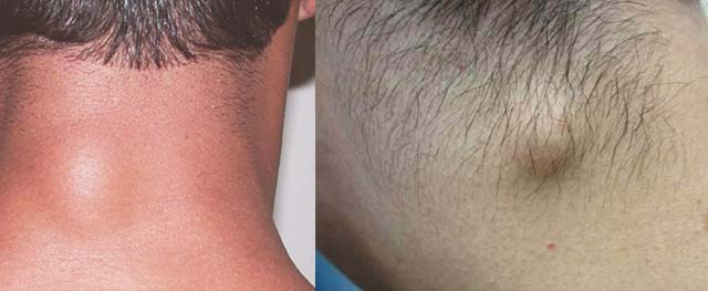
Depending on the etiology, a lump on the neck can be localized anywhere - back, side, front
Muscle tissue is located mainly at the back and sides. On the side there are also lymph nodes that follow each other. If several lumps are felt at once, inflammation has most likely begun in the lymph nodes.
If the lump on the neck is located at the back, the cause may be:
- wen or lipoma;
- lymphogranulomatosis;
- atheroma;
- furuncle;
- myositis - compaction in muscle tissue.
Also, protrusion is sometimes observed with cervical osteochondrosis.
If a lump appears on the side of your neck, it is possible:
- purulent inflammation;
- lymph nodes have enlarged;
- fibroma, less often lipoma;
- neoplasm of a neurogenic nature;
- allergy.
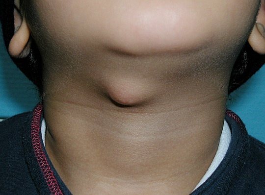
If the lump is located in the front, the thyroid gland or lymph nodes are most likely enlarged, since there are most of them in the front of the neck.
Some tumors are painless and can move under the skin upon palpation. The pain most often comes from an inflamed lymph node, an abscess or a lump in a muscle. Depending on the origin of the lump, treatment is prescribed.
Size of tumor
Large boil on the neck
Lymph nodes do not grow large: as soon as the inflammation passes, they return to their previous size. A lipoma is not a dangerous tumor, but it can grow into a giant lump, so it is better to remove it so that it does not leave a large scar in a visible place.
Boils also ripen quickly and rupture, followed by the discharge of pus. To prevent re-infection, it is recommended to treat the wound in the hospital and take a course of antibiotics for prevention.
The most dangerous diseases are lymphogranulomatosis and malignant thyroid tumor, as they can give metastases if not detected in time and treated late.
Lump under the arm, which doctor should I contact?
As soon as you notice such a formation under your arm, then you urgently need to contact a specialist. The surgeon deals with this issue
. When you come to see him, he will examine you, carefully examine the lump and give you all the necessary directions for testing. After such a diagnosis, the doctor will prescribe you treatment. If the tumor is benign, then surgery is not necessary. It can be cured with medications. If there is a large accumulation of pus inside the tumor, then most likely the surgeon will make a small incision and all the pus along with the blood will come out. If the tumor is malignant, the specialist will perform the operation under general anesthesia.
If the lump can be cured with medications, then you will not have to undergo any surgery.
One of the main reasons for the occurrence of such formation is inflammation of the sebaceous glands, usually purulent. The causative agent of such an infection will be the staphylococcus bacterium, which penetrates the glands and begins to activate there.
Other causes of a bulge in the armpit area can be many diseases that have not even been diagnosed in the patient. For example, mumps, AIDS and many other diseases.
As soon as you find a swelling under your arm that is starting to hurt, you don’t need to apply a warm cloth or anything else to it, immediately make an appointment with a surgeon.
VASHE ZDOROVIE / 06.21.2015
An armpit lump is a lump, in most cases painful, in the armpit. Such a bump, in most cases, constantly brings pain and discomfort. This is probably due to poor personal hygiene, uncomfortable tight underwear, or a razor cut.
This seal has a round or irregular shape. Depending on the circumstances of the appearance of the lump, the color is different - white, greenish (which indicates the purulent environment of the lump), red. Its sizes also vary, but much more often the lump is clearly visible; it stands out large above the surface of the armpit.
If you have a lump under your armpit, this indicates the presence of inflammation in this area. As a result, it looks inflamed, as evidenced by the reddish color of the skin and increased temperature in the area of the lump, and the general condition worsens. There may be only one lump, or a couple of lumps may appear. A lump is a nodule, abscess, or tumor.
Possible causes of pathology
In order not to panic in vain before seeing a doctor, it is recommended to understand how certain tumors on the neck behave, what symptoms they cause, how dangerous they are in the future or at the moment.
Lipoma
Wen does not pose a danger, but it interferes from an aesthetic point of view
The cause of lipoma is most often a violation of metabolic processes in the body. This is the most harmless tumor that does not require specific treatment. It is removed for aesthetic reasons, since the ball on the neck under the skin can quickly increase in size.
The fatty tissue is removed urgently if it is located near large arteries and blocks blood flow or partially impairs respiratory function.
A distinctive feature of a lipoma is its painlessness and mobility under the skin.
Atheroma
Atheroma is similar in appearance to lipoma, but its appearance is caused by blockage of the sebaceous gland duct. The lump can fester over time, which poses a health risk. It is most often located at the back, closer to the hair.
As the atheroma grows, it turns into a cyst filled with adipose tissue, epithelial cells, and fluid. The problem is that when it ruptures, purulent contents leak out, but the capsule itself can cause the lump to re-form. It must be removed in a hospital setting and the wound treated with antiseptics.
Fibroma
Fibroma grows slowly, has a round or oval shape with clear outlines
Fibroma is determined by its shape: it is round, regular in shape, dense in consistency, and has clear outlines.
A fibrous tissue tumor grows slowly. In most cases, the predisposition to connective tissue growth is inherited. It very rarely degenerates into a malignant tumor. A fibroma is operated on if it quickly increases in size and causes aesthetic discomfort.
Tumors of neurogenic origin
Neurogenic tumors develop on the nerve process of the parasympathetic trunk. These are cysts that come in different origins:
- neuroma;
- ganglioneuroma;
- neurofibroma.
Tumors of neurogenic origin are painless, most often safe, but there are malignant neoplasms that are removed surgically, then chemotherapy and radiation are prescribed. The closer the tumor is to the spine, the more likely it is that it is a cyst of nervous tissue.
Boils
A boil forms for several reasons:
- poor personal hygiene, clogging of skin pores with dirt, sweat and fat;
- prolonged exposure to drafts or hypothermia.
A boil is manifested by redness of the skin, itching and pain. Then it increases in size, matures and bursts. The danger of furunculosis lies in the possible infection of the blood through the network of capillaries, so self-medication is not recommended.
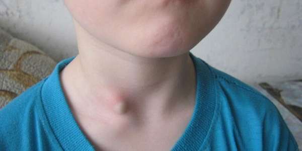
Enlarged lymph nodes

An enlarged lymph node may be due to a cold or viral infection
Inflammation in the lymph nodes accompanies almost every cold or viral infection, since it is in the lymph nodes that microorganisms are recognized and the toxic load on the body is analyzed.
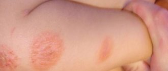
Inflamed lymph nodes are located on the side of the neck - closer to the ears or under the lower jaw. Sometimes several painful lumps may appear as the nodes are located along the lymphatic vessel in a chain. This phenomenon is especially common in children, since their immune system is poorly developed and reacts more sharply to pathogenic microorganisms.
If an inflamed and enlarged lymph node does not hurt, this may indicate a malignant process - lymphoma or lymphogranulomatosis.
Lymphangioma
Lymphangioma is a benign tumor whose cells grow from the walls of lymphatic vessels. Most often, the tumor is congenital and requires treatment by sclerotherapy, since surgical removal is ineffective.
Cystic lymphangiomas quickly grow to enormous sizes, while others grow slowly.
In adults, benign tumors are the result of a disease of the lymphatic system. They are formed in places where lymphoid tissue accumulates, one of which is the neck area.
Myositis or myogelosis
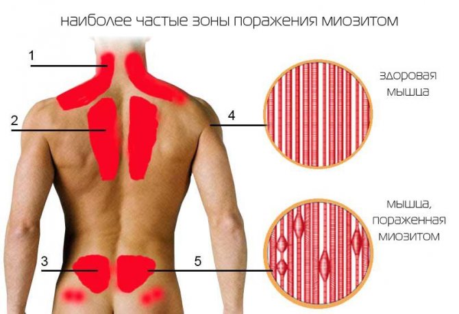
Myositis develops gradually: first there is pain in the neck, then the mobility of this area is limited. Later, upon palpation, you can feel small lumps, which are very painful. Myositis can be caused by hypothermia, prolonged sitting with a bowed head, or sleeping in an uncomfortable position. Cervical myositis can disrupt the functioning of the larynx and pharynx. The person has difficulty breathing or swallowing.
Myogelosis causes compaction of muscle tissue throughout its entire volume, which impairs blood flow due to compression of the arteries. As a result, a painful lump may appear on the neck, and then the pain will spread to the shoulders, arms, and spine. Severe headaches are possible.
Lump after injury
A lump on the neck may appear due to injury. Such bruises are especially dangerous, since large arteries supplying blood to the brain, as well as lymphatic vessels and nodes, are located nearby. When struck, lymph fluid leaks out, forming a swelling. The swelling may last for several days. Possible internal bleeding or arterial thrombosis.
If a person has received a blow to the neck area, he needs urgent medical assistance to relieve swelling and check the integrity of blood vessels.
Osteochondrosis
Osteochondrosis is a degenerative disease of the spine associated with a sedentary lifestyle and destruction of the intervertebral disc. The diagnosis can be confirmed even in young people who are overweight and have flabby muscles surrounding the spine. The cervical region is the most mobile, so the risk of developing osteochondrosis is higher in the neck area.
Sometimes, when the vertebrae rub against each other and bone tissue grows, osteophytes are formed. The largest of them can protrude above the surface of the skin. Osteophytes are an advanced stage of the disease. With cervical osteochondrosis, the brain suffers because blood flow in the vertebral artery passing through the neck area is disrupted.
Thyroid diseases

Tumor in the front of the neck due to an enlarged thyroid gland
A lump in the front of the neck may indicate a problem with the thyroid gland. Diffuse goiter is an increase in the size of the entire gland evenly. Nodal involves the presence of several dense areas.
The most common benign tumors of the thyroid gland are caused by a lack of iodine in the diet. By increasing its amount through a special diet or taking iodine-containing medications, it is possible to eliminate seals. Sometimes, in advanced stages, it is necessary to resort to surgical removal of the nodes.
A painless, hard lump or tumor is a cause for concern, as it most often indicates cancer.
What is a lump under the skin
The etiology of the appearance of lumps under the skin is very diverse. This may be a hygroma, boil, atheroma, abscess, etc. Often a lump under the skin in the form of a ball on the arm is associated with professional activities.
Due to the anatomical and physiological characteristics of the upper extremities, the joints are often deformed, salts accumulate in them, which cause such formations. The process of salt accumulation is accompanied by pain. It is also worth noting that there are other types of neoplasms, the causes of which are significantly different.
Hygroma
A dense ball is clearly visible, which can be easily felt upon palpation. Most often it is localized on the hands and wrist joints, does not differ in mobility, does not hurt, and does not in any way affect the general state of health. The main cause of concern is cosmetic discomfort.
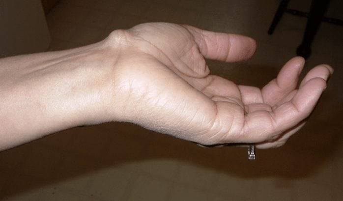
Appearance of hygroma of the wrist joint
Lipoma
It is a movable ball under the skin on your arm. This tumor is often called a lipoma. It is easily palpable, painless, and has clear outlines. The skin above it folds easily and has a natural color. Most often, lipomas are localized on the outside of the arm, where there is more hair. As the size increases, it may cause some discomfort. The lipoma does not penetrate into the surrounding tissue, so it is easily removed surgically.
Abscess
An abscess forms a hot, painful lump under the skin. In this case, there is a general malaise, sometimes the body temperature rises to 38-40 ° C. This type of tumor can occur as a result of injury, blows, or injections.
Folliculitis
This is an inflammation of the hair follicles. The disease can be triggered by infection, physical or chemical irritants. This pathology manifests itself in the form of hair pustules. Most often, folliculitis is diagnosed in patients with weakened immune systems, obesity or diabetes.
Cherry angioma
This problem is most often encountered by people over 40 years of age. In most cases, the disease does not require treatment. If tumors prevent a person from feeling attractive or bleed, they can be removed using a laser or electrocoagulation.
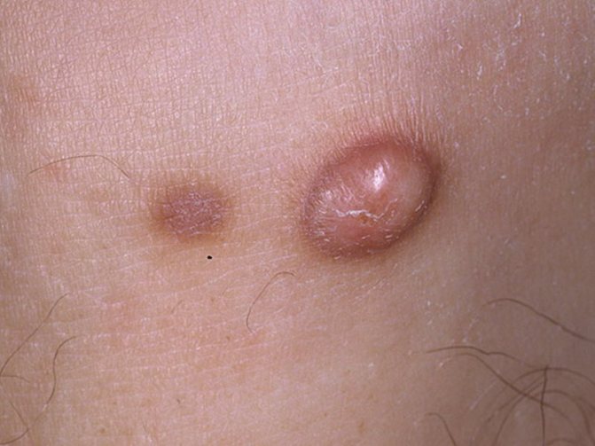
Dermatofibroma
A harmless small red-brown ball under the skin that is made of fibrous tissue. Over time it can change its size and color. Sometimes during its growth, pain and itching are felt, although there may not be any painful sensations. There is no need for treatment. If desired, this tumor can be removed surgically.
Neurofibroma
A soft, fleshy ball under the skin. This is a harmless tumor, which in rare cases can turn into a malignant neoplasm. If the growth does not cause any symptoms, no treatment is required.
Atheroma
Most often localized in the scalp, back, face, neck. It is a hard compaction under the skin that does not cause any discomfort, has clear boundaries and a rounded shape. The skin over the atheroma cannot be folded; sometimes the surface of the skin takes on a bluish color; when pressed, a fat-like substance can be released from the center of the atheroma. Atheroma can become inflamed and fester. If necessary, it is removed surgically.
Important! If you suspect a malignant tumor, you should immediately contact an oncologist

Collection of anamnestic data
Diagnosis of seals on the neck
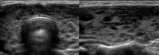
Thyroid nodule on ultrasound
When visiting a doctor, the patient will be recommended to undergo several types of diagnostics. First of all, this is a urine and blood test for an inflammatory process - acute or latent. This is a common procedure for all patients. If there is a purulent process (furunculosis), the analysis will determine the type of pathogen in order to correctly select antibiotics and auxiliary drugs.
If the lump on the neck is a benign tumor, it is first examined by ultrasound. If a malignant process is suspected, an MRI is prescribed, thanks to which layer-by-layer sections of soft tissue are obtained. If the oncological process is confirmed, the doctor prescribes a histological examination of the cells, for which a piece of tissue is taken and examined under a microscope. This allows you to determine the type of tumor, its tendency to metastasize and rapid growth.
For diseases of the thyroid gland, blood is drawn to determine the amount of hormones, as well as tissue diagnostics using ultrasound.
X-rays are done for myositis or myogelosis. It is advisable to perform angiography of the neck vessels to determine the speed and volume of blood flow in the vertebral artery.
There are several types of subcutaneous seals.
If they are small, white, move well and painlessly, then these are wen or lipomas
. They are completely harmless and develop due to the high activity of fat cells. Lipoma never turns into cancer. It can form in any part of the body. Usually, because they spoil the appearance, they are removed. Wen can sometimes grow very large.
Atheroma is sometimes confused with a wen
. Atheroma is also based on fat. But it accumulates in the sebaceous gland, clogging it. Unlike a wen, atheroma can fester and become inflamed. Sometimes the skin over the atheroma turns blue, and a blocked duct appears.
More dangerous than wen and atheroma is a hernia
. Hernia can be umbilical, inguinal and other organs. It can appear during heavy physical exertion; the skin protrudes like a small ball. It is not visible if you take a horizontal position. The hernia can go away on its own, or it can be removed surgically, depending on where it is located. In any case, you need to consult a doctor. If the subcutaneous lump is painful and is located on the neck, groin, under the arms, elbows and knees, then this is inflammation of the lymph nodes - lymphadenitis
.
The reasons may be different. Most often these are colds - sore throat, otitis media, flu and others. In this case, having cured the disease, lymphadenitis will disappear on its own. If the skin over the lymph node turns red and it is very painful, then lymphadenitis has developed - suppuration of the lymph node itself. But be careful: if the lymph nodes are enlarged for a long time, this is an alarm bell - this is one of the symptoms of the development of infection in the body. Furuncle - formation scheme The skin itself sometimes becomes inflamed in the form of a carbuncle and furuncle
.
They appear when the sebaceous glands and hair follicles become inflamed and can greatly increase in size, causing pain. This inflammation is accompanied by an increase in temperature, the appearance of redness and even swelling. If treatment is delayed for a long time, you will have to resort to surgery. But sometimes medication is enough. Follicle - varieties Small white balls with pus inside - this is folliculitis
. They develop on the face, back, chest, and genitals. Sometimes they become inflamed and a red rim forms around them. Their appearance is associated with insufficient hygiene and some infections.
Sometimes after surgery, cosmetic procedures, or piercings, lumps remain under the skin. They are harmless, do not become inflamed and, with proper care, go away over time. Almost all people have moles on their skin, some have warts and papillomas
. These are small brown or red growths on the skin called polyps. They are usually harmless, but sometimes appear and grow on the body. This is a reason to contact a specialist.
Once I noticed blue swollen balls on the ankles of one woman - this is a consequence of wearing tight, uncomfortable shoes, high-heeled shoes. Hygroma and fibroma If a stationary watery ball is located on the wrist or palm, then it is a (benign cyst)
It forms when fluid accumulates between tendons. It does not hurt and is harmless, it only interferes with the work of the hands. Therefore it is removed.
A fleshy deep lump that is immobile and located deep under the skin is a neurofibroma.
. It is dangerous because it can become malignant.
If you suddenly see at least one subcutaneous lump, do not panic, but simply consult a general practitioner, dermatologist or surgeon
. Only a doctor can give an accurate diagnosis. And don’t even think about getting rid of the hard ball under the skin on your own. It cannot be cauterized, squeezed out, or used folk remedies. This not only promotes inflammation, but can also leave an unsightly scar.
Lymphogranulomatosis (LGM)
Of all the reasons for the appearance of bumps on the neck, this is the most dangerous. It is characterized as a malignant disease of the lymphatic system, caused by the emergence of tumor tissue from lymphocytes.
From a medical point of view, this disease is not cancer. The reasons for this opinion are the relative ease of treatment and a certain systemic distribution of the tumor.
The appearance of lumps may be preceded by a number of signs, such as weakness, general malaise, high fever, sweating, joint pain, itching.
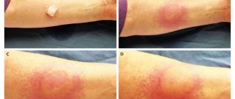
External manifestations usually begin with the appearance of small tumors on the lymph nodes of the neck, above the collarbone, sometimes under the arms and in the groin. At the initial stage, palpation of the lymph nodes does not cause pain, and they remain mobile. Later, their adhesion and compaction, as well as local redness of the skin, may be observed.
It is treated with chemotherapy and radiation therapy, as well as their combination. At the initial stage, only radiation therapy is sufficient. There is also a method of aggressive chemotherapy followed by bone marrow transplantation.
Enlarged lymph nodes (lymphadenopathy)
Lymph nodes are located in groups in the neck, under the lower jaw, above and below the collarbones, in the armpits, in the elbows and knees, in the groin and other parts of the body. These are components of the immune system that, like a filter, pass interstitial fluid through themselves, clearing it of infection, foreign inclusions and damaged cells, including tumor cells.
An increase in the size of the lymph nodes (lymphadenopathy), which become painful when palpated, usually accompanies infectious diseases: sore throat, otitis media, flux, panaritium, as well as wounds and burns. Treatment of the underlying disease leads to a reduction in the node.
If the skin over the lymph node turns red, and palpation becomes sharply painful, the development of lymphadenitis is likely - a purulent lesion of the node itself. In this case, you need to contact a surgeon. Minor surgery may be required, and early treatment can sometimes clear up the infection with antibiotics.
If a dense, tuberous formation is felt under the skin, and the skin above it cannot be folded, the node is likely damaged by a malignant tumor. In this case, consult an oncologist as soon as possible. Read more about other causes of swollen lymph nodes.
Cyst and fistula
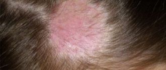
- In a child, a tumor in different parts of the neck may appear when the cyst suppurates. A similar developmental defect occurs in almost 5% of all newborns who have maxillofacial disorders.
- Cysts are distinguished by localization into lateral and medial. The middle ones are more common.
- Their appearance is associated with some anomaly in the development of the fetus in the first months of intrauterine growth. The thyroid gland, which has just begun its formation, moves from the area under the tongue to the neck. The skin canal disappears over time, and if it remains after development, it forms a cyst. In the future, it can turn into a fistula if it has a way out.
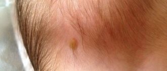
- Such fistulas are usually located in the central part of the neck and are small, painless lumps, but if the necessary measures are not taken, the inflammatory process will begin to grow. In case of advanced suppuration, the surgeon makes an incision, cleans the area of pus and installs drainage for complete outflow of pus. After the fistula has healed, the cyst is excised. Lateral cysts are removed in the same way.
- Most often, this operation is performed under local anesthesia for children over three years old, but if the fistula is regularly inflamed, the removal process can be performed earlier.
- Balls on the necks of children occur for a variety of reasons, most often for those described above, but even if they seem completely harmless, it is forbidden to neglect the examination. Because harmless formations can turn out to be malignant tumors. To treat such tumors, chemotherapy, radiation therapy, or surgical removal are used.
- A number of tests, such as ultrasound, MRI, allow you to see the cause of the problem and choose the right treatment. It is always easier to find and neutralize pathology at an early stage than to live and wait every day for something worse, while the tumor continues to grow.
When a person discovers some kind of neoplasm on his body, it does not cause any positive emotions. Some people, having discovered a lump on their neck, mentally prepare for the worst and try to get to the doctor as soon as possible. Others are optimistic that this is nonsense that will go away on its own (sometimes this is exactly what happens). However, most often you still have to go to the doctor, but with more serious consequences.
Most people take action when there is already a problem (few people will go to the hospital with a small lump on their neck unless it hurts). But it is precisely in such uncertain cases that it is better to be prompt. If it is really a small thing, then examination by a doctor will not turn it into a malignant tumor. When the matter is really serious, a timely visit can play a decisive role in the further development of the disease.
What are the most common reasons for the appearance of such neoplasms on the human neck? We will consider further.
Oncological process
A lump in the front of the neck may also indicate the presence of an oncological process in the human body. The causes of the appearance of neoplasms may be alcohol or smoking abuse, chewing tobacco, Epstein-Barr virus, cytomegalovirus, low doses of radiation cosmetic therapy, and exposure to radiation.
Malignant tumors of the neck, first of all, spread to the nearest lymph nodes, which facilitates their rapid detection. Similar hidden tumors are neoplasms of the thyroid gland, laryngeal cancer, and lymphogranulomatosis.
Characteristic manifestations, in addition to a lump on the neck, will be low-grade fever, possible pain in the growth area, a gradual and noticeable increase in size of the lump, weight loss, difficulty breathing or swallowing, hoarseness of the voice. In such cases, you need to contact a therapist or dermatologist.
Diagnosis of diseases is carried out by external examination, questioning, cytological examination, ultrasound, if necessary, using biopsy, computed tomography or magnetic resonance imaging, radiography, laryngoscopy or pharyngoscopy.
Treatment includes radiation and chemotherapy, and in some cases surgery, but much depends directly on the stage of the disease and the extent of the process, as well as on the person’s age and the presence of other concomitant diseases.
Diseases associated with damage to skin structures
The most common diseases in which a lump forms under the skin on the neck are skin cysts, folliculitis, lipomas and atheromas.
Cyst
Skin cysts are benign skin growths that look like small cavities or sacs filled with fluid (blood plasma, pus). Cysts are formed when the skin glands (sebaceous, sweat) are blocked, near foreign bodies and after an infectious process. They are often located in the superficial layers of the skin and are easily palpable. They are usually painless to the touch. Often resemble small peas or soft balls.
Such lumps under the skin may disappear on their own. In very rare cases, you can try to crush or puncture them to extract the contents. When the cyst becomes inflamed, it is removed surgically. Such procedures are carried out only in a hospital setting.
Folliculitis
Inflammation of the hair follicles is also a very common cause of small, red and painful bumps on and under the skin. Treatment is carried out depending on the etiology of the disease with antibiotics or antifungal drugs as prescribed by the general practitioner. If you have folliculitis yourself, you can lubricate the skin with an alcohol solution, hydrogen peroxide solution or chlorhexidine. You should also protect the cones from various mechanical damage and friction against clothing, so as not to aggravate the inflammation process.
Lipoma
Benign soft tissue tumors that develop painlessly and slowly are lipomas (fat tumors). They are usually elastic, soft and painless to the touch. Fats are removed surgically, under local anesthesia, only if they cause cosmetic or physical discomfort.
Atheroma
They most often form on the neck due to blockage of the duct in the sebaceous gland or swelling of the hair follicle (follicle). They are a hard compaction under the skin, with clear rounded boundaries and without pain. Atheromas with inflammation, trauma or excessive enlargement are removed surgically.
Lipoma
A subcutaneous benign formation formed from adipose tissue (in simple language - wen). Perhaps one of the most harmless bumps that can appear on the neck.
What are lipomas characterized by:
- Externally, at the initial stage they are small in size (1-2 cm) and regular in shape (reminiscent of a bean grain).
- Most often this is easily movable fat formation.
- They do not cause pain.
- They have a relatively soft and elastic consistency.
- They grow slowly. You can live with them for a long time without experiencing any inconvenience.
- It is considered harmless and not subject to malignant degeneration.
The causes of lipomas are not exactly known, here are some of them:
- Atypical localization of adipose tissue cells formed during embryonic development.
- Metabolic changes in the body associated with enzyme deficiency in combination with local blocking of fat breakdown pathways at the tissue level.
- The appearance of lipomas may be associated with hereditary factors, which usually appear at an early age.
Lipomas arising in the neck area practically do not cause complications, but in very rare cases the following may occur:
- As a result of inflammation, some pain is possible, as well as local redness and an increase in size.
- In very rare cases, lipomas can trigger the development of liposarcoma (a malignant neoplasm of connective tissue).
Treatment for lipomas involves their surgical removal. Thanks to the development of modern medicine, removal methods are very diverse and are selected by the doctor individually depending on the characteristics of each specific case (size, location, condition and age of the patient).