What diseases cause skin changes
When it comes to infectious dermatitis, it is necessary to differentiate it from a large number of other diseases that can cause changes in the condition of the skin.
These include:
- acne is one of the most common skin diseases, the essence of which is inflammatory processes that affect the sebaceous glands;
- acne - pimples that appear mainly under the influence of hormonal factors, also a very common pathology;
- atopic dermatitis - a chronic disease of an allergic nature, aggravated in response to the body’s contact with an allergen, usually manifests itself in early childhood;
- herpes is a viral disease;
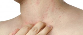
- eczema – is formed under the influence of a large number of reasons and is characterized as inflammation affecting the superficial layers of the skin.
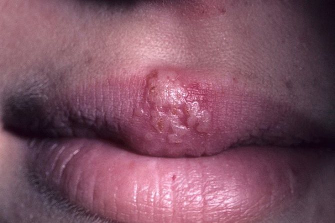
Photo: Herpes
In addition to common pathologies that cause skin changes, you can find a number of diseases that people encounter somewhat less frequently:
- bedsores;
- scabies;
- melanoma;
- shingles;
- warts, etc.
Features of rash localization
Infectious dermatitis is characterized by various locations of rashes on the skin. The location of the rash is highly dependent on what kind of infection caused the dermatitis.
For example:
- if a person gets measles, the rash will mainly be located on the skin of the face;
- with rubella, the rash begins to appear on the cheeks and neck, gradually spreading throughout the body;
- with scarlet fever, the rashes are localized in skin folds, such as inguinal folds, elbow and knee bends, and armpits;
- with enterovirus infection, a spotty rash appears all over the body, localized on the back, stomach, legs and face;
- with chickenpox, the rash consists of small pimples that begin to appear on the head and then cover the entire body;
- if a person is sick with typhus, the rash will be localized in the abdominal area, as well as on the elbows;
- the feet, palms and abdomen are affected by a scabies rash;
- Syphilitic rashes usually cover the entire body.
A doctor cannot make a diagnosis based on just one location of the rash.
Symptoms
External signs of the manifestation of a dermatological disease depend on what pathogenic source it was provoked. Let us consider in more detail the characteristic symptoms of viral dermatitis, which manifests itself in connection with the presence of the following infectious diseases, namely:
- Measles. The whole body is covered with a spotty rash, which, like tubercles, rises above the general level of the skin. After some time, the spots begin to merge into one single inflamed formation that resembles a pigment. The skin is very itchy and flaky.
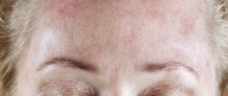
- Rubella. Infectious dermatitis of this origin is characterized by a small rash of bright pink color. The main localization of acne is the surface of the face and neck. Body temperature rises to 39 degrees and above, lymph nodes in the area of the lower jaw and collarbones enlarge.
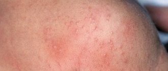
- Scarlet fever. Viral dermatitis, which occurs against the background of this infectious disease, manifests itself in the form of multiple rashes in the groin area, causing swelling of the mucous membrane of the genital organs in men and women. From the groin area, skin damage spreads to the abdomen, back and armpits. After 5 days from the onset of the disease, profuse peeling of the skin begins. The rash is very itchy, and the patient suffers from fever and fever.
- Enterovirus infection. The rash spreads completely over the entire surface of the body. They look like voluminous spots that resemble in appearance an allergic reaction like urticaria. The more successful the fight against enterovirus infection is, the dermatological symptoms will fade proportionally.
- Chicken pox. This type of infectious dermatitis manifests itself in the form of large large pink pimples that concentrate on the surface of the head and facial disc. After 3 days, the inflammatory formations transform into blisters with sanguineous fluid, which burst and turn into a weeping ulcer. As the ichor is released, a pink crust forms on the wound.
- Typhus. Viral dermatitis of this type begins with the formation of a small red rash on the elbows, spreads over the entire surface of the arm, and moves to the chest and abdomen. The disease is characterized by severe itching of the skin and a temperature that rises to 40 degrees.
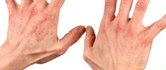
- Syphilis. Red spots appear on the epidermal tissues, which feel like nodules to the touch. As they mature, they increase in volume, and the inflammatory process becomes more acute. The final stage of the development of infectious dermatitis is the destruction of the surface of the epithelium over the inflamed node and the appearance of an extensive wound, the edges of which are inflamed, the epidermal tissue disintegrates, and the ulcer itself constantly increases in diameter. The patient experiences severe pain, and the body temperature is elevated to 40 degrees and lasts for several days.
- Staphylococcus aureus or streptococcal infection. It is characterized by the formation of small watery blisters that originate in the circumference of the oral cavity and in the navel area and spread throughout the body. After 3 days from the moment of appearance, the bubbles burst, leaving behind wound surfaces. The ulcerative formations are so deep and voluminous that they resemble a 2nd degree thermal burn. In addition to dermatological damage, the patient also encounters symptoms such as swelling of the mucous membrane of the genital organs and wounds in the oral cavity.
These are the most common types of viral dermatitis that occur against the background of infection with certain strains of bacterial infections. There are also other more severe dermatological diseases that are practically never encountered in medical practice. This category includes epidemiological outbreaks of anthrax and other mass destruction infections.
Types of infectious dermatitis
Infectious dermatitis, depending on its pathogens, is divided into several main types.
Viral
Skin lesions that develop under the influence of any viral influence are called viral dermatitis. In this case, dermatitis is not an independent pathology, but serves as a symptom of the development of the disease.
The main reason for the development of viral dermatitis is the entry of the virus into the child’s body.
There may also be other reasons why the viral type is formed:
- childhood viral diseases;
- surgical operations after which it was not possible to prevent the development of complications.
The main pathologies in which viral dermatitis develops are included in the group of childhood diseases.
These include:
- chickenpox (chickenpox);
- rubella;
- measles;
- herpes (not included in the group of childhood diseases).
Viral dermatitis also includes dermatitis that develops in children during scarlet fever, although the disease itself is the result of infection not by a virus, but by a bacterium.
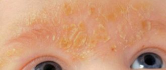
Photo: Rubella
Bacterial
Bacterial dermatitis develops due to the active reproduction of bacterial agents on the skin.
The most common disease from this group is impetigo. Children are more likely to develop this pathology, and it can be caused by various types of bacteria.
It is worth noting that the development of bacterial dermatitis is rarely influenced by factors such as poor body hygiene.
Pyoderma
A very common skin disease that develops due to the fact that pyogenic bacteria of the cocci type enter the skin.
Pyoderma can be primary or secondary. If we are talking about a primary pathology, then initially healthy skin is affected, but if it is a secondary pathology, then the infection is the result of other skin diseases that are accompanied by severe itching.
There are a number of factors that predispose to pyoderma pathologies:
- the presence of various minor injuries to the skin;
- failure to comply with hygiene standards;
- exposure to too high or too low temperatures;
- malfunction of any internal organs;
- pathologies of the central nervous system;
- metabolic pathologies;
- genetic predisposition;
- individual instability to pyogenic microorganisms.
Symptoms of the disease
The symptoms of dermatitis directly depend on the type of disease that caused it. The most common infections that cause symptoms of dermatitis are: 1. RUBELLA. In this condition, rashes are most often localized on the neck and face, with further spread throughout the body, especially on the inside of the limbs, back and buttocks. Dermatitis as a result of rubella is often accompanied by hyperthermia, lymphadenitis and a rash, which in the initial stage of the disease manifests itself as small oval spots. They fade after 2 days, and by the third they are practically gone.
2. MEASLES. The initial symptoms of dermatitis in the form of a rash appear in the ears, bridge of the nose, on the face, spreading over the upper chest. After 48 hours, the rash may appear on the arms, followed by lesions in the legs. The rash at the beginning of the disease is characterized by pink papules, merging in places. Subsequently, the color of the rash becomes bright red. After a day, viral dermatitis loses its papularity, the rashes become brown, peel, and pigmentation may appear in their place. In some cases, dermatitis, which is caused by measles, occurs atypically (at first the rash is purple, and during pigmentation the rash can turn green and then acquire a brownish tint).
3. ENTEROVIRAL INFECTION. Dermatitis due to enteroviral diseases is most often localized on the arms, body and lower extremities. There are no stages of rash. Skin symptoms disappear after 72 hours, without being accompanied by pigmentation or peeling.
4. SCARLATINA. With this disease, the rash is quite thick, hyperemic, consisting of many small papules, sometimes bluish in color. At the very beginning, the rashes are localized in places of high humidity (groin, armpits), and then they spread to the back, elbows, inner abdomen and thighs. Quite rarely, infectious dermatitis with the development of scarlet fever can be accompanied by hemorrhage (subcutaneous hemorrhages) and purulent rashes.
5. CHICKEN POX. In this case, dermatitis occurs in children, characterized by the appearance of a rash on the head, chest, and face. Subsequently appearing in the area of the feet and hands. Rashes are bright red in color with transparent contents in papules. Drying crusts gradually appear. Sometimes infectious dermatitis in children due to the smallpox virus is shingles. In this case, a monomorphic rash is noted, localized in areas of nerve damage.
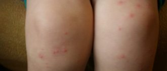
6. SCABIES. Such symptoms are most often observed on the arms, in the gluteal region, on the folds of the forearm, in the abdomen, and on the inner surfaces of the thigh. If pyoderma appears, the rash can spread throughout the body. A characteristic symptom that is not present in any disease is scabies between the fingers.
Clinical manifestations
Symptoms of infectious dermatitis can vary greatly. This diversity is explained by a large number of factors that provoke the development of this disease.
If we talk about measles, then in the early stages the rash will mainly cover the face and head, gradually spreading throughout the body.
In this case, there may be an increase in temperature and moodiness in the child. On the third or fourth day of illness, the rash usually covers the body completely, and then the symptoms subside.
If we are talking about rashes due to scarlet fever, then the rash will be bubbly in nature, and within 2-5 days it will begin to peel.
The chickenpox rash is mainly red spots, which are then covered with small pimples and then a small crust. With chickenpox, there is often a rise in body temperature.
How to determine
The symptoms are completely different. It will only depend on the reason for the appearance. Depending on what infection gave rise to infectious dermatitis, there will be symptoms.
- The first reason is rubella. The rash is localized mainly in the cervical region and facial part. Soon it spreads throughout the body. Favorite places: limbs, back, buttocks. Such rashes (caused by rubella) are accompanied by hyperthermia and lymphadenitis. The rashes are initially small and oval in shape. After two days they begin to fade and disappear.
- Measles rash begins its development in the auricle. Smoothly passes onto the face in the area of the bridge of the nose, then across the entire face and down to the chest. Within two days, you can notice dermatitis on the legs and arms. The first stage of this type of barnacle is pinkish in color and resembles papules. They combine into one large one, after which it turns red and acquires a bright color. Within 24 hours after redness, the color changes to dark red and begins to peel off. But, there are also deviations from the norm: purple spots, the appearance of greenery or the acquisition of a brown color for 3-4 days.
- Dermatitis on the legs can also be caused by enterovirus infection. Dermatitis develops on the legs and arms and only then spreads to the body, covering it completely. Pigmentation occurs quickly, then peeling begins.
- Many people have heard about such a disease as scarlet fever. But you don't see her that often. The rashes are very thick and extensive. They look like a small rash, the color is bluish. The first place where scarlet fever will manifest itself will be the armpits and groin, as there is more moisture there. Scarlet fever causes an increase in body temperature. The appearance of sweat gives rise to rashes throughout the body of a child and an adult: the back, elbows, abdomen, etc. The rash during scarlet fever can be purulent. Subcutaneous hemorrhage is also observed. Dermatitis on the legs with such a disease appears last.
- The familiar chicken pox is an infectious disease. The rash associated with this disease is also infectious in nature and is transmitted by airborne droplets. The pimples on the child's body explode, and the fluid gets not only further onto the body, but also onto the bodies of other people. You can only get this disease once in your life, in childhood. Rashes can be found throughout the body without exception: in the ears, on the head, in the groin area and on the fingertips. The rash tries to affect the nervous system and provokes herpes zoster.
- The causes of scabies have long been known to everyone: dirt and poor hygiene. The disease begins with the appearance of a rash on the hand. When scratched, it spreads to the entire body. The only place where the disease does not affect is the back. Particles of the epidermis, when scratched, instantly fall into the food and clothing of another person. It develops rapidly, but the treatment of viral dermatitis of this type does not last more than three days. There is no temperature.
Chicken pox covers the child's entire body
How to make a diagnosis
Diagnosis of infectious dermatitis begins with an examination by a dermatologist.
It is a consultation with him that first allows us to suspect the presence of this disease.
If it is necessary to determine which pathogen caused the development of dermatitis, bacterial scraping is performed from the affected areas. The material obtained after scraping is then examined under a microscope.
They also resort to the methods of serology - the study of antibodies or infectious agents.
If diagnosis and identification of the root cause of the disease is a problem, a histological examination may be performed.
The diagnostic stage is important in the further treatment of the disease, since depending on the identified causes, treatment tactics may differ significantly.
Diagnostic approach
One common reason for treatment failure for ALD is failure to isolate primary or persistent factors. Related and underlying factors of this problem are often not taken into account. The doctor prescribes conventional local or systemic treatment without understanding the relationship between the development of the disease and its further progression. Although ALD can be considered a psychogenic phenomenon, for diagnostic purposes it is necessary to conduct tests that exclude the main causes of chronic licking and self-traumatization. The initial evaluation of focal or multifocal lesions should include confirmation of location and size. To determine the size and shape of the lesion, you can use a compass. To control the size of the lesion, it is convenient to use acetate film or cellophane kitchen wrapping paper and a permanent marker. The resulting record can be kept for comparison throughout the treatment process. It is necessary to conduct hematological studies (clinical and biochemical tests), urine analysis, skin scrapings and culture for dermatophytes. Identifying the presence of an underlying disease should influence the direction of diagnosis and treatment of a specific animal pathology. The presence of cases of allergies in the medical history should direct the attending physician to identify the allergen that caused the disease.
Skin biopsy The results of biopsy of lesions caused by ALD are controversial among some dermatologists, although it is considered part of our diagnostic workup. This is the most appropriate way to rule out neoplasia or fungal infections that have similar skin manifestations. Mast cell tumors, histiocytoma, and squamous cell carcinoma are examples of tumors that arise from compulsive licking of an area. In addition, this procedure eliminates all bacterial diseases, including superficial keratinophilic fungi (dermatophytosis), deep mycoses (blastomycosis) and sporotrichosis. Biopsy material can also be sent for microbiological analysis (isolation of bacterial or fungal cultures and antibiotic sensitivity testing). Typically, impression smears are made from biopsy samples to rule out neoplasia.
X-ray examination During the initial diagnosis, depending on the chronic course of the disease and the progression of the lesions, an X-ray examination can be performed. This study is definitely indicated for chronic and extensive lesions or in the presence of underlying arthropathy. Radiography is very helpful in predicting the development of the disease. Diseases in which X-rays show extensive bone damage are unlikely to have a favorable prognosis.
Fine needle biopsy and cytology Fine needle aspiration should be performed early in the diagnostic workup if biopsy specimens are not available. This allows early diagnosis of cutaneous neoplasia, although lack of data does not exclude its presence. Cytological examination of samples obtained from a typical ALD lesion shows few cells other than the existing inflammatory cells. Imprint smears of superficial exudate contain many different bacteria and leukocytes, which indicates colonization of opportunistic microorganisms in the ulcerated lesion.
Bacterial culture and antibiotic susceptibility testing Diagnostic testing often involves bacterial culture of material from the affected area, but the test must be performed with a dermatome under aseptic conditions to obtain reliable results. Six-millimeter samples obtained using a dermatome are delivered to the microbiology laboratory for maceration, inoculation and identification of microorganisms. An intermediate nutrient medium should be used if samples cannot be immediately transported to the laboratory. Stewart's medium, commercially available, is suitable for this purpose. Identification of microorganisms using bacterial culture on a nutrient medium helps with chronic lesions, especially in the presence of gram-negative microorganisms. Sowing a culture from a superficial lesion is of little help in diagnosis, either for the selection of antibiotics or for understanding the relationship of constant factors. Some clinicians use a culture test to sample the defect left by the dermatome, but in our experience this method is not reliable for identifying the underlying organisms.
Electrodiagnostic testing Electrodiagnostic testing consists of electromyography and measurement of motor sensory nerve conduction velocity to rule out the presence of neuropathy or nerve root damage. The results of these studies are not of general significance except in situations in which peripheral neuropathy may occur as a result of motor vehicle collisions and other types of trauma.
Allergy testing Allergy testing is considered part of the diagnostic workup if the dog's medical history and clinical signs suggest atopy. To identify allergens for the purpose of prescribing antigen therapy, it is advisable to conduct intradermal allergy tests or invitro allergy tests. Although this will not have an immediate effect on an active lesion, it may help prevent new lesions. Intradermal allergy testing should be postponed if a second course of intralesional glucocorticoid injections is being performed. Of course, if constant factors predominate, treatment of the primary disease may not be effective. Animals suspected of being allergic to food ingredients are usually tested for food allergens, especially the Golden Retriever, Labrador Retriever, Shar Pei, Dalmatian, and German Shepherd breeds.
Which doctor should I contact?
This disease is a symptom of some kind of viral or bacterial infection, so often this disease should be treated not only by a dermatologist.
If a person has developed infectious dermatitis, it is also worth consulting a therapist and, if necessary, visiting a virologist.
If infectious dermatitis appears in a child, you should consult a pediatrician.
It is worth noting that most childhood infections are treated at home, and in this case the child is hospitalized only if the disease is very severe.
Treatment options
Treatment of infectious dermatitis is often difficult. This is mainly due to the fact that the root cause of the pathology is not so easy to establish.
Patients should keep in mind that self-diagnosis and self-treatment in cases of infectious dermatitis can lead to the development of unforeseen complications, and simply to aggravation of the course of the disease.
Find out what types of bullous dermatitis there are. What to do with dermatitis on the body? Details here.
Medication
Drug treatment of pathology is based on the use of complex therapy. Treating only the symptoms without trying to deal with the cause is pointless.
Depending on the cause of the development of dermatitis, the doctor may prescribe the following groups of medications:
- antibacterial;
- antifungal;
- anti-inflammatory;
These groups of drugs are aimed mainly at combating the causes of dermatitis. To get rid of dermatitis itself and relieve its main symptoms, such as itching and redness, topical preparations are used.
These include:
- antiseptics;
- glucocorticoids;
- antibacterial drugs in ointments.
It is necessary to completely get rid of the symptoms of dermatitis on the skin to prevent episodes of reinfection.
Folk remedies
If the patient needs to get rid of only the local symptoms of infectious dermatitis or simply alleviate his condition without the use of local medications, then he can turn to traditional medicine.
The choice of traditional medicine is best done together with a doctor.
The most common means are:
- raw potatoes, grated, are applied to the rash for 15-30 minutes, and then washed off with warm water;
- butter with a decoction of St. John's wort, which is given the consistency of an ointment and applied to the affected areas.
Prevention
Preventing the development of the disease is a complex task, and it is not always possible to achieve it, but it is still worth following a number of simple rules that will contribute to this:
- observe the rules of personal hygiene;
- avoid contact with sick people;
- be attentive to your health;
- consult doctors if necessary;
- carry out activities aimed at strengthening the body’s immune forces;
- get rid of addiction to smoking and alcohol;
- follow a diet as recommended by your doctor and try to eat healthy foods.
These recommendations will not completely protect against the disease, but will significantly reduce the risk of its occurrence.
Complications
Infectious dermatitis, if treated incorrectly or untimely, can turn from an acute pathology into a chronic one. In this case, the disease will have periods of remission with sudden exacerbations.
Other complications that may develop as a result of an incorrect approach to treating this disease may include:
- scars on the skin;
- areas of excessive skin pigmentation;
- areas with severely reduced skin pigmentation.
These complications are not life-threatening, but greatly affect a person’s self-esteem, especially in cases where the spots are located on the face or other open areas.
See what to apply to weeping dermatitis. The causes of dermatitis duhring herpetiformis, read on.
To learn about scalp dermatitis, go here.
Is this disease contagious?
Infectious dermatitis itself is not contagious, since other forms of this disease do not belong to the group of infectious diseases at all.
Another thing is that this disease is often a symptom of some infectious disease, from which no one is protected.
If a person has been in contact with a patient with an infection that causes infectious dermatitis, but does not have a vaccination or has not had this disease, nothing will protect him from infection and the development of dermatitis as a symptom.
Previous article: Bullous dermatitis Next article: Pathogenesis of bronchial asthma
Symptoms of dermatitis and localization of the rash
As already mentioned, the symptoms of the disease directly depend on the pathogen. Infectious dermatitis mainly develops against the background of various diseases:
- active development of scarlet fever;
- ongoing scabies disease;
- chicken pox;
- rubella;
- measles;
- infection with staphylococcal infection;
- streptococcal infection;
- erysipelas of the skin;
- the course of syphilis in the body.
It is worth noting that the rash is usually localized in the axillary and groin areas.