Modern treatment methods provide quick and effective elimination of almost any skin formations and age spots. Sometimes after removal of a mole you may notice a dark spot, sometimes a thickening that increases in size. Let's consider the reasons for their appearance, whether they mean a recurrence of the nevus, requiring consultation with a doctor.
Recurrence of the nevus after its removal is quite possible
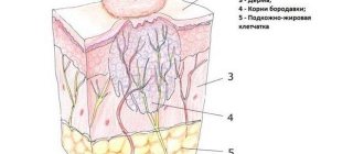
Moles are various types of pigmented formations that stand out against the background of the surrounding skin in color. Sometimes they can be flat, in other cases they can rise above the surface of the skin, have the shape of a tubercle or a pea. All types of moles are designated by the medical term “nevus”.
In what cases is it necessary to remove pigment formations?
The need to remove moles arises for various reasons:
- if they are located on the face and cause discomfort as a cosmetic defect;
- in the case when the mole is subject to constant friction against clothing;
- with mechanical damage to the nevus;
- for medical reasons - if there is a high probability of degeneration into a malignant neoplasm.
Usually, doctors recommend removing large nevi due to the high risk of degeneration of their cells, which can result in the appearance of a low-quality tumor in this place. Long exposure to direct sunlight and frequent visits to the solarium can cause the transformation of tissue into cancer. You should also avoid injuring the mole with a washcloth with hard fibers when taking a bath or shower.
Warning symptoms include an increase in the size of the pigment spot, a change in its color, and the appearance of cracks or depressions covered with scales on the surface. You should immediately consult a doctor if blood or light liquid comes out of the mole, or if you feel a tingling or burning sensation in this place.
A hard washcloth can irritate the site of the removed nevus and provoke a relapse
Do moles need to be removed?
For the most part, moles appear throughout life and do not bother a person in any way. Even among doctors, there is a point of view that removing moles does not lead to anything good, that this may disrupt a certain balance in the body, and, ultimately, more serious diseases may appear. This point of view has no rational basis. On the other hand, suspicious moles that are susceptible to injury should be removed before they degenerate into melanoma. Besides, not all moles add charm. Some significantly change their appearance and cause rejection among others. For cosmetic purposes, nevi located on the face and open areas of the body are most often removed. Even if a mole does not bother you, causing only aesthetic discomfort, you can get rid of it once and for all. In recent years, this procedure has become increasingly popular. Medical centers specializing in benign skin lesions are ready to provide the most modern equipment for removal. Under no circumstances should you remove moles yourself! When trying to get rid of a nevus at home, there is a high risk of leaving a piece of formation in the skin. In this case, trauma to the mole will occur, which can lead to the remaining piece degenerating into a dangerous melanoma. That is why treatment should be carried out by a qualified doctor who can completely remove the mole without leaving a single piece. And also, to distinguish benign formation from melanoma. It is best to remove suspicious moles using a scalpel, also cutting out a small area of adjacent skin. If a mole has a typical structure, it is possible to remove it with a laser, electrocoagulator, or radio wave method.

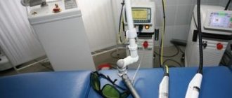
Laser mole removal is perhaps the most popular. In this case, tissue evaporation occurs.
The photo shows the removal of a mole using a loop of a radio wave device. Less popular than laser removal.
The course of the postoperative period
Careful selection of the medical institution where the procedure will be performed will help minimize the possibility of complications after mole removal. You should trust clinics that have been providing services in this area of cosmetology for a long time, equipped with modern equipment. Contacting an experienced specialist will significantly reduce the risk of nevus recurrence after removal.
After completing the procedure, you must follow all medical recommendations when caring for the skin in the surgical area. A brown crust will soon appear at the wound site. It must be protected from contact with soap solution, shampoo, cosmetics - cream, powder, etc. Do not allow the wound to come into contact with ordinary water - the microorganisms it contains can cause inflammation of tissues not covered with a protective layer of epithelium.
It is impossible to remove the crust that has healed over the wound: a new skin epidermis is formed under it. If it is torn off before it falls off on its own, a rough scar may appear in this place.
After tightening the area where the mole previously grew with new skin, the crust easily comes off on its own. Since at first the skin is very delicate and thin, it must be protected from damage and solar radiation.
Face powder should never be applied to the site of a removed mole.
Reasons for the appearance of spots after mole removal
Spots at the site of a removed mole are formed due to manipulations carried out with the skin, and do not depend in any way on the method used. The factors causing pigmentation are:
- Scale and depth of education. The appearance of the spot is directly related to the size of the mole; the larger it is, the more likely it is that after getting rid of it, a scar or pigmentation will form.
- A way to get rid of a mole. If removal is performed with a laser, then the likelihood of pigmentation formation is lower, since the procedure does not affect the deep layers of the skin. If surgical intervention is used, then there is a high possibility of red spots appearing, which are often eliminated within 6 months.
- Improper care of remote education. After removing a mole, there are a number of prohibitions that the patient must observe. For example, you need to avoid contact of the wound and sunlight until the area of the former formation heals and returns to its normal skin color. If the principles of care are not followed, a pigmented or white spot may appear at the wound site.
Features of the wound healing process
The duration of tissue healing at the site of mole removal depends on the area of the damaged area of skin and the individual characteristics of the body. On average, the duration of the process is 20 days; discomfort may be felt at the removal site for another 2–3 weeks. It is recommended to continue special skin care in this area until it acquires the same shade as the surrounding skin.
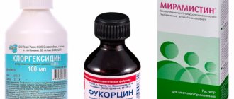
The wound heals better not when open, but when a bandage is applied with pre-treatment with brilliant green or a solution of potassium permanganate. The attending physician may also prescribe special ointments to apply to the wound. When using medications, specialists take into account a number of factors, including the size of the mole and what removal method was used.
After surgery, nevus tissue is sent for histological analysis. At the next visit to the clinic, the patient is informed about the results of the study. If the mole was on the face, the sutures are removed 3–6 days after surgery. For other localizations of the nevus, the sutures are removed after 7–20 days. The doctor gives recommendations on how to care for the wound surface to prevent infection from entering it. It is necessary to protect it from contamination, damage, and squeezing.
Zelenka will help in wound healing
The good and the bad
Moles can be very different - light or dark, flat or “rising”, superficial or embedded in the thickness of the skin, in a word, all sorts: both beautiful and ugly. What about the danger of their degeneration, which of them are the “bad”?
Doctors believe that the potential for degeneration into a malignant formation is much higher not in those moles with which we are born, but in those that appear on the skin later. And these are not necessarily raised or hairy moles, which most of us consider the most “bad” ones. Most often, it is flat formations that can be dangerous.
The size of moles also matters: medium, large and giant moles can more often degenerate into melanoma. Therefore, even if such moles “live” in an inconspicuous place and do not cause much trouble, you still need to consult dermatologists-oncologists about them once a year.
Possible complications
After surgery, you need to carefully monitor all changes occurring in the wound. As a rule, the postoperative period proceeds well, but sometimes complications may occur. If unusual symptoms appear or you suspect that the healing process is not going well, you should consult a doctor. An examination by a specialist will help determine whether there really are grounds for concern or whether the concern is caused by imaginary reasons.
It is necessary to visit a doctor if:
- severe pain is felt at the site of the postoperative scar;
- the wound bleeds or discharges liquid with an unpleasant odor;
- body temperature increased significantly;
- a lump appeared at the site of the removed mole.
Consultation with a specialist is required in cases where even the use of painkillers does not help eliminate the pain.
It must be remembered that immediately after surgery you should not take Aspirin and Ibuprofen, as these drugs thin the blood, increasing the risk of bleeding.
High temperature is evidence of an inflammatory process
What may cause the appearance of a tubercle?
Sometimes it seems that a new mole has appeared due to tissue swelling as a result of damage to the lymphatic vessel during excision of the nevus. Usually their condition recovers within a few days. If an infection gets into the wound, an inflammatory process may begin, accompanied by an increase in temperature to 39 °C or more. To eliminate it, antibiotics are prescribed.
A dense tubercle may also grow if the removed nevus has degenerated into a malignant tumor. In this case, examination and treatment by an oncologist is necessary. If necessary, a new operation is performed. The nature of the neoplasm can be confirmed by histological examination of its tissues.
It also happens that the mole has grown due to the fact that during the operation the deeply located melanocyte cells that make up the nevus were not removed.
As they grew, they caused pigmentation to appear in the same place. The reason for incomplete removal of mole tissue is sometimes the choice of an insufficiently effective procedure method - the use of laser radiation, cryodestruction. To correct the error, the spot is re-excised using a scalpel. Not only the tissue of the mole is cut out, but also the adjacent layers of the epidermis are captured at a short distance (3–4 mm). After healing, a scar may remain at the surgical site.
How to prevent the appearance of a new mole
First of all, you need to carefully select a method for eliminating pigment spots together with your doctor. Proceed with the procedure only after making sure that all cells included in the nevus can be reliably removed. After surgery, avoid infection of the wound, follow all doctor's instructions for treating its surface. Under no circumstances should you remove or scratch the crust covering the healing tissue.
Until the wound has completely healed, you should not be in the bright sun or visit a solarium.
The delicate area of skin that opens after the crust falls off must be protected from exposure to ultraviolet rays. To do this, you can cover it with clothes when you are outside or lubricate it with a special cream with an SPF factor of 50 or more.
There are observations when foci of pigmentation appear in the area of nevus removal - the nevus begins to grow after removal. In the WHO classification, such moles are classified into the group “persistent melanocytic naevus”. The characteristics of the growth of moles in patients with different types of removed nevi after removal were studied, while the translation as “continued growth” is more adequate, since the term “relapse” is less consistent with the histogenesis of this process.
The histological picture of the growth of a mole correlates with clinical signs: against the background of scar changes in the pigmentation zone, nevus cells remaining after removal, producing the melanin pigment, are identified, i.e. containing dark brown granules of melanin pigment in the cytoplasm.
Depending on the location and nature of the pigment-producing cells, a histological diagnosis of nevus growth after removal can be made, indicating the variant of its structure. Most often, in the zone of continued growth, simple intradermal nevi are detected, sometimes with fibrotization, as well as blue nevi.
The continued growth of a mole after removal coincides with the manifestations of a pre-existing nevus. The cells of a simple mole do not show signs of dysplasia, so a simple nevus is not dangerous for malignancy. A simple mole is usually removed using diathermocoagulation with preliminary excision for. In this case, the main reason for the growth is the incomplete removal of pigment-producing nevus cells that lie deep in the dermis in the intradermal version of a simple mole. Of course, to prevent this process, deeper coagulation is necessary, but there is a danger of the formation of a postoperative hypertrophic scar.
Sometimes the growth of nevi after removal in the form of the appearance of pigmented areas in the scar area is detected in the postoperative period during dermabrasion and surgical excision of giant nevi. Histological examination reveals clusters of pigmented nevus cells, characteristic of a simple intradermal nevus, or clusters of spindle-shaped pigment cells of a blue birthmark among the coarse fibrous scar tissue.
When analyzing the possibility of continued growth of both simple and giant birthmarks after treatment, the connection of this process with the histological structure of moles and the method of their removal is revealed. As a rule, primary pigmented nevi have the structure of an intradermal or blue nevus. It is these two variants that are distinguished by the deep intradermal location of pigment-producing cells, and a blue mole can also be found in the subcutaneous fatty tissue.
What to do when moles appear on the body?
When many moles appear, people begin to wonder if they are dangerous and what needs to be done about them. In fact, everything will depend on what caused their appearance and only a dermatologist can figure it out.
Read more Methods of infection with HIV and AIDS
In the future, you need to maintain your lifestyle and follow some rules:
- Try to visit solariums as little as possible, it is harmful to the skin and overall health.
- Direct sunlight is also dangerous, and first of all this affects the health of your epidermis.
- Before going outside, do not forget to wear hats and use sunscreen.
- It is not recommended to go out in the sun between ten in the morning and four in the evening.
- Constantly monitor your health, avoid colds and infectious diseases.
If newly appeared moles do not cause you trouble or bother you, then there is no need to worry. But of course, for your own safety, you should still see a doctor.
Remember that only a doctor can decide which nevi need removal and which are completely safe.
Risk of malignant transformation after removal
Thus, the possibility of continued growth depends on the completeness of excision of these variants of moles and is significantly reduced with their deep surgical removal, especially the blue nevus. The continued growth of these nevi after removal is not associated with their malignant transformation and cannot be regarded as a sign of malignant growth.
As for the melanoma-dangerous dysplastic nevus, the features of its histological structure also determine the possibility and nature of continued growth.
Dysplastic nevus is represented by melanocytes with dysplasia (“dis” - “disorder”, “plasia” - “maturation”) and therefore belongs to pre-melanoma nevi. The localization of these melanocytes is characteristic - they are located only in the epidermis, therefore, to completely remove this birthmark, it is not necessary to strive for deep excision. Continued growth may occur, but not due to the depth of germination, but due to the radial epidermal spread of the dysplastic birthmark lesions. Since a dysplastic mole is a highly pigmented neoplasm, its boundaries are usually clearly defined and excision is carried out within healthy tissue. Isolated observations of the growth of a dysplastic nevus after removal have been noted, but, as a rule, in these observations the characteristic signs are localized at the border of the scar and the surrounding skin. It is known that a dysplastic mole can not only transform into a malignant one, but also form a simple intradermal nevus. Macroscopically, against the background of a dark brown spot, an elastic plaque with a pronounced skin pattern on the surface of a less intense brownish color is revealed. In these cases, the depth of excision depends on the depth of distribution of nevus cells in the dermis, i.e. the same as with a simple intradermal birthmark. The continued growth of such neoplasms is also determined by the intradermal component of a simple nevus and the completeness of its removal. The article was prepared and edited by: surgeon
What is surgical excision of a nevus? Is it worth removing this formation? It is best to discuss such questions with your doctor, since the answers to them depend on the characteristics of the formation itself, which only a specialist can study.
A nevus is a growth on human skin. Its other name is mole. Nevi can be either congenital or acquired.
The result of nevus removal is impressive!
- if excision of the mole is recommended by a medical report;
- if the nevus causes physical discomfort to a person and there is a threat of damage.
Today, the most effective method of removing a nevus is with a laser, but such an operation is performed only if there is indications from a dermatologist.
Getting rid of nevi on your own at home can result in cancer. If the formation brings discomfort, as, for example, if it is located in the lip area, then you need to find a specialist who will conduct an examination and prescribe removal of the nevus.
Removal of nevus by surgical and radio wave methods
Surgical removal of nevus is the oldest method. This method helps with deep and large formations on the skin. However, this procedure has a drawback: it leaves scars and scars on the skin.
When resection takes place, the growth is cut out, including healthy skin. The manipulation itself takes place under local anesthesia and lasts 30 minutes. Once the formation is removed, the wound is sutured so that the scar is less noticeable.
After the operation, the person goes through a recovery period. The main thing is to care for the place where the manipulation was performed. It should be treated with anti-inflammatory drugs for a week after the stitches are removed.
Completing the process of surgical removal of a mole
Surgical removal of the growth is an outdated method; it has been replaced by more modern options. For example, removing a mole with Surgitron. Surgitron is a radio wave surgical device for getting rid of tumors. Its use is considered the safest to date. The procedure takes place without the electrode coming into contact with the skin. The main advantage is that there are practically no postoperative complications, and the rehabilitation period is easy.
This method is well suited for removing nevi, for example, in the lip area, since there are no scars after it. If you remove a mole near the lips surgically, you will be left with an unsightly scar that causes aesthetic discomfort.
After surgery, you should avoid bright sun for a couple of weeks. It is not recommended to visit the sauna and swimming pool for 3 days. Wound care is necessary. The area where the birthmark was removed should be treated with an anti-inflammatory agent for a week. This time will be enough for complete recovery after surgery.
Disadvantages of the radio wave method
Despite the huge number of advantages, this technique also has some disadvantages that you also need to know about.
- The cost of removing a mole using the radio wave method is much higher than other methods, so not every patient can pay for it.
- A radio knife is an expensive piece of equipment that is not available in every clinic. This is especially true for small cities.
- This method is not suitable for removing large moles.
- There are some contraindications, but every method of mole removal has them.
Nevus removal with laser
One of the most effective methods for eliminating moles is also laser removal. The laser removal surgery is performed under local anesthesia. This method is also one of the safest.
The procedure takes no more than 20 minutes, and healing occurs within 2 weeks. After this time, any traces that indicate the operation being performed disappear, there are no scars. This is very important if the operation is performed on the face, for example in the same lip area.
One of the features of the laser procedure is that the likelihood of the nevus reappearing is excluded.
It is also possible to remove a mole with a neodymium laser. This method is used only in aesthetic medicine and cosmetology. It is effective if a mole needs to be removed from the face, such as the eyelids, nose, lips or cheeks.
Despite the fact that neodymium laser is safe, it has a number of contraindications. It should not be used in certain cases:
- during pregnancy and breastfeeding;
- for purulent diseases;
- if the moles are malignant;
- if there are cracks, scratches or cuts at the impact site.
Advantages of the radio wave method
This technique has many advantages. Let's look at them.
- The procedure does not take much time. Usually its duration is no more than 20 minutes.
- The risk of complications after mole removal is minimized. Problems such as swelling, inflammation, and redness are extremely rare. For most patients, everything heals very quickly.
- The radio knife does not come into direct contact with the skin, but works at a distance. Therefore, the risk of infection getting into the wound is eliminated.
- The device affects only the nevus, leaving the surrounding tissue intact.
- Postoperative care is minimal. The patient does not need to follow any complex recommendations. It is enough to simply keep the wound clean and protect it from injury.
- The procedure is performed on an outpatient basis. The patient does not need to go to the hospital or take sick leave. After the operation, the patient immediately goes home and continues to lead his normal lifestyle.
- The procedure itself is virtually painless. After it, no unpleasant sensations are observed either.
- The radio knife delicately removes moles, so it can be used on the most delicate areas of the body - on the face, neck, ears, armpits, and intimate area.
- During the procedure, the doctor controls the depth of exposure to radio waves and the area of the treated area.
- The tissues are not damaged mechanically, so they heal quickly. In addition, they do not char.
- The removed nevus can be sent for histological examination if there is a suspicion of its malignant nature.
- After removing a mole, there are no scars left on the skin.
- Using a radio knife, you can completely remove even a convex mole.
- Local anesthetics (lidocaine, novocaine) are used for the procedure. Patients with allergies to these drugs can undergo manipulations without anesthesia, since the discomfort is minimal.
Consequences of nevus removal
Sometimes, after removal, a nevus recurs, that is, it reappears. This happens if the nevus cells were not completely removed during surgery. At the site of excision, when the crust falls off, a small dark spot may appear. It is worth knowing that this does not lead to cancer, but medical supervision is necessary. If the patient wishes, the nevus can be removed again. But in this case, mandatory histological examination will be required.
The person decides for himself whether to remove a mole or not. And if a decision is made about surgical intervention, it is necessary to take care of choosing a specialist who will carry out this manipulation.
The main thing is that the doctor has experience in carrying out such manipulations. You should always remember that removal that was carried out incorrectly can cause serious consequences, in particular, the risk of developing cancer increases. And if a mole does not bother a person and does not cause him aesthetic or physical discomfort, it should not be removed.
If a mole still bothers you, be sure to consult a doctor. Don't try to remove it yourself. The specialist, after examining the formation, will suggest surgical, laser or radio wave removal. Be sure to listen to the recommendations.
Mole removal
There are moles on every person's body, some have more, some have less. They do not always decorate the body with their presence. Their ability to degenerate into malignant tumors leads to severe cancer. Therefore, when they discover new birthmarks or convex growths, people run to the doctor, demanding that the suspicious mark be removed.
Removing moles is a serious procedure that requires a high level of professionalism from the doctor. A mole needs to be removed if it bothers a person, for example: it has increased in size, hurts when touched, changes color and structure. If the tumor is located in a place that is often subject to friction or clinging, then it is better to remove it too, so as not to damage it or cause infection.
THIS IS REALLY IMPORTANT! An effective and affordable remedy for acne and blackheads. find out what kind of anger >>
Brown dot - what is it?
Sometimes, some time after the mole has been successfully removed and the skin has healed, a dot appears in the same place. Most likely, such a spot is a symptom of a relapse - the reappearance of a nevus. It may take 1-2 months from the moment of surgical removal to the visual appearance of a new mole, sometimes more.
When you carefully examine the brown spot that appears at the site of the removed mole, under a microscope you can see cells that are typical for all moles. They are capable of producing a brown pigment known as melanin. It is thanks to him that the nevus turns brown.
Why did it appear again?
The main reason for the reappearance of a mole after removal lies in the insufficient quality of surgical intervention. If a previously existing nevus was not completely removed, part of its cells (a microscopic fraction is enough) will remain in the skin, and after some time will begin to produce brown pigment.
However, some doctors claim that even when a mole is removed, including a significant amount of healthy surrounding tissue, there is a risk of relapse. In this case, it is not yet possible to determine its causes.
What to do if it is not completely removed?
If some time after the mole was removed, it suddenly grows back, do not panic. The first thing you need to remember is how the nevus was eliminated. If it was removed according to all the rules with a histological examination of the tissues, and the tests did not find any pathological (cancerous cells), you can calm down and not worry. Of course, it would be a good idea to show a new (recurrent) mole to a doctor and make sure it is safe.
When is pigmentation dangerous?
A benign nevus, which for some reason appears again, is considered a benign phenomenon. However, there are typical symptoms that require special attention. They are, in principle, similar to similar signs of malignancy of any mole and include:
- Increase in size. If a new nevus grows before your eyes, you should immediately show it to a dermatologist-oncologist.
- Ulceration (appearance of cracks, erosions on the surface of the nevus).
- Bleeding.
- Soreness.
- Permanent injury.
It is worth noting that the risk of developing melanoma after incomplete removal of a benign mole is quite small. Nevertheless, this possibility is worth considering.
Recurrence of dysplastic nevus
Dysplastic nevus has an increased tendency to malignancy. Such a mole can have different sizes, bizarre borders and heterogeneous coloring. Sometimes it rises above the skin level. Doctors strongly recommend removing such single moles with mandatory histological examination. But even after surgical resection, they can appear again, which is considered a rather alarming symptom.
If a dysplastic nevus recurs, it is better not to hesitate and go to the office of a dermatologist-oncologist. Most likely, such a mole will need to be excised again, followed by a comprehensive examination.
What if there was no histology?
If the nevus was removed in a regular beauty salon or evaporated by a laser (electric current, etc.) without a histological analysis, there is already something to worry about. After all, there is a high risk that malignant processes of degeneration have already begun in the removed mole, and accordingly, the tumor may continue to develop. Moreover, the time for timely diagnosis and early treatment may already be lost. Tumors that develop in the skin after a mole is removed can grow especially quickly and metastasize.
If the nevus recurs, it is best to remove it again and conduct a histological examination. Only in this case will you be able to remain calm about your health.
Is it necessary to remove it again?
Diagnostic methods allow us to objectively study the condition of a person’s skin. Effective options are:
- examination in a specialized clinic;
- performing dermatoscopy;
- digital analysis of tumor growth (in dynamics);
- formation of a “map” of structures at a specific location;
- performing a skin biopsy.
Read more How much discharge will there be after childbirth
Dermatoscopy is the process of examining the skin using a specialized apparatus. A physician can examine the internal structure of the growth and classify it. Afterwards, an effective treatment plan is selected. An equally informative diagnostic method is a puncture biopsy. The research mechanism involves manipulation of a special needle. The material is collected under sterile conditions using painkillers. Diagnostics allows you to study the state of neoplasms. If melanoma or tumor is detected, you should immediately consult a doctor.
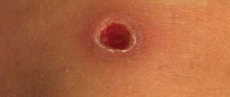
Signs of a mole healing after removal are:
- a dry dark crust remains on the operated area;
- the size of the keratinized structure is 1 mm;
- after a couple of weeks the growth will peel off from the skin;
- the formation of a small red pit, the depression does not exceed 1-2 mm;
- after 28 days, the bottom of the fossa gradually rises to the epithelial level;
- 90 days after the intervention, the inflammation disappears, and an invisible unevenness or scar remains from the mole.
If a light-colored liquid flows out from under the crust, this is a good sign, which means the treatment is going according to plan. To keep complications to a minimum, follow all doctor's instructions. The incisions are treated with high-quality antiseptics.
Oncologists and dermatologists agree that recurrence of nevus is only in rare cases dangerous to human health. The risk of melanoma formation increases markedly if the operation was performed without dermatoscopy. Taking a scraping or puncture will allow you to clearly determine the causes of the development of the pathology. Qualitative histological examination in this case is the main indicator of a person’s health status.
It is not recommended to remove tumors using cosmetic surgery. Unskilled impact on the structure leads to complications. It will be a shame when a person faces cancer due to negligence or inattention. It is better to perform procedures in specialized institutions. Before the mole removal process, a high-quality skin diagnosis should be performed. Otherwise, you increase the likelihood of additional complications.
There is nothing wrong with the appearance of moles on the human body. The process of mole formation is a mechanism that depends on various factors - internal and external. A qualified doctor will be able to determine the clear reasons for the formation of pathology. The specialist will identify nevi that need to be removed. In the future, a person must adhere to a number of rules and lead a healthy lifestyle.
Repeated excision
Usually, to remove a recurrent mole, doctors resort to excision of such a formation with a scalpel. It is believed that this method allows one to reliably eliminate all nevus cells, capturing the surrounding healthy skin, and also obtain suitable material for histological examination. Of course, using a scalpel is considered a rather traumatic method of removal, and it leaves scars on the skin.
In some cases, it is possible to remove a mole using a laser, electric current or radio wave method. But at the same time, the doctor should not evaporate the nevus, but cut it off in order to again obtain material for histological analysis.
Removal methods
There are many methods for removing birthmarks. Each method has certain pros and cons. To remove growths, use a scalpel, laser beam, radio or electric knife. The doctor decides which method to remove the tumor.
Surgical method
Surgery is the only way to qualitatively remove large nevi with roots deeply penetrated into the epithelial tissue. Using a scalpel, the surgeon excises moles from areas of the body covered by clothing.
After surgery, a scar remains on the skin . The size of the scar is influenced by the diameter of the excised tumor and the degree of damage to surrounding tissue.
Surgical removal allows you to qualitatively remove the nevus along with the stem and root. After surgery, the tissue is sent to the laboratory for histological examination. Analysis of the biomaterial allows us to determine the nature of the pigment spot: benign or malignant.
Laser
Laser removal of birthmarks is an effective method that gets rid of pathological growths on the skin. The tumor is removed during 1 session. The doctor aims the laser. Mole tissue evaporates under the influence of a powerful light beam, while healthy epithelial tissue is not affected.
After removing nevi with a laser beam, there are no scars left. The wounds do not bleed because the capillaries are clogged during the procedure. Over time, the light spot acquires the shade of the skin and merges with it. The method is used to remove birthmarks on the head, nose, eyelids and other open areas of the skin.
Electrocoagulation
When eliminating moles using electrocoagulation, the lesion is subjected to heat treatment. An electric current entering the platinum loop heats it up to 200 °C. The doctor, bringing a red-hot loop to the tumor, cauterizes the pathological tissue without touching healthy areas.
A small wound remains at the treatment site, which heals quickly without leaving marks.
Radio wave method
The radio wave method is a non-contact treatment of moles. The growths are removed using a radio knife - an electrode capable of generating energy. Under the influence of radio waves, pathological tissues heat up and evaporate. The mole is destroyed.
Radio wave removal of tumors is a minimally invasive technique. After the procedure, no traces remain on the skin. Wound healing is not a hassle. The damage heals within 7 days and does not produce undesirable consequences.
The Surgitron device effectively removes moles. Many leading clinics in different countries of the world are equipped with this device. Wound surfaces after removal of birthmarks do not bleed, heal quickly, and infectious and inflammatory processes do not develop in them.
The red spot does not go away
Sometimes, even after successful removal of a mole by laser or other methods, a red or pinkish spot remains on the skin, which is not a recurrence of the nevus. This mark may appear:
- If the skin healing process is still ongoing. It may take about six months or even more until the color of the epidermis is completely evened out.
- Soon after the crust falls off. In this case, a pinkish spot is observed on the skin - an area of young, delicate epidermis.
- If you tear off the crust ahead of time (accidentally or intentionally). Such a dark spot at the site of a removed mole can remain for life; to get rid of it, you must definitely use means to activate regenerative processes (silicone ointments and patches, Contractubex, Solcoseryl, etc.).
- With improper skin care. In particular, reddish spots may appear when the young epidermis is exposed to sunlight. In this case, they indicate a skin burn and can subsequently lead to pigmentation of such an area.
- Due to the penetration of infectious agents. In this case, the problem area may swell, itch, and even be noticeably painful. The formation of an abscess is also possible.
If strange spots appear on the skin after removing a mole, it is better to play it safe and consult a specialist. This will help reduce the risk of developing melanoma, a skin cancer.
After removal, a mole has grown again - this is a cause for concern. Occurs due to the partial removal of a formation that germinates and increases in size. It is necessary to understand the nature of the phenomenon and take the necessary measures.
Indications for removal are pain when touched, changes in size, color, and shape. If the growth often catches or is injured, the growth should be removed to avoid infection. Moles are removed from the body using the following methods:
- Surgical - allows you to get rid of moles along with the root. If, after excision, a tumor grows again in the area, the root has been partially removed.
- Cryotherapy involves freezing the mole with liquid nitrogen. A painless method, the risk of a new mole remaining remains.
- Electrocoagulation - consists of the use of high-frequency current that burns out the nevus. After the procedure, a slight scar is left that disappears over time.
- Laser is effective and leaves no traces after the procedure. The action is aimed directly at education and does not affect healthy skin.
- The radio wave method helps remove moles without leaving any traces, without leaving minor scars after the procedure. Healthy skin is not exposed to radiation and does not become infected.
After eliminating the problematic mole, you may notice that it has grown again. Relapse after mole removal is quite real and occurs for the following reasons:
- During surgery, errors were made in determining the depth of pigment spots, and the excision was partial.
- A dangerous method of eliminating growths has been chosen. Freezing with liquid nitrogen brings a lot of trouble. The procedure results in a burn that does not heal well and becomes infected.
- Improper care of the wound after surgery or failure to comply with a doctor’s prescription leads to infection.
- During the process of removal, the mole degenerates into melanoma.
- Removing a nevus at home without observing all sterility conditions is fraught with the penetration of pathogenic bacteria. Partial removal is a potential threat of recurrence of the nevus.
Reasons for the appearance of spots after mole removal
A common method is laser removal. It is performed in a short period of time and without pain. After the procedure, no scars remain at the site of formation, and the surrounding tissues remain undamaged.
After eliminating the formations, no difficulties arise. If the intervention was successful, a red spot appears at the site of the mole. This indicates that the healing process is proceeding properly. After complete healing of the wound, no traces remain.
Black
A black spot appears at the site of the nevus. This is not affected by the chosen technique. The dark spot formed after the removal of a mole acts as a protective crust until new skin forms, then disappears.
We suggest you read: Is it possible to pull hair out of a mole and why does it grow?
Pink
The pink spot is newly formed skin that forms under the crust. The epidermis is sensitive and delicate. But with the normalization of all processes in the tissue, the pink tint goes away and the cover acquires a natural color.
Reds
A red spot appears if the crust did not fall off on its own, but was torn off. A red tint indicates an infection of the skin area.
Pigmented
A pigment spot may appear at the site of the removed nevus if the area where the operation was performed is exposed to ultraviolet radiation.
Brown
If a brown tint remains on the damaged area, the removal was not complete. Seek advice from a specialist who will prescribe a course of treatment or send you for a repeat procedure.
Burgundy
Situations rarely arise when burgundy spots appear at the excision site; they may also have a purple color. This is a signal that an inflammatory process is taking place as the wound heals. If the spots are dark in color, consult an oncologist; there is a risk of a malignant tumor forming from an incompletely removed mole.
White spots
White spots at the site of the mole indicate that the nevus was removed using a laser technique. The white color disappears within 3 months, but may remain for the rest of life.
Contact specialists; in the initial stages of complications, it is easier to eliminate them.
The color of the suture may change, pus will begin to ooze, and patients will notice that the wound formed after the removal of the mole becomes wet. If the depression, lump or bulge begins to itch and ooze, there is a possibility that recovery does not occur and a new growth appears. Redness can be observed in the hair or hands, these areas are often in an open state and may become irritated and itchy.
Experts do not recommend getting the area of skin wet for two weeks, not peeling off the crust, and not being in the sun. Lubricate the epidermis with protective sunscreen in summer and winter.
If there is a point, hole or wound left after surgery, it may become wet.
Dangerous changes are considered:
- the crust does not fall off for a long time;
- the wound gets wet;
- the process of suppuration is noticeable.
If you notice the beginning of the formation of a keloid scar, use Contractubex. The ointment slows down the process of scar development.
The spot that remains at the site of the mole after removal can have different sizes and shades:
- Red spot. It forms when the wound has not had time to heal, but the crust has already been torn off from it. With this type of spot, infection of the damaged skin is possible, which is fraught with serious complications.
- White spot. It is observed if a mole was removed with a laser. For most people, the white spots disappear on their own within six months, but in some patients they remain for life.
- Pink spot. Appears at the moment when the crust falls off the wound. The skin becomes natural color 3 months after the formation is eliminated.
- Black or dark spot. It is observed after removal manipulations and does not depend on the method of getting rid of the nevus. The mark is a crust that performs a protective function and protects the wound from infection. After the removal site heals, the dark scab falls off on its own.
- Brown spot. This indicates that during the procedure the formation was not completely removed. In such a situation, it is important to consult a doctor in order to prescribe another mole removal if necessary.
- Pigment spot. It forms at the site of a disappeared nevus when the principles of caring for moles after removal are not followed. Often the cause of the appearance is the rays of the sun that hit the wound during the healing process.
- Purple pigmentation. Most often it is observed if an infection gets into the wound and an inflammatory process begins under the skin. Sometimes the area of injury darkens even when the nevus was not completely removed and because of this, a malignant tumor develops. In this case, the person should immediately visit a doctor.
We invite you to familiarize yourself with the radiosurgical method of removing papillomas - treatment
What to do if a mole grows back after removal
After removing the growth, a lump or dark dot appears; do not do anything on your own. You should examine the area on your skin daily. If a scar or scar appears, visit a specialist. He will examine and prescribe medications that accelerate the healing process and resorption of the skin defect.
Before the procedure, think carefully and weigh all the pros and cons. Improper removal or introduction of infection will lead to serious consequences and the development of cancer. Consider contraindications: the presence of chronic diseases, cardiovascular disorders and problem skin.
Contraindications for specific methods:
- Surgical is contraindicated in case of exacerbation of herpes, inflammatory or infectious diseases.
- Cryotherapy is not recommended for malignant tumors.
- Laser removal cannot be carried out:
- if you are allergic to the sun;
- if the tumor is malignant;
- during exacerbation of herpes;
- during pregnancy;
- during menstruation in women;
- increased body temperature;
- for inflammatory diseases.
When exposed to radio waves, there are the following contraindications:
- during the degeneration of education;
- during pregnancy or while breastfeeding a baby;
- diabetes;
- presence of a pacemaker.
If nevi appear on the body in large numbers or reappear after getting rid of it, this is a reason to undergo examination. Timely identification of the problem will help to avoid complications.
Signs of a malignant neoplasm are a cause for concern:
- the growth of a removed mole causes pain;
- presence of severe itching;
- the surface of the skin becomes very dry and becomes covered with small cracks;
- the presence of ichor from under the growth;
- change in the size of the nevus (increases);
- change in color and structure of formation.
If signs appear, you should immediately consult a doctor. After the studies, the degree of malignancy of the tumor will be determined. If cancer is detected in the early stages, timely treatment will be successful. If the tumor has grown and metastasized, affecting vital organs, treatment will be difficult.
Contraindications for removal
It is not always possible to remove moles on the human body, because there are certain contraindications that must be taken into account. These include many chronic diseases, heart disease, and unhealthy skin. In addition, specific treatment methods may have contraindications; for example, surgical excision cannot be performed in the following cases:
- Herpes is in the acute stage.
- Diseases of an inflammatory or infectious nature.
All of the above problems prohibit removal with liquid nitrogen, and also if the formations are malignant. Laser removal also has certain contraindications, including the following:
- Allergy to the sun, that is, photodermatosis.
- There are suspicions that the formations are malignant.
- Herpes is in the acute stage.
- A woman is carrying a baby.
- Treatment is prohibited on the day when menstruation occurs.
- Body temperature is increased, even if only slightly.
- Inflammatory diseases.
If you have these problems, under no circumstances should you remove moles with a laser. As for radio wave treatment, the following contraindications can be identified:
- The process of degeneration of moles occurs.
- A woman is pregnant or breastfeeding her baby.
- Diabetes.
- The patient has a pacemaker.
It is imperative to take these contraindications into account, otherwise consequences cannot be avoided.
If you notice that you have a lot of moles on your body, or they appear again after treatment, you need to undergo an examination. Remember that any growth on the skin can cause you to develop cancer. Therefore, the sooner the problem is identified, the fewer complications it will bring you.
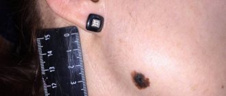
My daughter had a nevus mole removed on her leg, and two months after removal it appeared again. Is he dangerous? Histology showed that it was an intradermal pigmented nevus. I am attaching his photo.
Marina
(Arkhangelsk), 36 years old
Relapse Prevention
After diagnosis and confirmation of the benign quality of the formation, you should consult a doctor with experience in this field. The country's leading dermatologist-oncologist Igor Evgenievich Sinelnikov faces similar problems every day. He offers various ways to remove the growth without complications. To avoid consequences and re-growth, you must follow these rules:
- In the first week, do not allow water to enter the area after removing the growth.
- Read the instructions for proper wound treatment. Wash your hands thoroughly before handling and monitor the area for two weeks to avoid infection. Will help avoid inflammatory processes and relapse.
- After the procedure, a crust will appear on the wound; it is not recommended to remove it yourself. Over time, it will disappear, leaving healthy pink skin.
- It is not recommended to sunbathe in the sun or in a solarium. Ultraviolet radiation negatively affects the skin, stimulates the growth of old and new formations, and the appearance of birthmarks. If you cannot hide from the sun, cover the area with an adhesive plaster or cover it with clothing.
Simply following the rules and carefully caring for the skin area is the key to a quick recovery. preventing relapse. The use of medicinal ointments and creams will hide cosmetic defects.