17.04.2017
Dermatofibroma is classified as a benign skin formation; it does not pose a potential danger and has no tendency to degenerate.
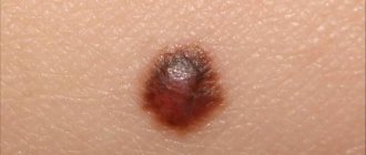
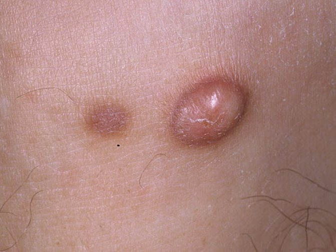
Outwardly it resembles a convex mole or wart. It is round in shape, gray or pale pink in color, with a smooth surface.
Doctors note that the main part of the tumor is located under the skin, with a small fragment on the surface. The tumor is characterized by slow growth. Dermatofibroma is diagnosed only by a highly qualified dermatologist, and treatment is carried out in several ways, including surgical excision and removal of skin fibroma with laser (radio waves).
Causes of skin tumors
The main set of reasons has not yet been sufficiently studied by modern medicine. The list of main factors including the mechanism of formation and development of such a tumor includes:
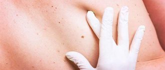
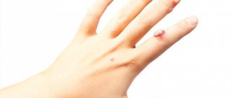
Fibroids can form anywhere
How dermatofibroma includes:
- Genetic disorders, family history, hormonal and age-related changes in the postmenopausal period or other disorders.
- Various stages of pregnancy, pathology in the formation of hormones in the blood.
- The appearance and stimulation of the development of such a neoplasm as skin fibroma tends to form against the background of diabetes mellitus, acromegaly, and other diseases of the endocrine system, which becomes predetermining the causes of subsequent pathology.
It is also important to pay attention to provoking factors. Benign skin neoplasms arise as a consequence of friction with clothing elements, with increased sweating, manifestations of excessive insolation or regular hypothermia, frequent inflammatory processes, errors in diet and routine.
Soft fibroma: risks of appearance
Fibroid tumors involve the occurrence of neoplasms on the skin, which are formed from fibrous and connective tissue. A soft fibroma is an elastic growth of the skin layer. It may have a round or oval shape. Soft fibroids can be flesh-colored, tan, or tan in color.
Soft fibroma most often occurs in older people, overweight people and pregnant women.
To date, doctors do not have an exact answer to the question of why fibroids form. But they can indicate risk factors for the disease. Soft fibroma can occur due to frequent mechanical damage to the skin.
Risks of developing soft fibroids:
- Chronic skin irritations;
- Age-related changes;
- Improper functioning of the endocrine system;
- Papilloma infection.
Soft fibroma almost never transforms into malignant formations. Fibroids are not life-threatening, but the tumor itself can cause physical and aesthetic discomfort. Removal of fibroids is possible through surgery.
Pathological types of disease
The characteristics of the growth of neoplasms allow them to be classified as follows:
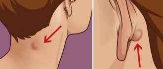
The main causes of fibroids are not well understood
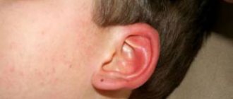
- A form of limited disease. Fibroids usually form under the skin in the neck, back of the head and behind the ears. Sometimes it may resemble a dripping drop on a thin stem.
- Fibroma of diffuse or aggressive type. It does not have growth-limiting capsules and has the ability to grow into adjacent tissues. With a deep location, invasion into the vascular bed and regional organs is possible.
Consistency also has a huge impact on classification. It is divided into soft skin fibroma and hard type benign tumor. In soft ones, adipose tissue predominates; they can be elastic and mobile. Solid fibromas are more dense upon palpation, do not divide, and are located on any organ, but most often on the mucosa.
Types of skin fibroids
There are clinical and histological classifications of tumors. Taking into account the structure, several types of dermatofibroma are distinguished:
- Fibrous variety, in which multidirectional young and mature collagen fibers, as well as mature cells (fibrocytes), are found in the tissues, the content of other elements is in small quantities;
- The cellular type is characterized by a predominance of cells and a small number of tender and loose collagen fibers. Many fibroblasts and nests of histiocytes are found inside the tissues;
- Fibroxanthoma is a neoplasm, in the structure of which multinucleated Touton cells are distinguished, including typical xanthoma cells;
- Sclerosing hemangioma is formed when there are a large number of vessels of different sizes.
Clinical classification is based on the appearance of the lesion. Soft skin fibroma is a lobulated, protruding formation that is soft to the touch. It is usually located on the face, torso, or in the shoulder girdle.
Solid dermatofibroma consists of many dense lobules and is found on any part of the body. Lenticular fibroma is a collection of small nodules of varying sizes, usually appearing in areas of minor trauma.
Atypical types may occur, such as cutaneous angiofibroma, with uneven color, heterogeneous consistency or changes in the surface.
Symptoms and signs
The symptoms of fibroids are quite vague and do not manifest as painful moments. Neoplasms of this type are characterized by the following:
- Flesh or light yellow in color, round, oval, dome-shaped, a nodule with clear boundaries with a thin or wide stalk.
- Often additional elements appear in the form of additional tubercles, irregularities, dents, and depressions.
- The place on the skin of fibroids can be indicated by pigment spots; the soft form is small in size - from 0.5 to 3.0 cm.
- The surface may be thin and wrinkled, with slight violations of integrity causing minor pain.
- Tumors of a similar nature on the extremities, back or facial skin are formed subcutaneously, do not move during palpation, and have a clearly defined shape.
Prognosis and prevention
Fibroma is not a dangerous neoplasm, therefore, subject to timely and adequate treatment, the prognosis is favorable. In this case, the formations are eliminated before any complications develop, thereby preventing the likelihood of malignancy. The pathology does not cause harm to health, and if laser or radio wave treatment is used during therapy, the risk of relapse is almost completely eliminated.
There are no specific ways to prevent the appearance of fibroids in different areas of the skin. You can reduce the risk of developing such a pathology if you follow some rules:
- lead a healthy lifestyle;
- monitor skin condition;
- adhere to a healthy diet;
- timely treatment of chronic skin diseases;
- undergo regular medical examination.
To keep your skin healthy, you should include dairy products, as well as fruits and vegetables, in your daily diet. Apples, viburnum, cucumbers and tomatoes are especially beneficial for the skin.
Diagnosis and treatment methods
Symptoms of fibroids are painless
According to established tradition, diagnosis is carried out by examination by a qualified specialist. To refute all possible versions of a malignant etiology, the doctor prescribes a tissue biopsy; for this, the dermatofibroma is sent for histological examination. If necessary, an ultrasound scan of the affected area and consultation with specialists in the field of gynecology and mammology are prescribed.
When making a preliminary diagnosis, the obtained data should be compared with the characteristics of hygroma, wen, skin atheroma, senile keratoma, manifestations of nevi, papillomas, and various types of molluscum contagiosum.
Folk remedies
Is it possible to treat fibroids on the leg with folk remedies? In some cases, alternative medicine recipes actually turn out to be quite effective, but their use is allowed only with the permission of a doctor.
MORE ABOUT: Hysteroscopy in Balashikha. Compare prices, buy consumer goods on the marketplace
So, what methods will be effective? Let's look at a short list:
- lubricating the tumor with fresh celandine juice 2 - 3 times a day for 2 - 4 weeks,
- treating the growth with potato juice,
- lubricating fibropapilloma with camphor alcohol three times a day,
- the use of lotions or compresses from a decoction of oak bark, marigolds,
- a mixture of iodine and alcohol tincture of aloe, which is used to lubricate the growth, or make applications on its surface.
But you shouldn’t rely too much on these methods of fighting fibroids. If the tumor is still very small and has just begun to form, these recipes may help. But with large skin tumors, you will only waste time performing such procedures.
Thus, despite the fact that the answer to the question whether fibroids can turn into cancer will be negative, you cannot turn a blind eye to the problem. This tumor can cause pain and discomfort, increase in size and become infected. Therefore, it is better not to wait for such complications, but to remove the growth as quickly as possible.
Loading…
Types of treatment
In modern conditions, dermatofibroma can be cured by injections of 0.20 ml. diprospan solution. In addition, surgical or other techniques are used.
Fibroids can be removed surgically
- Excision using cryodestruction using liquid nitrogen, after which scars and cicatrices may appear.
- Application of electrocoagulation. The discharge of electric current causes tissue destruction.
- The traditional method of surgical excision is acceptable for tumors of impressive size located deep in the skin. Local anesthesia is used and there may be residual scarring.
- Destruction of the tumor using a laser beam using local anesthesia.
- The method of radio wave coagulation is no less popular. Leaves no consequences for distorting the appearance of patients.
After any operation to remove fibroids, a mandatory histological examination of the tissues is performed for the presence of malignant cells.
Leading clinics in Israel
Solid fibroma, or dermatofibroma, is the most common form. Most often it forms on the face, back, limbs (on the arm in the area of the shoulder and forearm, on the leg under the skin in the area of the foot, lower leg), and also affects the mucous membranes (fibrohemangioma).
It has a dense structure and smooth surface. The neoplasm can occur both on the skin and under it (flat fibroma). Localized under the skin, the lump grows slowly. The disease occurs in both men and women. In children and adolescents, fibroblastoma is more common than fibroma.
If there is a faster growth of the formation, this may indicate fibromatosis. This neoplasm affects not only the skin, but also the muscles. Forms on the feet, sometimes accompanied by pain. Occurs due to infections, leg injuries, tendon tumors. Fibromatosis is mainly treated with ointments. Wearing special shoe insoles is also recommended.
Who is more susceptible to fibroids?
At risk are:
- Pregnant women due to hormonal changes occurring in the body;
- Women over 40 years old;
- Overweight people;
- Patients with diabetes mellitus.
What is it and is it dangerous?
Fibroma is a benign, highly differentiated tumor that is formed from connective tissue fibers. It is not prone to rapid growth and increase in size. The neoplasm is not dangerous if it is not injured.
Fibroids can develop both on the surface of the body and in internal organs. Its most typical formation is in the uterus and mammary glands.
The tumor can also grow and mix with other tissues, for example, with:
- Muscular (fibromyoma).
- Glandular (fibroadenoma).
- Vascular (angiofibroma).
- Fatty (fibrolipoma).
What does fibroblastoma look like?
It should be understood that fibroblastoma, unlike fibroma, is a malignant tumor. Externally, it looks like a single node on or under the skin, with unclear boundaries.
Fibroblastoma is typical for children and adolescents, since the tumor is formed during the period of intrauterine development of the fetus.
Fibroma looks different, depending on its type.
There are hard and soft tumors.
Symptoms of fibroids
Fibroma, depending on the degree of tissue growth, can manifest itself in the form of soft and hard formations, each of which differs in size, structure and symptoms. A solid fibroma is a tumor with limited mobility. Most often it appears in the deep muscle layers, gradually growing, slightly rising above the healthy skin. Upon palpation, you can feel a dense formation that has a homogeneous structure.
Initially, the diameter of such a tumor is no more than 5-10 millimeters. However, over time, without proper treatment, it increases to 10 centimeters.
The vessels are compressed, blood flow is disrupted and associated symptoms appear.
Soft fibromas on the leg under the skin are wrinkled neoplasms with a heterogeneous loose structure. Such tumors appear exclusively in the upper layer of the epidermis.
A soft fibroma usually has a wide, flat base with a thin stalk, consisting of connective tissue.
The size of the tumor will be individual, from 1 to 4 centimeters in width and length. Soft tumors are painless, but due to their shape and location they can cause a lot of inconvenience when walking, rubbing their legs, which forces most patients to see a doctor for treatment and removal of the tumors.