General description of the disease
Cutaneous horn on the forehead or face (ICD 10 code - L57.0) is a disease that is not very pleasant in appearance. The growth is formed by the outer cells of the layers of the epidermis.
This neoplasm is similar to the horny process of an animal, which is why the disease is popularly called a horn. Most often, the cutaneous horn appears independently and is a benign neoplasm. In rare cases, this may be the initial stage of oncology - squamous cell carcinoma, which is considered one of the more aggressive malignant diseases. To rule out the worst, when this disease appears, it is necessary to immediately conduct a histological examination of the cells that make up this formation.
We also recommend reading:
Places of horn formation
The horn, which has a cylindrical shape, is distinguished by its pronounced density. The color of this formation can range from light beige to brown with a yellow tint. The size of the horn can be impressive or very small.
This disease brings a lot of trouble to the patient. But the great danger of this pathology lies in the possibility of the neoplasm degenerating into a malignant one.
Most often the disease occurs among women. Localization locations can be as follows:
- ears, cheeks;
- scalp;
- rarely - mucous and semi-mucous membranes (border of the lips, etc.).
The growth looks like a tapering and tapering upward formation on a wide base. Most often, single keratomas occur. Multiple neoplasms of this type can be found very rarely.
Stages of keratoma development
Horny cutaneous keratomas occur in two main forms:
- Primary. This species is harmless, although currently poorly studied. The formation of the growth occurs gradually: darkening of the color and compaction are observed. In this form there are no inflammatory processes. The transformation of a benign primary form into a malignant one is possible to a lesser extent, but it is still necessary to carefully monitor the entire process of its development.
- Secondary. With it, the neoplasm transforms into malignant, so this stage of keratoma is very dangerous for human life. The secondary form can even occur due to the development of inflammation in other neoplasms on the body, degeneration or proliferation of papillomas and warts, and skin injuries.
Both forms of the disease have pronounced differences, but the patient with the primary stage of the disease still has a greater chance of cure.
Causes of the disease
The essence of this pathology is the enlargement and proliferation of the skin, in particular the upper layers of the epidermis. This can happen due to the progression of severe keratosis, as well as due to damage to the tissue of the papilloma or wart.
The provoking factors of this disease may be the following:
- too much exposure to sunlight without using special sunscreens;
- long course of a disease of viral etiology;
- injury to the skin and introduction of the virus there.
Experts also include manifestations of tuberculosis and lupus erythematosus as risk factors.
Even children can develop such a horn. The causes are most often skin injuries or incompletely cured infectious diseases.
Clinical picture of the disease
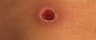
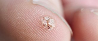
The manifestation of such benign neoplasms most often begins with the appearance of a small growth on the surface of the skin, which gradually grows and thickens, and numerous longitudinal grooves appear along the length of the neoplasm. The skin tone then turns light yellow and eventually brown. The formation gradually takes the shape of a horn, the upper part of which hardens. A red, inflamed stripe can be seen around the base. The main focus of inflammation is located in the upper part of the skin cone.
The horn may grow slowly, but most often there is a rapid increase in the growth. Based on the size of the horn, a preliminary diagnosis can be identified:
- if the length of the growth is less than 1 cm, it is diagnosed as a keratoma or papilloma;
- more than 1 cm - as keratinizing papilloma, seborrheic wart with a high percentage of probability of degeneration into a horn.
What is cutaneous horn?
Representing a not very aesthetic disease formed by the outer cells of the upper layer of the epidermis, the cutaneous horn externally resembles the horny process of animals - it is for this reason that the disease has a similar name.
Most often, this condition appears independently and is considered or is the initial stage of oncology, which is considered one of the most aggressive diseases.
Therefore, when it appears, a histological examination of the cells that make up this formation should immediately be carried out.
Where can this cylindrical skin growth occur and what varieties can it be?
Localization
Having a cylindrical shape, the skin horn has a distinct dense or compact consistency, its color ranges from light beige to brown with a yellowish tint, and its size can be very small and very pronounced.
Resembling an animal horn, this disease brings a lot of trouble to the person who suffers from it. But this condition is of great danger due to the possibility of its degeneration into a malignant neoplasm.
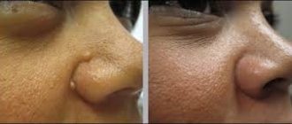
This disease occurs somewhat more often in women; the place of its localization is mainly the area of the face (on the skin of the ears, on the cheeks) and the head (the scalp), occasionally this condition is observed on semi-mucous and mucous membranes.
Photograph of a cutaneous horn on the facial eyelid
Externally, this new formation has a pronounced base of significant area, which sharpens and noticeably narrows towards the upper part. Cases of its formation in single copies are more frequent; multiple neoplasms of this type are relatively rare.
Kinds
The neoplasm in question is benign. However, under the influence of certain factors and even without apparent reasons, its character may change, and the neoplasm may become malignant.
Cutaneous horn can occur in two main types:
- primary form
- this type of disease is not dangerous, but has not been sufficiently studied at present. The formation of the horn itself occurs, its compaction and gradual darkening of color; in this form, no inflammatory processes or other lesions are observed on it.
The transition from the benign nature of this form of the disease to a malignant one is possible to a lesser extent, however, when the growth of such a formation begins, the process of its development should be closely monitored;
- secondary form
- at this stage it becomes more malignant and for this reason this stage of the cutaneous horn is considered very dangerous. It can occur with the development of an inflammatory process in other neoplasms on the body, and the course of such a process is often characterized by a chronic course.
Also, the appearance of a secondary form of this condition can be caused by skin injuries, proliferation or degeneration (usually malignant) of warts and papillomas.
Both forms have pronounced differences; the earliest stage of the disease has a greater chance of complete cure.
Traditional methods of treating keratoma
Modern medicine allows you to get rid of this unpleasant disease quite quickly. Removal of growths can be carried out both in cosmetology centers and in oncology treatment institutions.
After such an operation, a small scar or scar may remain on the surface of the skin.
Modern radiosurgery and its advantages
Various growths on the skin of the face and body are removed in cosmetology clinics and clinics using the Surgitron device. Its actions are aimed at evaporating cells in the affected tissues using repeated point exposure of the electrode.
During treatment with such a radio knife, layer-by-layer evaporation of skin tumors is observed. The main feature of this device is the ability to use electrocoagulation and electrocurettage modes when necessary.
The main advantages of radiosurgical removal of formations are as follows:
- rapid epithelization of the skin;
- low probability of relapse;
- decent cosmetic result.
Method of electrocoagulation and curettage
Experts call this method of removing skin horns the method of choice when treating growths with a diameter of 0.5 to 2 cm.
Local anesthesia is first administered. Specialists use sharp dermal curettes for curettage, after which they treat the edges and bottom of the removed tumor with an electrocoagulator. After 2-3 treatments, an antibacterial ointment and bandage are applied to the wound.
The disadvantages of electrocoagulation and curettage are as follows:
- subsequent formation of keloid scars - in 20% of cases;
- the probability of relapse is almost 50% of cases;
- the removal method is not suitable for large formations;
- not used for patients with pacemakers.
Cryosurgery and its features
Modern cryosurgeons use various cooling substances to influence the affected areas of the body. By applying liquid nitrogen with a swab or cotton swab to papillomas, warts or other skin formations, the skin temperature drops to -20 C, which causes the destruction of proteins. To get rid of a skin horn, including a malignant form of a neoplasm, you will need a temperature below -50 C.
But cryodestruction also has disadvantages:
- when a skin area freezes and then thaws, the patient feels pain;
- Removal of skin growths is most often performed in several sessions.
But if the horny keratoma is small, then the pain after the procedure is quite tolerable. During cryodestruction, local anesthesia is administered.
During removal of skin tags from sensitive and thin skin, swelling may occur. This is often observed when treating the area around the lips and eyes with cryogen. To get rid of such side effects, hormonal ointments are used, as well as other medications with steroids.
Another unpleasant consequence can be blisters with bloody contents. When treating them, specialists open the bladder and apply a dry bandage to the affected area.
Among the consequences of cryodestruction, hyperpigmentation and hypopigmentation are possible. It is not difficult to prevent this by using cream and clothing on sunny days that protect the skin from ultraviolet radiation. In addition, the skin must be protected from exposure to various aggressive chemicals.
Treatment methods
The achievements of modern medicine make it possible to quickly cope with this unpleasant disease. Treatment of cutaneous horns can be carried out both in cosmetic and cosmetology centers and in oncology treatment institutions.
After surgery to remove a skin growth, a scar or small scar may remain on the surface of the skin.
The most commonly used methods include:
- if the tumor has reached a large size, then surgical excision of the skin growth is used - after the operation, a suture is applied to this place;
- cryodestruction - carried out by exposing the emerging neoplasm to liquid nitrogen, the operation is carried out quickly, and there is often no visible trace or scar left at the site of skin growth;
- The most modern and least destructive methods of removing skin horns include exposure to a laser beam - the operation takes very little time, and after it is not required a significant rehabilitation period.
This method - laser removal is considered the least painful, and the removal of overgrown tissue is carried out by the action of light.
Traditional medicine also offers its own methods of treating and stopping the growth of this tumor, but in terms of effectiveness they cannot be compared with removing the growth.
Cutaneous horn
Cutaneous horn is a local hyperplasia of the epithelium, consisting of horny masses, resembling a cylinder or horn of wild animals, having clear boundaries and an inflammatory process at the base. There are no gender or age differences. There is no information about the prevalence or endemicity of the process. The question of the independence of the disease is controversial. In dermatology, the concept of “cutaneous horn” is considered to be a collective one, since it can be a consequence of many tumor processes, but more often it serves as a marker of precarcinous proliferation of the epidermis, developing as a result of actinic, senile keratosis, degenerating into squamous cell skin cancer.
Many representatives of various dermatological schools studied the cutaneous horn. German dermatologists represented by Korting and Denk suggested that the basis of the cutaneous horn is changes in collagen at the molecular level, followed by a failure in the differentiation of dermal and epidermal cells. The Greek physician Higamenakis noted a hereditary predisposition to the development of the disease, which, in combination with provoking factors, leads to the appearance of a cutaneous horn. However, today there is no consensus on this matter. The relevance of the problem lies in the close connection of such processes of hyperkeratinization with neoplastic, malignant neoplasms of the skin, a significant violation of the quality of life of patients, and defects that are disfiguring from an aesthetic point of view.
Cutaneous horn: what is it?
— The secondary form is a dangerous stage of malignancy of the skin horn. It may be accompanied by an inflammatory process at the base of the tumor, rapid growth and pain. This condition can develop against the background of degeneration of warts or papillomas, which were the basis of the cutaneous horn. When the first symptoms of inflammation appear, it is necessary to consult a doctor and completely remove the skin horn.
It is worth noting that only a specialist can tell about the condition of the neoplasm, the degree of its danger to health, as well as whether the skin horn needs to be removed. Self-medication is highly discouraged, as this can provoke the development of more complex, difficult-to-treat skin pathologies.
Treatment methods for cutaneous horn
You can diagnose cutaneous horn at your first visit to a dermatologist.
Thanks to the achievements of modern medicine, it is possible to quickly and easily remove pathology at any stage of development without a trace. Treatment can be prescribed in one of the following ways:
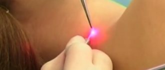
- Laser removal of cutaneous horn
- This is the most modern and safe method. The laser beam completely destroys the tumor, and the procedure lasts no more than 10 minutes, and after it there is no rehabilitation period required. The treatment is painless because the patient is given local anesthesia before the operation.
Surgical removal is used if the skin horn has reached a sufficiently large size. After the operation, sutures are placed at the removal site; the rehabilitation period takes up to 2 weeks. In most cases, after removal with a scalpel, a scar remains on the skin.
— Cryodestruction is the destruction of the skin horn using cold. For this procedure, liquid nitrogen is used at a temperature of 195 degrees; when exposed to cells, the neoplasm begins to die. However, after cryodestruction, a small scar may remain on the body.
What to do after removal?
After removing the skin horn, experts recommend having all tumors on the body examined once a year. This can be done using RTM diagnostics. In 95% of cases, removal of horny keratoma is safe and does not cause relapses. Several preventive measures after removal of the tumor:
- Try to avoid being in direct sunlight for long periods of time, especially at lunchtime when the sun is most active;
- Protect the tumor removal site from injury;
- Add more vitamins and nutrients to your diet to strengthen your immune system.
Causes of cutaneous horn
To date, the exact cause of the formation of a cutaneous horn has not been identified. It is believed that the basis for such a powerful proliferation of the epidermis is a violation of cell kinetics, which consists of several components: a sharp acceleration of mitotic cell division, accelerated migration of cells overflowing with keratin to the skin surface, acceleration of genetically programmed physiological cell death. Each individual component can cause adaptive changes in the skin in the form of proliferative acanthosis (an increase in the number of epidermal cells due to their excessive formation). The combination of processes causes proliferative hyperkeratosis, an independent skin disease known as “cutaneous horn”.
The development of such a process can be provoked by both internal (endocrine pathology, tumors, viral infection) and external (ultraviolet radiation, trauma) factors. The essence of the changes in the layers of the skin lies in the growth of the spinous, granular and stratum corneum of the epidermis, which become loose, the number of desmosomes (intercellular contacts) in them increases, having lost contact with tonofilaments (the protein filaments of the desmosome framework and the cytoplasm of keratinocytes), “soft” ones appear. zones accessible for “pushing through” them with the dermis. The adjacent unchanged areas of the epidermis are compensatoryly introduced into the thickness of the dermis. This creates a solid basis for horn formation. Subsequently, more and more products of disrupted cellular kinetics are layered on this platform, and a primary cutaneous horn appears.
In the case of secondary hyperkeratosis, inflammation occurs, the severity of which determines the extent of the cutaneous horn and its ability to transform. Exogenous-endogenous causes damage the skin (alteration), causing the release of mediators from the cells into the surrounding tissues - biologically active substances (serotonin, histamine), which shift the acid-base balance of the dermis to the acidic side. Acidosis increases the permeability of the walls of blood vessels, the fluid from which enters the tissues located around the base of the primary horn, triggering the process of exudation, to which the vessels react according to the feedback principle: the more fluid sweats into the dermal tissue, the narrower the vessels become, causing disruption of metabolic processes.
Ischemia is replaced by vasodilation with an increase in blood flow speed and an increase in metabolic processes in the source of inflammation. This process involves arteries, veins and lymphatic vessels, when overfilled, stasis occurs (stopping blood and lymph flow), that is, the area of inflammation is separated from healthy tissue. Thus, blood continues to flow to the platform on which the horn is formed, and there is no outflow back. The accumulating exudate contains cells that kill pathogens (cells of the lymphoid series). The spread of the further process depends on their activity, since they initiate proliferation. If the degree of cell proliferation is excessive, it becomes a provoking moment of genetic failure in the cell division program in favor of abnormally changed cells. The patient does not experience any subjective sensations. We emphasize that this is only one of the most common theories of cutaneous horn formation.
Classification of cutaneous horn
Dermatologists distinguish two types of the disease, each of which can be in the form of a single formation or multiple elements of the skin horn:
- Primary, or benign – appears on the skin spontaneously, the causes are unknown, the course is benign, there is no inflammatory component. The localization is random, depending on this, the primary cutaneous horn can cause cosmetic or practical inconvenience, disrupting the quality of life. Depending on the size of the formation, the patient is monitored (malignancy cannot be ruled out) or radical removal of the skin horn is offered.
- Secondary (false), or malignant, is a consequence of the transformation of the primary formation as a result of exogenous-endogenous causes with the development of an inflammatory process at the base of the horn. This is the most dangerous type of pathology; it is prone to rapid malignancy of the process, so therapy should be started as soon as possible.
Forms of cutaneous horn
The factors that provoke the development of cutaneous horn have not yet been sufficiently studied. Dermatologists suggest that they may be due to the following reasons:
- long-term viral disease;
- frequent skin injury;
- infection of wounds or microtraumas with a virus;
- damage to warts or papillomas;
- progression of senile keratosis;
- lupus erythematosus;
- tuberculous lupus.
Experts distinguish two main forms of cutaneous horn:
- Primary . This form of neoplasm rarely degenerates into a malignant tumor. The cutaneous horn appears on healthy skin and goes through the following stages in its formation and development: formation, thickening and darkening. Its course is not accompanied by an inflammatory process or other pathological changes.
- Secondary . At this stage, the neoplasm is accompanied by the development of an inflammatory process, which is caused by injury, proliferation, increased insolation, or the degeneration of warts or papillomas into cancerous tumors. This stage of cutaneous horn development is dangerous for the patient and requires urgent treatment.
Symptoms of cutaneous horn
Clinically, the horny growths of the epidermis have clear boundaries, they are dense to the touch, indefinitely dark in color, with a smooth or striated surface, with an erythematous inflammatory rim around the base. The size of the neoplasm varies and can reach ten centimeters or more. They can be cylindrical in shape, sometimes the base is many times larger in diameter than the top. Older people are more likely to suffer from the disease. The cutaneous horn is predominantly a single element, but it can also be multiple.
Overwhelmingly, the appearance of the dermal horn resembles biker spikes in its size and cone-shaped shape. Horny hyperkeratosis is localized mainly on the skin, but can also affect mucous membranes, especially the red border of the lips against the background of leukoplakia (keratinization of the mucosal epithelium in smokers). The height of the skin horn is a prognostic sign of malignancy: small formations tend to degenerate into squamous cell skin cancer. If a cutaneous horn is a symptom of a disease, then its course and prognosis depend entirely on the primary pathology.
Cutaneous horn has two clinical features associated with danger to the patient's life. The first, of course, is spontaneous malignancy, and the second is a serious violation of the quality of life due to the localization of the pathological process on the limbs, face, scalp, gluteal region, and eyelids.
Manifestations of cutaneous horn
Clinically, the horny growths of the epidermis have clear boundaries, they are dense to the touch, indefinitely dark in color, with a smooth or striated surface, with an erythematous inflammatory rim around the base. The size of the neoplasm varies and can reach ten centimeters or more. They can be cylindrical in shape, sometimes the base is many times larger in diameter than the top. Older people are more likely to suffer from the disease. The cutaneous horn is predominantly a single element, but it can also be multiple.
Overwhelmingly, the appearance of the dermal horn resembles biker spikes in its size and cone-shaped shape. Horny hyperkeratosis is localized mainly on the skin, but can also affect mucous membranes, especially the red border of the lips against the background of leukoplakia (keratinization of the mucosal epithelium in smokers). The height of the skin horn is a prognostic sign of malignancy: small formations tend to degenerate into squamous cell skin cancer. If a cutaneous horn is a symptom of a disease, then its course and prognosis depend entirely on the primary pathology.
Neoplasms on the human body are a common occurrence. And there are a great many of them; you can figure out which one has appeared and whether it poses a danger only at an appointment with a specialist. There are also growths on the skin that are easy to confuse with each other, since they are very similar in appearance. Today we will talk about how to distinguish a wart from a skin horn.
Diagnosis and treatment of cutaneous horn
The clinical manifestations of the disease are typical, but a cutaneous horn is diagnosed with mandatory histological examination to exclude malignancy of the process. A distinctive feature of histology is papillomatosis, a pronounced hyperkeratosis, which is based on acanthosis of the granular layer of the epidermis. When the process becomes malignant, a large number of pathological mitoses are noted. Differential diagnosis is carried out with other types of keratosis, keratoderma, warts, calluses, dermatofibromas, warty nevus, psoriasis, lichen planus, angiokeratoma, skin tuberculosis.
Treatment consists of radical removal of the horny masses. The patient is operated on after consultation with a dermatologist, cosmetologist and surgeon. After removal, a small mark in the form of a scar is possible (depending on the treatment method and the size of the tumor). In modern dermatology, both conventional surgery with suturing and cryodestruction, laser coagulation, and radiosurgery are used. The prognosis is favorable with timely diagnosis and treatment. If the cutaneous horn causes the slightest suspicion regarding possible malignancy, the patient is taken to the dispensary for observation in the postoperative period (relapse, metastases).
Getting rid of skin horn
But, despite the gloomy forecasts, it itself causes rather aesthetic inconvenience: few people will be happy to watch it on their lips or face. Therefore, on the website ko6e4ka.ru we have collected all the methods that modern medicine and cosmetology offer for removing skin horns.
Firstly, this is excision of the cutaneous horn using surgical methods. This method is used quite rarely and is used only in cases where it has already reached quite a significant size. In general, surgical removal is usually performed under local anesthesia, but despite this, it is rarely used.
If you have had your skin horn removed using cryodestruction, then you have chosen the fastest and most cosmetically successful way to get rid of it. This treatment usually involves exposing it to very cold temperatures or liquid nitrogen. Thus, all vital activity in the cells that form it ceases, and the horn dies. After removal of the cutaneous horn by cryodestruction, there are usually no traces left on the skin. But despite this, this procedure remains quite expensive and usually requires prior consultation with a doctor.
The most popular and widespread method is laser removal of skin horns. Firstly, compared to, for example, the previous method, this is a slightly more inexpensive procedure. In addition, it is completely painless, and the laser completely eliminates any discomfort. Equipment for laser removal is used much more often and safer, and therefore you can get rid of it using this method in any beauty salon, and not just at the doctor’s.
In conclusion, it is worth noting that cutaneous horn itself, with careful observation and a timely decision on how to treat it, is not the most serious disease for both adults and children. Despite the fact that when it appears, you should check with your doctor how carefully you should monitor your health further, usually after removal complications develop in no more than 5% of those operated on. As for relapses, they are even more rare - only 1%. Therefore, when deleting it, you can be calm and have no doubt that it will not appear on you again.
The article was checked by a practicing family doctor, Elizaveta Anatolyevna Krizhanovskaya, 5 years of experience More about the author
(you can vote for the article)
Tags: skin diseases, cutaneous horn, treatment
- Related Posts
- What is hand dyshidrosis and how to cure it?
- How can a woman stop drinking beer?
- Which doctor should I go to to cure insomnia?
Characteristics of etiology and pathogenesis
A favorable background for the appearance of this neoplasm is precancerous proliferation of the epidermis and senile keratosis. It is almost impossible to determine the nature of the occurrence of cutaneous horn without histological examination. It may be one of the variants of actinic keratoma, which can mask a squamous cell cancer of the skin. It should be noted: the concept of “cutaneous horn” should only be a preliminary diagnosis, which characterizes the features of the clinical manifestation of this pathological formation. Many leading dermatologists in the world, trying to understand the factors that provoke the occurrence of such a strange phenomenon, indicate a genetic mutational predisposition, which is provoked by microtrauma of apparently healthy skin, damage to the epidermal layers of an actinic nature, or special types of viral infections.
Clinical manifestations
Clinical manifestations of “cutaneous horn” in a patient are horny masses, which have a typical growth similar to an animal’s horn. The shape of the new growth is conical, the color is dark yellow-brown, the consistency is dense. The surface layer is smooth with the possible presence of multiple longitudinal grooves. The presence of inflammatory phenomena is noted exclusively at the base of the neoplasm, which may look like an erythematous corolla. The sizes can be very different, sometimes reaching a length of thirty centimeters. The base of such a keratinized growth may be crowned with seborrheic warts, keratinizing papillomas or keratoacanthomas. In cases where the lower lip area is affected by a horny formation, the skin horn, as a rule, does not grow more than a centimeter in height. The cause of damage to the lip area can be a history of lupus erythematosus or tuberculosis, leukoplakia and other similar ailments. The cutaneous horn, as a rule, is not a multiple type formation and appears in a single copy. The development of this pathology is noted mainly in female patients of the older and elderly age category, forming in the area of the cheeks or ears. If skin horn occurs in a child, it is only due to the development of a hyperkeratotic nevus. The favorite place of dislocation is the head in the part of the hairline. Occasionally, cases have been identified where the presence of such a horny growth was recorded on mucous membranes. Despite all its pathology, the skin horn growing in any part of the body except the head under the hairline was a benign formation. The growth of the cutaneous horn occurs slowly. In most cases, only growth of the horny mass was observed in a limited range, which can rise above the surface of the skin by no more than one and a half centimeters, resembling a miniature animal horn. Sometimes the overgrown keratinized masses could resemble claws similar to those of animals or birds. The detection of cancer during the study of a neoplasm is usually due to its occurrence in areas of tumor location.
When diagnosing a cutaneous horn, a neoplasm should be clearly differentiated from such formations as a wart, fibroma, callus, as well as verrucous nevus or verrucous psoriasis and other similar ailments.
Therapeutic measures
Treatment of cutaneous horn is indicated exclusively by surgical methods. After removal of the tumor, it is possible to use diathermocoagulation of its base. The patient is recommended to undergo constant monitoring by a dermatologist with periodic consultations with an oncology specialist.
Cutaneous horn (horny keratoma, senile horn) is a rather rare dermatological disease that affects the skin. This is a benign neoplasm consisting of horn cells. Externally, it resembles an animal’s horn; inside, it is formed by the cells of the spinous layer of the epidermis. A skin horn may appear on its own or as a result of other benign formations (mole, wart, papilloma, birthmark). Very rarely it malignizes in the initial stage of squamous cell skin cancer.
Diagnosis and treatment
Manifestations of the disease are typical, but to exclude the occurrence of malignant cells, it is necessary to conduct a histological examination, which will reveal papillomatosis and severe hyperkeratosis.
In the case of malignancy of the process, many pathological mitoses are detected.
The cutaneous horn is differentiated with diseases such as:
- other types of keratosis;
- calluses;
- keratoderma;
- warts;
- dermatofibromas;
- verrucous nevus;
- psoriasis;
- angiokeratoma;
- lupus.
The disease is treated only by surgical intervention by removing the horny masses. Before surgery, consultation with specialists such as a dermatologist, cosmetologist and surgeon is required.
Depending on the size of the formation and the chosen method (radiosurgery, cryodestruction, laser photocoagulation or surgery performed with sutures), a scar may appear after its removal.
Doctors give a favorable prognosis if timely diagnosis and treatment are carried out. In case of the slightest suspicion of the possibility of malignancy, the patient is registered at a dispensary.
Description
Cutaneous horn (xanthelasma or xanthoma) - what is it? Cutaneous horn refers to neoplasms that are keratinized growths on the skin. At an early stage, the growth looks like a smooth yellowish or grayish plaque, which, as it grows, forms a cone-shaped bulge and rises above the skin. Typically, xanthoma forms in folds of skin that form wrinkles.
This neoplasm appears as a result of excessive proliferation of epidermal skin cells. Active and dead cells accumulate and subsequently merge into a single formation of a specific shape. This collection of cells grows in length and does not usually affect healthy tissue. In most cases, the neoplasm is 1–3 centimeters in size, but sometimes growth up to tens of centimeters is observed. The largest record cutaneous horn reached a length of 30 centimeters.
Horny keratoma usually forms in older people, and in most cases in women. In men, this neoplasm is rare, and, as a rule, among those working in harmful and dangerous conditions (drivers, stokers, miners, sailors, industrial workers). The International Classification of Diseases classifies this neoplasm in the section “Other specified epidermal thickenings.” The ICD 10 code is L85.
Medical scientists doubt the independence of this pathology; the pathogenesis of horny keratoma has not been fully studied. Modern dermatology considers the term “cutaneous horn” to be a collective term, since it can be the result of many tumor processes, including as a condition of an optional precancer.
Cutaneous horn is dangerous because in 5% of cases it becomes malignant. The neoplasm degenerates into squamous cell carcinoma of the epidermis. Therefore, if such a formation is detected, it is necessary to undergo a histology analysis.
Keratoma keratoma occurs extremely rarely in children. This neoplasm is formed in a child due to various damage to the skin or infections that have entered the skin. One of the reasons for the appearance of such pathology in adolescents is considered to be changes in hormonal levels. Horny keratoma can arise from hormonal imbalance and changes in the body. Such processes occur both in adolescence and old age, or during pregnancy.
Reasons for appearance and varieties
Skin horn on the face appears as a result of excessive growth of epidermal cells. There is an accumulation of active and already dead cells, which merge into a single whole, provoking the development of the formation of a specific form. The following factors may also be the cause:
- skin injuries with infection;
- penetration of viral infection into the skin layers;
- prolonged contact with direct sunlight.
In rare cases, a neoplasm may appear as a result of a person becoming infected with lupus erythematosus or tuberculosis.
Experts distinguish two types of cutaneous horn - primary and secondary. The primary form appears on the skin for no apparent reason. In this case, there are no inflammatory processes, the skin is not damaged. As a rule, the prognosis is benign, but it is not recommended to relax and let the disease take its course, since the process of evolution of this particular form has not been fully studied.
The secondary form results from the transformation of papillomas or warts, as well as trauma to an area of skin with an inflammatory process. This type of horny keratoma is more dangerous because it can develop into a malignant form. This suggests that treatment should be started as early as possible.
Causes
Modern medicine has still not fully elucidated the etiology of cutaneous horn. Most often, such neoplasms form in older people. The reason for this is age-related metabolic disorders, which leads to abnormal proliferation of epidermal cells. In the presence of harmful factors, this disease can occur even in infants.
Scientists believe that several processes serve as prerequisites for the development of cutaneous horn:
- acceleration of mitotic division of skin cells;
- acceleration of migration to the surface of skin cells with excess keratin;
- acceleration of physiological death of skin cells;
- accumulation of keratinized skin cells, which leads to growth in length.
Some internal factors are considered to be the causes of keratoma keratoma:
- genetic predisposition;
- obesity;
- hypovitaminosis (lack of vitamins in the body);
- digestive disorders;
- a history of cancer;
- hormonal imbalance;
- disruption of intracellular metabolism in the body;
- alcoholism and drug addiction;
- disorders in the body's immune system;
- nervous overstrain.
Possible external causes of the appearance of a cutaneous horn are:
- excessive insolation (long-term exposure to solar radiation);
- skin infections (for example, cutaneus larva migrans - a rare disease transmitted to humans from cats and dogs);
- damage to the skin;
- viruses (human papillomavirus, feline viral leukemia, etc.);
- failure to maintain personal hygiene;
- aggressive environment (contact with acids and alkalis);
- poor circulation from wearing tight clothes.
In addition, many diseases can cause the formation of horny keratoma:
- warts;
- papillomas;
- keratoacanthoma - a benign tumor of the hair follicles;
- actinic keratoma (keratosis) – benign skin formation;
- leukoplakia – a disease of the mucous membranes that causes keratinization;
- fibroma is a benign tumor of fibrous connective tissue;
- angiokeratoma is a dermatosis characterized by single or multiple benign vascular formations;
- Bowen's disease is a rare type of squamous cell carcinoma;
- fungal diseases;
- seborrhea;
- psoriasis;
- long-term dermatitis and allergic rash of various etiologies;
- lupus erythematosus and tuberculosis (in rare cases).
Many of the above factors serve as a stimulus for the appearance of hyperkeratosis - abnormal growth of the stratum corneum of the epidermis. Cutaneous horn is one of the histological signs of hyperkeratosis. In many cases, this disease is accompanied by other skin pathologies: acanthosis, keratoacanthoma, papillomatosis.
Concept of cutaneous horn
This is a benign skin formation 1-5 cm high. The shape is conical, similar to the horn of an animal. The color is dark or dark brown. The surface is smooth or covered with grooves. There are two types:
- primary (true),
- secondary (false).
Primary cutaneous horn is an age-related change in the skin for no apparent reason. Secondary cutaneous horn can accompany chronic inflammation of other skin neoplasms (papillomas, warts).
The location of the cutaneous horn is most often the face, sometimes the body and even the mucous membranes.
Varieties
What are the types of horny keratomas? In modern medicine, these tumors are divided into two types: primary and secondary. The primary form of the disease begins to develop on healthy skin without obvious prerequisites. Usually it is not preceded by any damage to the skin or inflammatory processes. As a rule, this type of cutaneous horn is benign. Despite this, one should not neglect this disease, since its origin and course are still not well understood.
A secondary (false) cutaneous horn is formed as a result of skin damage, an inflammatory process, or as a result of the degeneration of other formations (warts, papillomas, etc.). This form of the disease is considered more dangerous, since in this case there is a risk of transition to a malignant disease. Treatment of such a neoplasm must begin at the earliest stages of development.
The primary form of the cutaneous horn can turn into a secondary one. Therefore, if such a formation is detected, it is necessary to urgently contact a dermatovenerological clinic for consultation with a dermatologist.
Kinds
Doctors distinguish two forms of horny keratoma:
- Primary;
- Secondary.
The primary form of cutaneous horn develops on healthy skin for no apparent reason. Its appearance is not preceded by inflammatory processes or skin damage. The primary form of horny keratoma usually has a benign course. However, this disease should not be taken lightly, since its origin is not fully understood.
The secondary (false) form of horny keratoma develops as a consequence of injury, an inflammatory process, or as the degeneration of other neoplasms (warts, papillomas). The secondary form of the disease is considered more dangerous, since there is a high risk of transition to a malignant form. In this case, it is necessary to treat the cutaneous horn at the earliest stages.
Symptoms
Most often, the cutaneous horn has obvious external signs. In the early stages, the disease manifests itself as a yellow growth on the skin. As it develops, the tumor usually takes the shape of a cone or horn, tapering to a point. In some cases, it can have a variety of shapes: straight, curved or spirally twisted.
The cutaneous horn is characterized by very slow growth. The formation has clearly defined boundaries and a focus of inflammation at the thickened base. A small red rim is visible at the base, which begins to swell and grow over time. Patients often experience itching at the base of the horn.
During the growth process, grooves begin to form from the base, encircling the formation. At the foot of the cutaneous horn, metabolic processes are accelerated, and blood flows through the vessels to the base. But blood does not penetrate into the tumor, since blood flow and lymph flow in the tumor are stopped.
The tissues of the horny keratoma are similar in composition and density to a derivative of the epidermis - the nail plate. The color of the formation ranges from light yellow to dark brown, almost black. In most cases, the cutaneous horn is single; multiple formations are extremely rare. Most often, the patient does not experience any pain.
The surface layer of the horny keratoma can be smooth or rough, mottled with irregularities and grooves. The height of the tumor is considered a prognostic manifestation of malignancy: a small skin horn has a tendency to degenerate into cancer. If keratoma corneum is a manifestation of another disease, its development and prognosis will depend on the primary pathology.
Horny keratoma has two features that are life-threatening for the patient:
- spontaneous malignancy.
- deterioration in the patient’s quality of life due to discomfort in places where the pathology is localized: in the chest area, under the nail, on the face, in the intimate area (penis), on the buttocks, and so on.
Postoperative period
After removal, the tumor undergoes further analysis to determine the degree of benignity.
The laser removal procedure does not require a recovery period. A barely noticeable trace remains at the site of impact. It resolves in two to three weeks. Still, during the week it is better to avoid:
- mechanical impact,
- cosmetic procedures,
- solar radiation.
The laser method is ideal for removing skin horns. To date, this is the safest and most effective solution to this problem.
Diagnostics
Diagnosis of horny keratoma begins with a visual examination. During the examination, the doctor evaluates the external signs of the tumor. It should be cone-shaped and formed from dead skin (not have sensitivity at the end). An area of inflammation should be observed at the base of the cone. After the examination, a histological examination is prescribed.
Histology analysis allows you to accurately make a diagnosis and not confuse this neoplasm with other diseases. This method identifies all current processes: inflammatory, infectious, etc. If there are signs of a tumor degenerating into a malignant one, phenomena such as excessive development of abnormal epidermal cells and accelerated cell division (pathological mitosis) are noted. Then a biopsy is prescribed.
This method uses tissue samples taken from around the tumor and from its internal contents. Horny keratoma has a vulnerable spot at the base. It is in this place that pathological cells actively grow. For this reason, material for analysis is usually taken from the base of the tumor. A biopsy determines whether the growth is malignant or benign.
Other laboratory and instrumental examinations are prescribed as needed in each specific case.
When diagnosing a cutaneous horn, it is necessary to clearly differentiate this neoplasm from some diseases with similar symptoms or other types of keratosis:
- warts and calluses;
- verrucous nevus;
- atheroma (cyst or wen);
- basal cell carcinoma;
- verrucous psoriasis;
- dermatofibroma;
- lichen planus;
- keratoderma;
- angiokeratoma;
- lupus.
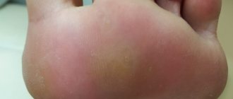
Need to know! To differentiate a cutaneous horn from a plantar wart or callus, it is necessary to cut off part of the thickened skin. If you damage a wart, then it bleeds, but the skin horn does not.
Diagnostic methods and treatment
A fully formed formation will not cause any difficulty for a specialist during diagnosis. As a rule, the disease is easily recognized only by the clinical picture without additional tests and studies. However, if a secondary form of horny keratoma has been diagnosed, it is necessary to find out the reason against which the formation appeared. For this purpose, a special histopathological examination is carried out. Its purpose is to collect cells from the base of the horn and then study it.
This will help identify all current processes: infectious, benign and malignant, inflammatory. If the doctor detects signs of transformation into a malignant tumor, he will note phenomena such as cell polymorphism (rapid development of affected skin cells) and pathological mitoses (excessively rapid cell division).
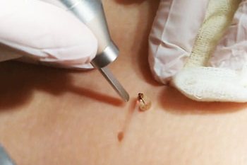
Treatment consists of eliminating the horny keratoma. Removal can be carried out by specialists in cosmetic or dermatological clinics, as well as oncology centers. There are several removal methods:
- excision - the formation is surgically cut out, after which sutures are placed on the skin. The method is practiced when removing a large horn or in cases where there are many keratomas;
- cryodestruction - cauterization of the tumor with liquid nitrogen. Cold stops all metabolic processes inside the skin horn, as a result of which the affected cells quickly die, and the formation disappears on its own. The good thing about this method is that it doesn’t leave any marks on the skin;
- laser therapy - removal of a formation by exposing it to a laser beam. Tissue destruction occurs due to high temperature.
There are no preventive measures to prevent the development of keratoma, but there is an opinion that cutaneous horn will not occur if you do not allow contamination of the injured skin and constantly maintain a high level of vitamin C in the body.
Benign neoplasms
Treatment
The traditional method of treating such a formation is surgical removal of the cutaneous horn. Surgery is used in difficult cases when the tumor has reached a fairly large size. The horny keratoma is removed with a scalpel and the tissue around the lesion is excised, then sutures are applied. As a rule, scars remain on the skin after surgery. The rehabilitation period lasts up to 15 days.
You can also remove the skin horn using cryodestruction - destroying the neoplasm with cold. During this procedure, liquid nitrogen is applied to the affected cells, causing the tumor to die. After exposure to nitrogen, a small scar remains on the skin.
The most modern and safe method of treating keratoma corneum is laser removal. The laser beam completely destroys the tumor, and the operation takes about ten minutes. The procedure is performed under local anesthesia and is completely painless. No rehabilitation is required after laser tumor removal.
In addition, methods for removing horny keratoma such as electrocoagulation and the radio wave method are used. These methods are contraindicated in patients with concomitant cancer diseases.
You cannot cut out a horny keratoma yourself at home. Despite its painlessness, this neoplasm can be the focus of a malignant process. Therefore, the tumor must be removed by a doctor in a medical facility.
Often patients with cutaneous horns resort to treatment with folk remedies. Traditional healers recommend applying products such as compresses made from onion peels, plantain, celandine, aloe and propolis tincture to the affected area of the skin. In addition, use an ointment that contains acids or is plant-based (for example, bay leaf).
Useful materials:
- Itching and odorless discharge Main causesBefore considering the factors that provoke the appearance of discharge that has a sour odor, it is necessary to immediately note...
- Normal temperature in animals Normal temperature in different types of animals Veterinary services Day hospital for animals Veterinary certificates Vaccination…
- Discharge in women What kind of discharge between menstruation is considered normal? Female discharge normally consists of mucus from the cervical canal, dead...
- Discharge like snot Where does discharge come from? The sources of mucous secretion that is released from the vagina are the following structures of the female genital organs:...
Removing the cutaneous horn using a scalpel
Surgical removal of a cutaneous horn is an operation using a conventional medical scalpel. It is the most effective of the currently available procedures, although it also has some disadvantages. Thus, many doctors prefer not to remove skin keratomas with a scalpel on the patient’s face, since such an operation may leave a scar. Despite the fact that the mark is always very small, not every patient, especially women, is satisfied with this result. A longer recovery period for the skin is also important, as is the need to remove sutures. At the same time, for the removal of large keratomas, when other methods are ineffective, this method of eliminating them remains the only acceptable option.
Diagnosis of cutaneous horn
When any type of neoplasm appears, it is necessary to conduct a full histological analysis, thanks to which it will be possible to determine the diagnosis and make a further prognosis of the disease.
This formation should not be confused with different types of papillomas, warts, and calluses. After differential diagnosis with histology, the squamous cell form of skin cancer is excluded or confirmed. Usually, for this purpose, specialists perform a biopsy of the tissue taken for analysis.
The most vulnerable area of these tumors is their base . It is there that many processes are observed that cause pathological tissue proliferation and horn formation. For a more detailed analysis, tissue is taken from the base and it is determined what type of disease it is - malignant or benign.
Types of cutaneous horn
For cutaneous horn, there are 2 main forms:
- primary;
- secondary.
Each type of cutaneous horn is characterized by its own characteristics that determine the method of treating the disease.
The primary form refers to the cutaneous horn as such. Its development on the skin occurs for no apparent reason, there are no inflammatory processes or damage. This form is less prone to malignancy, but it is dangerous due to the fact that the primary form of the cutaneous horn has not been fully studied.
The secondary form of cutaneous horn is the result of the development of the inflammatory process in other neoplasms that are chronic in nature. This form of the disease is very dangerous because it is characterized by pronounced malignancy and may develop into skin cancer.
Signs of cutaneous horn
Signs of cutaneous horn include the growth of horny masses that resemble animal horns. In most cases, these masses have a conical shape, are usually straight, yellowish-brownish or dark in color, and have a dense or compact consistency. The cutaneous horn has a smooth or uneven surface on which multiple grooves are formed. Detection of inflammatory phenomena is possible only in close proximity to the base of the horn, and they manifest themselves as a narrow erythematous rim. Horny neoplasms can reach very large sizes; formations that are short in length are rare. Larger areas grow over the surface, but the size of the top in this case is much narrower than the base.
As a rule, the cutaneous horn is single; multiple neoplasms are very rare. The development of the disease occurs somewhat more often in women, especially those at an older age; the place of their localization is mainly the face (ears, cheeks) and scalp. In rare cases, the cutaneous horn may be located on the mucous and semimucosal surfaces.
Treatment of cutaneous horn
For cutaneous horns, surgical excision is required, for which traditional surgery, cryodestruction or a laser device can be used. The operation itself is carried out either in beauty salons, or in an oncology center, or in venereology clinics. Today, the most effective and safe way to remove skin horns is the laser method, which allows painless and non-contact removal of the formation in a few minutes. After the operation, no rehabilitation period is required; a light mark remains at the site where the treatment was performed, which resolves within 2 weeks.