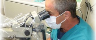
Skin scraping is a diagnostic method that involves obtaining skin samples for testing in a laboratory. The method helps determine the type of disease, etiology (cause), and also helps select and evaluate the success of treatment.
COST OF SOME DERMATOLOGIST SERVICES IN OUR CLINIC IN ST. PETERSBURG
| Price for a dermatologist appointment | 1000 rub. |
| Removing a mole with a radio knife | 500 rub. |
| Analyzes | from 300 rub. |
| Call free: 8-800-707-1560 *The clinic is licensed to provide these services | |
When is a skin scraping test prescribed?
The content of the article
The main function of the skin is to protect human internal organs from various pathologies. The variety of skin diseases is due to the peculiarities of its structure and the large number of factors influencing it. Almost always, skin diseases are the result of the influence of microbiological factors (viruses, fungi, parasites).
An experienced dermatologist can suggest a particular disease after a visual examination. However, in order to be confident in the diagnosis, it is necessary to use other diagnostic methods.
Skin scraping, as a diagnostic method, is prescribed in the following cases:
- If you suspect skin diseases;
- To clarify the diagnosis and select a treatment method;
- To evaluate the therapy performed.
Most often, scraping is prescribed if a fungus or demodicosis (a disease caused by a microscopic mite) is suspected.
Progress
How to take tests for demodex (what type of material and how to take it) is decided by the doctor, depending on the type of manifestations.
The following biomaterial can be taken as a research subject:
- eyelashes, hair from the head or eyebrows along with the follicle;
- epithelium (scraping). Scraping is usually taken from the facial skin in the area of the nose, eyebrows, chin and interlash space;
- contents of the papule (pimple).
As an additional test, a complete blood count is sometimes taken. The goal is to identify additional pathologies: anemia, allergies, parasitic lesions, bacterial infections.
Skin scraping
To detect subcutaneous mites on the face in the presence of acne, ulcers and papules, scraping is done. For the procedure, an eye spoon or scalpel is used, which is passed over the affected surface, collecting epithelial particles.
The process of collecting biomaterial for analysis is painless. The resulting preparation is not highly informative, since there may be no mites on the outside.
An analysis taken from the skin of the face is placed on top of an alkaline glass slide (a few drops of 10% alkali are added), which is then examined under a microscope.
Superficial and skin biopsy
Epithelium can be collected by superficial biopsy. The essence of the procedure:
- glue is dripped onto degreased glass: cyanoacrylate, sulfacrylate or BF-6;
- apply glass to the affected area for 1 minute;
- remove it along with the adhered biomaterial.
Instead of glue, the doctor can use regular tape (test tape), which is applied to the affected area, then removed and pasted onto the glass. An alkaline solution is dripped onto the resulting biomaterial, then examined under a microscope.
Skin biopsy is the removal of part of the epidermis or the contents of the sebaceous glands using a scalpel (excision method) or a puncture needle (punch method). The method is traumatic, but the resulting material is the most informative.
It is kept in 10% neutral formaldehyde for 24 hours and stained with eosin or hematoxylin.
Extrusion
Squeezing is used to extract demodex from the sebaceous gland. Disadvantages of the method: it is traumatic, it is impossible to take analysis from a large area.
When the skin around the sebaceous gland is squeezed, a thread-like white mass (fat) is released from its duct, which is immersed in a special solution for subsequent dissolution. The resulting suspension is then examined under a microscope.
Eyelash analysis
If there are signs of eyelid inflammation, 3-4 eyelashes are carefully pulled out from above and below to test for demodicosis (eyelash test). The doctor holds the area where the hair is taken, preventing damage. The method is traumatic, but it is the only one available for eyelashes, allowing one to study the contents of the follicle.
The resulting material is placed inside a transport medium (glycerin or alkali solution). Directly on the slide, the follicle is squeezed out or kneaded for a more complete view.
The biomaterial can be placed in an Eppendorf tube (Eppendorf tube), a disposable microcentrifuge container of a conical shape that ensures absolute sealing of the contents. The epithelium and eyelashes are placed into it without a transport medium.
What the analysis shows
Skin scraping shows the presence or absence of: fungi and subcutaneous parasites.
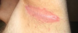
There are many types of fungal infections. It should be remembered that the fungus is contagious, and the carrier can be not only an infected person, but also an animal. Scraping the skin affected by the fungus will help identify the type of fungus and, accordingly, make it possible to choose the appropriate treatment.
Signs of skin damage by fungus:
- The affected areas differ in color, bumpiness, and peeling of the skin. In case of injury, the area may become larger in size and ulcers may appear. Itching bothers me;
- If the hands and feet are affected, severe itching and peeling appear; the skin becomes thinner and becomes covered with erosions. The nail plate may thicken in size and turn yellow;
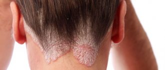
- Fungus on the scalp makes hair dull and unhealthy. Peeling, itching, and plaque appear on the hair. Hair may fall out.
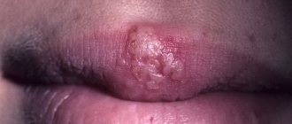
Demodex is a microscopic mite that parasitizes the sebaceous glands of the skin, hair follicles of humans and animals.
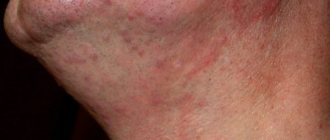
Signs of demodicosis can be confused with allergies and dermatitis. But there are such differences:
- The rash is accompanied by itching, burning, and discomfort. When the eyelids are affected, itching appears, the eyes become inflamed and swollen. Eyelashes become coated and fall out;
- There is an oily sheen on the face. The affected areas of the skin turn red, swell, and peeling and roughening may begin.
- Over time, scars appear. There is a feeling that something is moving under the skin.
It is impossible to see this mite without a microscope. Therefore, the dermatologist scrapes the skin, then evaluates the results and prescribes treatment.
Skin scraping will help identify scabies. Scabies is an infectious skin disease caused by the scabies mite. The tick can be seen with the naked eye (a white or yellow, small dot).
The tick can penetrate human skin very quickly - within 15 minutes. Scabies is highly contagious, with an incubation period of up to 14 days. The main route of infection is through touch.
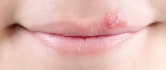
Symptoms of scabies:
- Itching, especially intense in the evening and at night;
- Rashes;
- General malaise, fever in children.
Indications
Scraping for demodex is prescribed when there are characteristic signs of infection with the parasite, but sometimes the disease is asymptomatic.
Factors that provoke active reproduction of mites and progression of dermatosis:
- reduced immunity (due to diabetes, HIV, lupus erythematosus);
- hormonal pathologies;
- gastrointestinal diseases;
- abuse of coffee, alcohol, solarium, bath;
- stress.
Demodex infection is manifested by the following symptoms (they are also indications for analysis):
- redness, bumpiness, peeling of facial skin;
- rosacea, papules, purulent pimples, ulcerations, enlarged pores;
- itching in the ears, on the eyelids;
- gray tint of facial skin (observed in certain areas);
- partial baldness;
- discomfort when blinking, blepharitis (acute or chronic inflammation of the eyelids);
- loss of eyebrows, eyelashes covered with light scales;
- slight increase in the size of the nose (rhinophyma);
- pain during facial movements;
- itching of the facial skin when using cosmetics, as well as in the cold.
How to do scraping
First of all, the doctor conducts a survey, identifies complaints and medical history.
Before taking a scraping, the dermatologist cleans the surface of the skin from dust and sebum. To carry out scraping, a scalpel, a special spatula, and scissors (to take a nail sample) are usually used. If a scraping needs to be taken from the scalp, the doctor will trim the affected area.
The procedure is absolutely painless. The material obtained during the analysis is delivered to the laboratory. Based on the results, the dermatologist selects a course of treatment.
If you find an error, please select a piece of text and press Ctrl+Enter
Preparation for collecting biospecimens for microscopic analysis
No special preparation is required for collecting skin scrapings for fungi. It is advisable not to take water procedures the day before the planned collection of biomaterial, this will increase the chances of identifying pathological microorganisms on the surface of the skin. Also, before the day of collecting biomaterial for analysis, it is recommended to refrain from using gels, ointments and various cosmetics that are applied to the skin; this will simplify microscopic examination, since there will be no foreign microelements in the biosamples being studied.
Where are fungal tests and cultures performed?
If the reader is interested in where to get tested for nail fungus, the information below may be useful. First of all, such studies are carried out in most city or regional budget clinics equipped with a laboratory. The attending physician may also recommend contacting one of the private laboratories. And if in the first case the diagnosis will be free or have a symbolic cost, then in private laboratories you can get tested for a more significant amount. However, it is worth remembering that in a private organization, the quality of the equipment is often much better, which means that the diagnosis can be many times more accurate.
Important: the patient has the right to decide for himself where to undergo such tests. Coercion on the part of the attending physician is unacceptable. It is worth paying attention to the fact that in a private laboratory the patient may be asked to perform a certain set of tests aimed at clarifying the diagnosis.
Examination of the skin and nail plates for superficial mycoses
[10-014] Examination of the skin and nail plates for superficial mycoses 800 rub.
Microscopic examination of scrapings from smooth skin and nail plates, used for the diagnosis of superficial mycoses of the skin and its appendages.
Synonyms Russian
Microscopy of skin scrapings and nail plates for fungi.
English synonyms
- Direct microscopy, Superficial mycoses
- KOH-test
Research method
Microscopy.
What biomaterial can be used for research?
Nails, scraping.
General information about the study
Superficial mycoses of the skin are a group of diseases of the skin and its appendages (nails and hair), the causative agents of which are fungi that are capable of superficial invasion of the epidermis, hair and hair follicles and the nail apparatus. Most often, superficial mycoses are caused by the presence of fungi of the genus Dermatophytes (in this case we speak of dermatophytosis), but the disease can also be caused by fungi of the genus Candida, Malassezia, Trichosporon and Hortaea.
Clinical examination may suggest the presence of smooth skin mycosis or nail mycosis (onychomycosis), but is not used as a definitive diagnostic method, since many other diseases may have a similar clinical picture (for example, nail psoriasis may resemble onychomycosis).
The main method for diagnosing this group of diseases is direct microscopy, in which the material under study (skin flakes obtained by scraping at the lesion, or fragments of the affected nail plate) is examined under a microscope after treatment with a solution of potassium hydroxide (KOH).
Microscopy makes it possible to identify hyphae/pseudohyphae and yeast cells of the fungus and characterize their morphology, on the basis of which the diagnosis of “superficial mycosis with clarification of its nature” can be confirmed. For example, dermatophyte fungi (Trichophyton, Microsporum and Epidermophyton) are represented by multiple tube-like structures with true septa and spore formation.
Fungi of the genus Candida, on the contrary, are represented by elongated yeast cells (pseudohyphae), and do not form true spores. Fungi of the genus Malassezia produce round yeast cells and elongated pseudohyphae.
The pathogens of superficial mycoses are in fact often part of the normal microbiota of the skin or temporarily colonize it (“healthy carriage”) and can be identified by microscopy of scrapings from normal skin or scrapings from the distal part of the nail plates in many healthy people.
For this reason, a positive test result in the absence of any clinical signs of mycosis has no clinical significance.
This statement, however, does not apply to the analysis of the subungual contents of the proximal nail plates, which normally do not contain any microorganisms, including fungi.
It should be noted that microscopy does not allow specifying the type of fungus: for these purposes, inoculation on a nutrient medium is used (microbiological method). A negative test result does not completely exclude superficial mycosis.
What is the research used for?
- For the diagnosis of superficial skin mycoses (dermatophytosis, candidiasis, pityriasis versicolor and others) and onychomycosis.
When is the study scheduled?
- With symptoms of superficial mycosis of the skin (single or multiple foci of peeling and hyperemia with a clear edge, accompanied by itching);
- in the presence of symptoms of onychomycosis: changes in color (yellow, whitish), thickness (subungual hyperkeratosis) and shape of the nail (cracks, onychogryphosis).
What do the results mean?
Reference values: negative.
Positive result:
- dermatophytosis, candidiasis or other superficial mycosis of the skin or nails;
- “healthy carrier”.
Negative result:
Important Notes
- Microscopy does not allow us to determine the specific type of fungus.
- Dermatophyte fungi persist in skin flakes for several months, and Candida fungi for several weeks.
Also recommended
Who orders the study?
Dermatovenerologist, mycologist, general practitioner.
Literature
- Burns T., Breathnach S., Cox N., Griffiths C. Rook's Texbook of Dermatology / T. Burns, S. Breathnach, N. Cox, C. Griffiths; 8th ed. – Wiley-Blackwell, 2010.
- Wolff K., Johnson RA Fitzpatrick's Color Atlas and Synopsis of Clinical Dermatology / K. Wolff, RA Johnson; 6th ed. – McGrawHill, 2009.
Source: https://helix.ru/kb/item/10-014
Fungal infections. Peculiarities.
Fungal diseases (mycoses) of the skin and nails are contagious diseases transmitted from person to person. Transmission of infection can occur through direct contact with an active lesion, when using someone else's shoes, as well as in public places - baths, showers, swimming pools. The penetration of infection is facilitated by damage to the integrity of the skin (cracks, sores, diaper rash, etc.). Detection of fungal structures in samples taken from affected areas of the skin or nails allows us to confirm the presence of a fungal infection. Differentiation of different types of fungi is not made in this study.
The importance of analysis
If the reader thinks that getting tested for fungus is not so important, and that a dermatologist can determine the type of disease by eye, then this opinion is fundamentally wrong. The fact is that if the diagnosis is incorrect and insufficient, the fungus on the nail continues to grow and progress. Over time, not only the plate will deteriorate. Cells of pathogenic flora will begin to negatively affect the entire body. This is why it is so important to accurately determine the type of mushroom. In addition, the importance of fungal testing is determined by the following additional factors:
- The risk of infection of relatives and people from the immediate environment with the fungus. Therefore, the patient should undergo a hemotest as soon as possible to determine the type of fungus;
- The risk of incorrect self-treatment without consulting a doctor. And this can lead, at a minimum, to a lack of treatment results, or, at maximum, to a significant deterioration of the situation.
At-risk groups
The most common types of fungal diseases include mycosis of the skin of the feet and onychomycosis of the nail plates. From the moment of infection to the appearance of the first clinical symptoms, it takes from several weeks to several months.
Men suffer from fungal diseases more often than others. Among women, those who constantly wear pointed-toed shoes, especially high heels, are more likely to develop the disease. In this case, the toes are constantly flattened, which leads to friction, small wounds, and abrasions, which are entry points for infection.
You can “catch” the fungus in a regular beauty salon during a pedicure procedure if the master used improperly processed tools. To remove spores and fungal fragments from the metal surfaces of cutters, scissors and tweezers, the instruments must be sterilized in a dry-heat oven. Not all salons have such equipment, so they limit themselves to “soaking” in a disinfectant solution and “drying” in ultraviolet boxes. This treatment does not fully protect against infection.
Frequent addition of a fungal infection may signal the development of diabetes. According to statistics, diabetics are three times more susceptible to mycosis. The fungus can also appear due to allergic skin lesions (itching, inflammation, weeping), scratching insect bites; while taking antibacterial drugs, corticosteroid hormones, antidepressants.